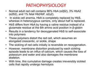
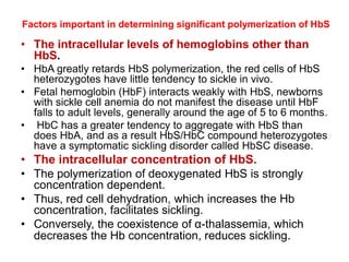
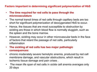
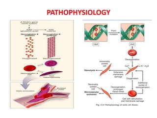
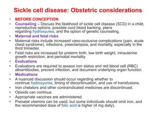
SICKLE CELL DISEASE
Dr G.VENKATA RAMANA
HOD FAMILY MEDICINE
RDT HOSPITAL BATHALAPALLI
HAEMOGLOBINOPATHIESoglobino
pathiesoglobino opathiesthies
• Caused by mutations affecting the genes encoding the
globin chains of haemoglobin
• Adult haemoglobin (HbA-αα/ββ)
• Fetal haemoglobin (HbF-αα/γγ)
• Haemoglobin A2 (HbA2-αα/δδ)
• Alpha globin chains are produced throughout life,
including in the fetus, so severe mutations in these may
cause intrauterine death.
• Disorders affecting the beta chains do not present until
after 6 months of age
• Qualitative abnormalities
• abnormal haemoglobins e.g. S, C, D or E
• Quantitative abnormalities
• Alpha-thalassemia
• Beta-thalassemia
• Mutations causing a reduced rate of
production of one or other of the globin
chains altering the ratio of alpha to non-
alpha chains
Hemoglobin patterns in common hemoglobinopathies
Older children (≥5 years), adolescents, and adults
Condition Hb A(%) HbA2(%) Hb F(%) HbS(%) HbC(%)
Normal 95 to98 2 to 3 <2 0 0
Beta thalassemia
trait
90 to 95 >3.5 1 to 3 0 0
Sickle cell trait 50 to 60 <3.5 <2 35 to 45 0
Homozygous sickle
cell (Hb SS) disease
0 <3.5 5 to 15 85 to 95 0
Sickle-
β0 thalassemia
0 >3.5 2 to 15 80 to 92 0
Sickle-
β+ thalassemia
3 to 30 >3.5 2 to 10 65 to 90 0
Hb SC disease 0 <3.5 1 to 5 45 to 50 45 to 50
Hb CC disease 0 <3.5 <2 0 95
Hb C traitΔ 50 to 60 <3.5 <2 0 40 to 50
PATHOPHYSIOLOGY
• Normal adult red cell contains 96% HbA (α2β2), 3% HbA2
(α2δ2), and 1% fetal Hb(HbF, α2γ2).
• In sickle cell anemia, HbA is completely replaced by HbS,
whereas in heterozygous carriers, only about half is replaced.
• HbS differs from HbA by having a valine residue instead of a
glutamate residue at the 6th amino acid position in β-globin
• Results in a tendency for deoxygenated HbS to self-associate
into polymers
• These polymers distort the red cell, which assumes an
elongated crescentic, or sickle, shape
• The sickling of red cells initially is reversible on reoxygenation.
• However, membrane distortion produced by each sickling
episode leads to an influx of calcium, which causes the loss of
potassium and water and also damages the membrane
skeleton.
• With time, this cumulative damage creates irreversibly sickled
cells that rapidly undergo hemolysis
Factors important in determining significant polymerization of HbS
• The intracellular levels of hemoglobins other than
HbS.
• HbA greatly retards HbS polymerization, the red cells of HbS
heterozygotes have little tendency to sickle in vivo.
• Fetal hemoglobin (HbF) interacts weakly with HbS, newborns
with sickle cell anemia do not manifest the disease until HbF
falls to adult levels, generally around the age of 5 to 6 months.
• HbC has a greater tendency to aggregate with HbS than
does HbA, and as a result HbS/HbC compound heterozygotes
have a symptomatic sickling disorder called HbSC disease.
• The intracellular concentration of HbS.
• The polymerization of deoxygenated HbS is strongly
concentration dependent.
• Thus, red cell dehydration, which increases the Hb
concentration, facilitates sickling.
• Conversely, the coexistence of α-thalassemia, which
decreases the Hb concentration, reduces sickling.
Factors important in determining significant polymerization of HbS
• The time required for red cells to pass through the
microvasculature
• The normal transit times of red cells through capillary beds are too
short for significant polymerization of deoxygenated HbS to occur.
• Hence, the tissues that are most susceptible to obstruction by
sickling are those in which blood flow is normally sluggish, such as
the spleen and the bone marrow.
• However, sickling may occur in other microvascular beds in the face
of factors that retard the passage of red cells, particularly
inflammation
• The sickling of red cells has two major pathologic
consequences:
• chronic moderately severe hemolytic anemia, produced by red cell
membrane damage, and vascular obstructions, which result in
ischemic tissue damage and pain crises .
• The mean life span of red cells in sickle cell anemia averages only
20 days
PATHOPHYSIOLOGY
GENETICS-AUTOSOMAL RECESSIVE INHERITANCE
SICKLE CELL TRAIT
• One abnormal allele of the hemoglobin beta gene
• HbA 60%,HbS 40%,HbF <2%
• Asymptomatic
• They act as carriers and transmit the disease to
their off springs
• Heavy exercise under extreme conditions may
provoke gross hematuria and complications
• Exercise-related rhabdomyolysis can result in
renal failure and death
• Exercise-associated sudden death-occurred
predominantly in foot ball players
SICKLE CELL DISEASE
• Acute manifestations
• Infection
• Anemia
• Splenic sequestration
• Acute vaso-occlusive pain
• Stroke
• Acute chest syndrome
• Kidney infarction or medication toxicity
• Dactylitis or bone infarction
• Myocardial infarction
• Complications related to pregnancy
• Priapism
• Venous thromboembolism
• Chronic manifestations :
• Pain
• Anemia, with transfusional iron overload
• Neurologic deficits or seizure disorder
• Pulmonary conditions including pulmonary
hypertension
• Impaired kidney function and hypertension
• Osteoporosis and complications of bone infarction
• Cardiomyopathy with diastolic dysfunction and
heart failure
• Liver injury and pigmented gallstones
• Delayed puberty and reduced growth
• Chronic leg ulcers
• Proliferative retinopathy
• Psychosocial stress
CLINICAL PRESENTATION
• Sickle cell hemoglobin C:
• Painless hematuria
• Aseptic necrosis of bone-less common
• Vaso-occlusive crises less common,occur late in life
• Pregnancy related problems:mild anemia(10-12g/dl)
• Sickle-β+ thalassemia
• Rare crises,milder severity than sickle cell disease because of
production of HbA
• Hb 10-14g/dl with microcytosis
• Sickle-β0 thalassemia or α thalassemia
• No Hb A production
• Severity similar to sickle cell anemia Hb 7-10g/dl with
microcytosis
DIAGNOSIS
• For children and adults
• HPLC-high performance liquid chromatography
• Hb electrophoresis
• ISF-Isoelectric focusing
• Sickledex or solubility testing is not adequate for
diagnosis.
• Peripheral blood smear - sickled red cells ,
polychromasia indicative of reticulocytosis, and
Howell-Jolly bodies reflecting hyposplenia
• Neonatal testing - obtain blood samples by heel stick
or cord blood and spot the sample onto filter paper for
stable transport and subsequent electrophoresis, thin-
layer isoelectric focusing, or HPLC
• Prenatal testing - obtaining fetal DNA samples by
chorionic villus sampling at 8 to 10 weeks gestation
HOWELL-JOLLY BODIES
PS IN SICKLE CELL ANEMIA
TREATMENT
• Prophylactic penicillin — Beginning by two
months of age and continuing until at least age
five years, all patients with Hb SS and Hb S-
β0 thalassemia should receive
prophylactic penicillin V potassium.
• Dosage:
• Age 2 months to <3 years – 125 mg orally twice
daily
• Age ≥3 years – 250 mg orally twice a day
• Erythromycin for penicillin-allergic patients.
• Folic acid — Folic acid (400 micrograms to 1 mg
daily) is often given to children with Hb SS and Hb
S-β0 thalassemia (and other hemolytic anemias)
• Hydroxyurea and other disease-modifying therapies
• It decreases the frequency and severity of vaso-occlusive
complications (eg, dactylitis, painful episodes, acute chest
syndrome [ACS]) and it may reduce the risk of organ damage
in individuals with Hb SS and Hb S-β0 thalassemia.
•
• Options for patients who cannot take hydroxyurea or who
have continued symptoms despite optimally dosed
hydroxyurea include L-glutamine, crizanlizumab,
and voxelotor.
• Education — The child's parents or caregivers should be
educated about acute and chronic complications of SCD
• Routine immunizations plus influenza, pneumococcal and
meningococcal vaccinations
HYDROXYUREA
• MECHANISM OF ACTION Inhibition of ribonucleotide reductase
• Increased Hb F production,reduce adhesion
• Increase in red cell size, lowers the intracellular Hb concentration
• Increase NO levels- vasodilator and inhibitor of platelet aggregation.
• Anti-inflammatory effect - inhibition of white cell production
• Baseline testing
• CBC with differential, platelet count, reticulocyte count, Hb F
percentage, tests of kidney and liver function, and pregnancy test for
females of childbearing potential.
• Dosing
• Infants and children 20mg/kg/day,Adults15-20mg/kg/day
• For creatinine clearance <60 mL/minute, reduce the dose by 1/2.
• The dose is titrated by 5 mg/kg/day every eight weeks to maximum
tolerated dose (MTD) using hematologic parameters
• Treatment is continued indefinitely if effective.
• Adverse effects
• Relatively nontoxic.
• Myelosuppression is the predictable, dose-limiting toxicity.
• Skin, hair, and nail changes and gastrointestinal upset may occur.
• Hematopoietic stem cell transplantation
• The only therapy that is curative
• Best candidates are :
• younger than 16 years of age
• With severe complications
• Have HLA-matched donors
• Risks-mortality,Graft rejection,and secondary malignancies
• Gene therapy is under investigation
• MANAGEMENT DURING HOSPITALIZATION
• Hydration oral fluids normal saline
• Incentive spirometry To reduce the risk of acute
chest syndrome.
• Thromboembolism prophylaxis For all adults (ie,
those >18 years)
Red blood cell transfusion in sickle cell disease
• ACUTE THERAPEUTIC
• Treatment of hemodynamic compromise, ACS, acute
cerebral infarct, transient ischemic attack, multiple
organ failure, or acute single organ failure, Acute
symptomatic anemia, A drop in baseline reticulocyte
count (relative reticulocytopenia) with symptoms of
acute hemodynamic compromise,Hepatic or splenic
sequestration
• PROPHYLACTIC (REGULARLY SCHEDULED)
TRANSFUSION
• Indications regular transfusions are used in the secondary
prevention of stroke, acute chest syndrome (ACS), painful
events, priapism, and pulmonary hypertension
• Preoperative –For those with Hb SS or Hb S-
beta0 thalassemia undergoing elective surgery, suggest
simple transfusion to increase the Hb to 10 g/dL rather than
exchange transfusion
Risk in subsequent pregnancies
• If both parents are heterozygous (with sickle cell trait or
other beta globin variant), the risk of having another
affected child is 25 percent. If either parent is
homozygous for a beta globin variant, the risk is 50
percent and, if both are homozygous, all children will be
affected
ROUTINE EVALUATIONS AND TREATMENTS
• screen for the following:
• Hypertension Blood pressure screening should be done at every
visit.
• TCD screening for stroke risk In children ≤16 years of age with
hemoglobin SS or hemoglobin S-beta thalassemia started at two
years of age and performed annually
• Screening for neurocognitive dysfunction
• Retinopathy Retinal evaluation is begun at 10 years of age and
continued routinely to detect early proliferative sickle retinopathy
• Bone health Vitamin D screening annually and bone density testing
every one to three years screening physical exam for avascular
necrosis
• Kidney disease start at age three to five
• Adults are monitored at regular visits, typically four to six times per
year (creatinine and a urine for proteinuria and/or albuminuria)
• Asthma/obstructive lung disease spirometry in asymptomatic
children in intervals of one to two years,
• Growth measure height and weight in children and adolescents,
and weight in adults
VASO-OCCLUSIVE PAIN
• Sickled red blood cells (RBCs) have a marked
reduction in deformability as well as other effects
including increased adhesion to vascular
endothelial cells, inflammation, and activation of
hemostatic mechanisms; all of these changes
synergize to cause vascular obstruction and vaso-
occlusion.
• Many patients have specific triggers for pain such
as cold, wind, low humidity, dehydration, stress,
alcohol, and menses, which they develop
strategies to minimize or avoid
• The sites of pain can include the back, chest,
extremities, and abdomen.
• In young children, dactylitis (acute pain in the
hands or feet) may be the most common site of
pain.
Wong baker FACES pain rating scale
Common sites of vaso-occlusive pain
• Most individuals will require opioids
• Initial dose is based on intensity of pain and previous effective doses
• If initial dose is unknown, use one of the following:
• Intravenous morphine (0.1 to 0.15 mg/kg; maximum single dose 10 mg)
• Intravenous hydromorphone (0.02 to 0.05 mg/kg; maximum single dose
1.5 mg)
• Intranasal fentanyl (1.5 mcg/kg, give up to two doses, 5 to 10 minutes
apart; maximum single dose 100 mcg)*
• Rapidly reassess for efficacy and repeat dose if needed at
appropriate interval (20 minutes for morphine, 30 minutes for
hydromorphone)
• Provide adjunctive therapies
• Ensure adequate hydration (encourage oral fluids, give intravenous
fluids if hypovolemic)
• Enlist family and other psychosocial supports
• Use heat packs if helpful
• Possible use of ketamine
• Treat insomnia and other conditions that interfere with adequate
sleep
• Therapies to avoid or use sparingly
• Never use placebo
• Avoid ice and cold compresses (may precipitate sickling)
• Avoid meperidine
• Avoid ketorolac in adults; if ketorolac is given, no more than one
dose should be used
• Use anxiolytics only if indicated for anxiety, not for pain
• Use NSAIDs cautiously as they may cause renal failure or bleeding
and may be ineffective in many cases
• Use oxygen only for hypoxia, not for routine pain
• Use transfusion only for indicated complications, not for
uncomplicated vaso-occlusive pain
• OPIOID SIDE EFFECTS
• Sedation
• Respiratory depression
• Gastrointestinal Nausea and constipation are common
• Pruritus Due to histamine release from skin mast cells
• Opioid use disorder
• Management of opioid side effects
• Use stimulant laxatives for constipation; osmotic agents are
not as effective in opioid-induced constipation
• Use nonsedating (H1-antagonist) antihistamines for pruritus if
needed
• Use antiemetics for nausea if needed; a selective 5-
HT3 receptor antagonist such as ondansetron should be the
first choice because of minimal CNS findings
• Treat insomnia with nonpharmacologic or pharmacologic
approaches as needed
ACUTE CHEST SYNDROME
• Pulmonary arterial circulation has low oxygen tension and low flow, both of which
facilitate sickling
• Syndrome of fever, chestpain, hypoxemia, wheezing, cough or respiratory distress in
the setting of a new pulmonary infiltrate
• Causes
• Infection,vaso occlusion, hypoventilation, atelectasis, thrombosis or
thromboembolism, and in some cases fat embolism
• Management-
• Analgesia
• Oxygen
• Incentive spirometry
• Bronchodilators
• Antibiotics (cephalopsorins and macrolides)
• Transfusion
• Preventive approach-
• Prophylactic antibiotics and immunizations during early child hood
• Hydroxyurea
• Blood transfusions for those who continue to have ACS episodes despite hydroxyurea
therapy
INFECTION
• Major cause of morbidity and mortality
• Mechanisms-Functional hyposplenism or asplenism, Impairment of
splenic function can occur in infants as young as 3months
• Altered humoral and cellular immunity
• Reduced tissue perfusion
• Presence of an indwelling catheter
• Splinting and hypoventilation
• Common sites-Bacteremia,Meningitis and Pulmonary infections
• common organisms-Encapsulated organisms (streptococcus
pneumoniae,Hemophilus influenzae)
• E.Coli, Staph aureus, Salmonella species
• Recommended antibiotic – Ceftriaxone
• Vancomycin- Reserved for children or adults with suspected meningitis
• Resiratory coverage-Azithromycin is added to ceftriaxone for acute
chest syndrome and/or pneumonia
• Oseltamivir –During influenza season
• Known or suspected cephalosporin allergy-Levofloxacin,Meropenem
NEUROLOGIC COMPLICATIONS
STROKE AND TIA
• Ischemic stroke is more common than Hemorrhagic
stroke in children and adolescents
• Hemorrhagic stroke is more common in adults
• Treatment-Exchange transfusion to reduce HbS to less
than 30% of total Hb
• Primary prevention-Regular transcranial doppler
measurements for risk stratification and selective
screening with magnetic resonance angiography
• Extra cranial internal carotid artery stenosis is common
• At risk children are treated with chronic prophylactic
transfusion
• Individuals who have had a stroke are treated with
chronic prophylactic transfusions to prevent recurrent
stroke
NEUROLOGIC COMPLICATIONS
SEIZURES AND PRES
• Seizures and epilepsy are 2-3 times more
common
• Male sex and dactylitis in childhood were
associated with an increased risk of epilepsy
• Posterior reversible encephalopathy
• Syndrome of confusion, headache, visual
symptoms and seizures
• PRES is less common than stroke in individuals
with SCD
MULTIORGAN FAILURE
• Acute multiorgan failure is a life-threatening
complication of SCD in which multiple organ
systems are affected by ischemia and/or infarction.
• It is typically seen in the setting of an acute painful
episode
• Complement and other factors have been
implicated.
• Some patients may present with a thrombotic
microangiopathy (TMA, such as thrombotic
thrombocytopenic purpura [TTP] or complement-
mediated hemolytic uremic syndrome [CM-HUS]
picture), which has led to anecdotal use of
plasmapheresis and complement therapy
• Management of multiorgan failure prompt and
aggressive exchange transfusion therapy
APLASTIC CRISIS
• An acute drop in hemoglobin level caused by a
transient arrest of erythropoiesis, leading to abrupt
reductions in red cell precursors in the bone marrow
and a markedly reduced number of reticulocytes in the
peripheral blood (typically, reticulocytes <1.0 percent,
absolute reticulocyte count <10,000 per microL).
• Cause -Infection
• Most cases in children follow infection with human
parvovirus B19, which specifically invades proliferating
erythroid progenitors.
• Other reported causes of transient aplasia are
infections by Streptococcus pneumoniae, Salmonella,
other streptococci, and Epstein-Barr virus.
• Aplastic crisis can result in a rapid and life-threatening
drop in hemoglobin level caused by chronic hemolysis
without the ability of the bone marrow to compensate.
• Management is with transfusion
The peripheral smear from this patient shows numerous sickled RBC's and
the absence of polychromasia. A high power view of the biopsy illustrates
the sinus plugging with sickled RBCs. A single nucleated erythroid precursor
is present (arrow).
SPLENIC SEQUESTRATION CRISIS
• Acute drop in hemoglobin level, typically two g/dL below
baseline.
• This occurs when RBCs are captured and pool within the
spleen.
• A large percentage of the total blood volume can
become sequestered in the spleen, leading to
hypovolemic shock and death.
• Present with a rapidly enlarging spleen and a marked
decrease in hemoglobin level despite persistent
reticulocytosis
• Treatment -Transfusions
• Splenectomy in certain patients to prevent recurrences
SKELETAL COMPLICATIONS
• Dactylitis
• Dactylitis is vaso-occlusive pain in the small bones of the
hands and feet that typically occurs in infants and children
with SCD up to approximately four years of age.
• Pain may be severe
• Repeated episodes of dactylitis will lead to a mottled
appearance of the small bones.
• Older children and adults may experience vaso-occlusive pain
episodes affecting the bones and joints as well
• Management
• Hydration,
• Analgesics and warm packs.
• Hydroxyurea therapy
• Hematopoietic stem cell
transplantation may be indicated.
OSTEOPOROSIS
• Chronic hemolytic anemia in SCD leads to a compensatory increase
in erythropoietic activity.
• The extension of hematopoietic bone marrow can lead to a number
of skeletal changes including chronic tower skull, bossing of the
forehead, and fish-mouth deformity of vertebrae.
• These effects in turn cause widening of the medullary space,
thinning of the trabeculae and cortices, and osteoporosis.
• As a result, individuals with SCD have a high rate of vitamin D
deficiency and osteoporosis .
• Orbital compression syndrome may occur in the setting of vaso-
occlusion in the periorbital bone marrow space, with subperiosteal
hemorrhage.
• Symptoms - headache, fever, and palpebral edema.
• Compression of the optic nerve may also occur and may require
surgical decompression
• Assess bone health including calcium and vitamin D intake,vitamin
D status at every visit, and measure bone density at 12 years of age.
• vitamin D screening annually and bone density testing every one to
three years
A, At 1 year old, expansion of diploe begins in the
posterior frontal area and is definitely identifiable.
B, At age 2 expansion is prominent. At ages 23 (C)
and 30 (D), false hair-on-end” sign appears.
Fish mouth vertebrae
Osteoprorosis of both knees
AVASCULAR NECROSIS
• Results from infarction of bone trabeculae.
• The femoral and humeral heads may be
affected.
• The femoral heads more commonly
undergo progressive joint destruction as a
result of chronic weight bearing.
• Avascular necrosis may be an underlying
cause of chronic pain.
• The changes are best detected by MRI
• Bone marrow infarction involving death of
hematopoietic cells can also occur, leading
to reduced RBC production and anemia
with reduced reticulocyte response.
• Some patients may have a
leukoerythroblastic blood picture or
pancytopenia.
• Bone marrow infarction may be associated
with life-threatening pulmonary fat
embolism
B/L Osteonecrosis of the hips
OSTEOMYELITIS
• Long bones are usually affected, often at
multiple sites, resulting from infection of
infarcted bone.
• The most common organisms
are Salmonella species.
• Staphylococcus aureus, the most common
organism in patients without SCD, accounts
for less than a quarter of cases.
• Articular infection is less common and is often
due to Streptococcus pneumoniae.
• It may be difficult to distinguish osteomyelitis
from vaso-occlusive events involving bone.
Bone imaging studies may be helpful, and
cultures are essential.
• Broad spectrum antibiotic -Ceftriaxone
Salmonella osteomyelitis
PRIAPISM
• Persistent painful penile erection not related to sexual interest or
desire
• Caused by sickling of the red cells producing venous stasis in the
erectile tissue of the penis
• The resulting stasis causes ischemia,hypoxia and pain
• Treatment
• Hydration,analgesics
• For priapism lasting >4hrs that doesnot respond to conservative
management,aspiration of blood from the corpus cavernosum with
or without saline irrigation,followed by injection of an alpha-
adrenergic agonist.
• if no response -Surgical shunt
• Prevention-Hydroxyurea
• Other options-Sildenafil,pseudoephedrine or hormonal therapy
• Penile prosthesis –repeated episodes of priapism with scar tissue
• Venous thromboembolism
• SCD is considered a hypercoagulable state, and patients are
considered to be at increased risk of venous thrombosis and
pulmonary embolism (PE), especially adults in the setting of an
indwelling catheter, immobility, infection, surgery, or pregnancy.
• Use thromboprophylaxis in hospitalized adults with SCD, and have a
low threshold for evaluating adults and children for
thromboembolism if they develop symptoms
• Growth and development
• Impaired growth and delayed puberty are common in children with
SCD.
• Most have detectable growth reduction that affects weight more than
height by the age of two years .
• Normal height is often achieved by adulthood but weight remains
lower than that of individuals without SCD.
• The pathogenesis is uncertain and may include primary
hypogonadism, hypopituitarism, and hypothalamic insufficiency.
• Children may have delayed sexual maturation and delayed
menarche
RETINOPATHY
• From retinal artery
occlusion and ischemia,
with associated proliferative
retinopathy, vitreal
hemorrhage, and retinal
detachment proliferative
retinopathy is more
common in hemoglobin SC
disease
• Evaluate children with an
ophthalmologic examination
(eg, dilated examination),
typically starting around age
10 years and continuing
annually through adulthood
• Management Laser
photocoagulation
LEG ULCERS
• Vaso-occlusion in the skin can produce
leg ulcers and myofascial syndromes
• May develop spontaneously or after trauma
• Typical sites medial and lateral malleolus
• Leg ulcers in SCD may become superinfected.
• Staphylococcus aureus, Pseudomonas species, Streptococci,
or Bacteroides species may be cultured.
• Rarely, they may lead to systemic infection, osteomyelitis, or tetanus
.
• The lesions can be slow to heal and often recur
• Treatment
• Antibiotics and other local and systemic therapies
• Preventive strategies well-fitting shoes and early, aggressive
treatment if signs of skin injury appear.
HEPATOBILIARY COMPLICATIONS
• Hepatic dysfunction due to Acute ischemia
• Cholestasis
• Hepatic sequestration crisis
• Transfusional iron overload
• Drug toxicity from iron chelators or other medications
• Hepatitis C virus (HCV) infection
• Autoimmune liver disease
• Fibrosis
• HCV infection pigment gall stones
• Since most adults with SCD receive repeated intermittent transfusions, annual HCV
screening is warranted, along with monitoring iron overload
• Pigment gallstones
• Gallstones can present with cholecystitis, pancreatitis, biliary colic, or acute
abdominal pain, which can be incorrectly attributed to vaso-occlusion.
• Ultrasonography - an effective screening test for gallstones
• Endoscopic retrograde cholangiopancreatography (ERCP) and/or magnetic
resonance cholangiopancreatography (MRCP) are required to identify common duct
stones.
• The French National Authority for Health recommends elective laparoscopic
cholecystectomy for children with SCD who have asymptomatic cholelithiasis
CARDIAC COMPLICATIONS
• Cardiomyopathy and heart failure
• Cardiomyopathy is increasingly being identified in individuals with SCD,
especially left-sided diastolic dysfunction, both with and without concomitant
pulmonary hypertension
• Potential contributing factors may include:
• Pulmonary hypertension
• Chronic anemia and hypoxemia with increased cardiac output, increased
left ventricular stroke volume, and dilation of the left ventricle
• Transfusional iron overload, especially in older individuals
• Hypertension
• Alterations in intravascular volume associated with vasculopathy and
chronic kidney disease
• Myocardial infarction, dysrhythmia, and sudden death
• Acute myocardial infarction in the absence of epicardial coronary artery
disease
• Conduction abnormalities such as QT prolongation, ventricular arrhythmias,
first-degree AV block, and nonspecific ST-T wave changes
• Sudden death is often multifactorial due to a combination of
cardiopulmonary dysfunction, pulmonary fat embolism, sudden pulmonary
hypertension, unexpected acute sequestration crisis, and/or intracranial
hemorrhage
KIDNEY COMPLICATIONS
• In young patients, hyperfiltration results in the GFR underestimating the
magnitude of kidney dysfunction
• Urinary concentrating defect with hyposthenuria; this may cause enuresis
and may lead to overestimation of the glomerular filtration rate
• Abnormalities in urinary acidification and potassium excretion associated
with distal renal tubular acidosis
• Acute kidney injury (AKI, rapid increase in serum creatinine; associated with
hospitalization)
• Painless hematuria due to papillary infarcts
• Proteinuria from albuminuria, which is often a precursor of progressive
kidney disease
• Contrast-induced nephropathy associated with intravenous iodinated
contrast material used for imaging studies, although this may be less of a
concern with second-generation low- and iso-osmolar contrast agents
• Medication toxicities, especially if glomerular filtration rate is overestimated
• Hypertension
KIDNEY COMPLICATIONS
• Kidney infarction, papillary necrosis, and renal colic
• Arginine vasopressin resistance (previously called nephrogenic diabetes
insipidus) with polyuria
• Focal segmental glomerulosclerosis that can lead to end-stage kidney
disease
• Renal medullary carcinoma (found almost exclusively in Black patients with
Hb SC disease or sickle cell trait)
• Avoid unnecessary exposure to nephrotoxic medications.
• Treat hypertension with agents other than diuretics (which may
cause volume depletion and precipitate vaso-occlusion), and
maintain adequate hydration during acute hospitalizations or
imaging studies that require contrast agents
• Screening - creatinine and a urine for protein and albumin, typically
done by age three to five years and no later than 10 years of age.
• Adults are monitored at regular visits, typically four to six times per
year.
• Individuals receiving an iron chelator or other nephrotoxic
medication or those with known chronic kidney disease may require
more frequent monitoring
• Hemodialysis and/or kidney transplantation are both reasonable
options for individuals with SCD who develop kidney failure,
although these patients are at increased mortality risk.
Sickle cell disease: Obstetric considerations
• BEFORE CONCEPTION
• Counseling – Discuss the likelihood of sickle cell disease (SCD) in a child,
reproductive options, possible cord blood banking, plans
regarding hydroxyurea, and the option of genetic counseling.
• Maternal and fetal risks
• Maternal risks include increased vaso-occlusive complications (pain, acute
chest syndrome), infections, preeclampsia, and mortality, especially in the
third trimester.
• Fetal risks are increased for preterm birth, low birth weight, intrauterine
growth restriction, and perinatal mortality.
• Evaluations
• Evaluations are required to assess iron status and red blood cell (RBC)
alloantibodies, prevent infection, and document underlying organ function
• Medications
• A nuanced discussion should occur regarding whether to
continue hydroxyurea, timing of discontinuation, and use of transfusions.
• Iron chelators and other contraindicated medicines are discontinued.
• Opioids can continue.
• Appropriate vaccines are administered.
• Prenatal vitamins can be used, but some individuals should omit iron, and
the recommended dose of folic acid is higher (4 mg daily).
• During pregnancy
• •Assessments – Review baseline health assessments and
counseling.
• Second- and third-trimester assessments include blood
pressure; urine protein; fetal screenings (including hemolytic
disease of the fetus and newborn (HDFN) in individuals with
alloantibodies or a history of alloantibodies); and, in addition
to routine prenatal testing, testing for asymptomatic
bacteriuria .
• Increase ultrasound screening for fetal growth restriction and
fetal assessment in the third trimester.
• Complications – Vaso-occlusive pain prevention involves
disease-modifying therapies (hydroxyurea or transfusions)
and avoiding dehydration, hypoxia, acidosis, infection, and
cold.
• Pain is treated promptly and adequately. Nonsteroidal
antiinflammatory drugs (NSAIDS) are avoided.
• Transfusions may be used to treat or prevent complications.
• Labor and delivery
• •Delivery – There are no medical contraindications to vaginal
delivery; cesarean birth is reserved for obstetric indications.
• Induction of labor at 37 to 39 weeks is reasonable (even
without maternal or fetal complications), with details
individualized to genotype and comorbidities.
• If time allows, simple transfusion prior to cesarean birth is
reasonable in patients at increased risk of complications.
• Continuous fetal heart rate monitoring is appropriate.
• The patient should be kept warm, hydrated, and well
oxygenated.
• Umbilical cord blood can be used for newborn screening and
harvested for hematopoietic stem cell transplant if desired.
• Venous thromboembolism (VTE) prophylaxis
• After cesarean or vaginal delivery, prophylactic dose low
molecular weight (LMW) heparin rather than no VTE
prophylaxis
• Postpartum
• •Breastfeeding and medications – Breastfeeding should be
encouraged and is possible in individuals taking hydroxyurea.
• If hydroxyurea was restarted, it may take up to six months for full
therapeutic effect. Iron status should be evaluated before restarting
chelation.
• Contraception – Progestin-only contraceptives or nonhormonal
methods are preferred.
• Estrogen-containing contraceptives and depot medroxyprogesterone
acetate (DMPA) increase the risk of VTE and generally are not the
best choice.
• Levonorgestrel intrauterine device (IUD) decreases menstrual
bleeding and is an excellent contraceptive
• Copper IUD increases bleeding, making it less desirable in
individuals with SCD.
• Infant – Hemoglobinopathy testing
• If the mother was receiving chronic opioid therapy, the neonate
should be monitored for opioid withdrawal.

SICKLE CELL DISEASE.pptx

  • 1.
    SICKLE CELL DISEASE DrG.VENKATA RAMANA HOD FAMILY MEDICINE RDT HOSPITAL BATHALAPALLI
  • 2.
    HAEMOGLOBINOPATHIESoglobino pathiesoglobino opathiesthies • Causedby mutations affecting the genes encoding the globin chains of haemoglobin • Adult haemoglobin (HbA-αα/ββ) • Fetal haemoglobin (HbF-αα/γγ) • Haemoglobin A2 (HbA2-αα/δδ) • Alpha globin chains are produced throughout life, including in the fetus, so severe mutations in these may cause intrauterine death. • Disorders affecting the beta chains do not present until after 6 months of age
  • 3.
    • Qualitative abnormalities •abnormal haemoglobins e.g. S, C, D or E • Quantitative abnormalities • Alpha-thalassemia • Beta-thalassemia • Mutations causing a reduced rate of production of one or other of the globin chains altering the ratio of alpha to non- alpha chains
  • 4.
    Hemoglobin patterns incommon hemoglobinopathies Older children (≥5 years), adolescents, and adults Condition Hb A(%) HbA2(%) Hb F(%) HbS(%) HbC(%) Normal 95 to98 2 to 3 <2 0 0 Beta thalassemia trait 90 to 95 >3.5 1 to 3 0 0 Sickle cell trait 50 to 60 <3.5 <2 35 to 45 0 Homozygous sickle cell (Hb SS) disease 0 <3.5 5 to 15 85 to 95 0 Sickle- β0 thalassemia 0 >3.5 2 to 15 80 to 92 0 Sickle- β+ thalassemia 3 to 30 >3.5 2 to 10 65 to 90 0 Hb SC disease 0 <3.5 1 to 5 45 to 50 45 to 50 Hb CC disease 0 <3.5 <2 0 95 Hb C traitΔ 50 to 60 <3.5 <2 0 40 to 50
  • 5.
    PATHOPHYSIOLOGY • Normal adultred cell contains 96% HbA (α2β2), 3% HbA2 (α2δ2), and 1% fetal Hb(HbF, α2γ2). • In sickle cell anemia, HbA is completely replaced by HbS, whereas in heterozygous carriers, only about half is replaced. • HbS differs from HbA by having a valine residue instead of a glutamate residue at the 6th amino acid position in β-globin • Results in a tendency for deoxygenated HbS to self-associate into polymers • These polymers distort the red cell, which assumes an elongated crescentic, or sickle, shape • The sickling of red cells initially is reversible on reoxygenation. • However, membrane distortion produced by each sickling episode leads to an influx of calcium, which causes the loss of potassium and water and also damages the membrane skeleton. • With time, this cumulative damage creates irreversibly sickled cells that rapidly undergo hemolysis
  • 6.
    Factors important indetermining significant polymerization of HbS • The intracellular levels of hemoglobins other than HbS. • HbA greatly retards HbS polymerization, the red cells of HbS heterozygotes have little tendency to sickle in vivo. • Fetal hemoglobin (HbF) interacts weakly with HbS, newborns with sickle cell anemia do not manifest the disease until HbF falls to adult levels, generally around the age of 5 to 6 months. • HbC has a greater tendency to aggregate with HbS than does HbA, and as a result HbS/HbC compound heterozygotes have a symptomatic sickling disorder called HbSC disease. • The intracellular concentration of HbS. • The polymerization of deoxygenated HbS is strongly concentration dependent. • Thus, red cell dehydration, which increases the Hb concentration, facilitates sickling. • Conversely, the coexistence of α-thalassemia, which decreases the Hb concentration, reduces sickling.
  • 7.
    Factors important indetermining significant polymerization of HbS • The time required for red cells to pass through the microvasculature • The normal transit times of red cells through capillary beds are too short for significant polymerization of deoxygenated HbS to occur. • Hence, the tissues that are most susceptible to obstruction by sickling are those in which blood flow is normally sluggish, such as the spleen and the bone marrow. • However, sickling may occur in other microvascular beds in the face of factors that retard the passage of red cells, particularly inflammation • The sickling of red cells has two major pathologic consequences: • chronic moderately severe hemolytic anemia, produced by red cell membrane damage, and vascular obstructions, which result in ischemic tissue damage and pain crises . • The mean life span of red cells in sickle cell anemia averages only 20 days
  • 8.
  • 9.
  • 10.
    SICKLE CELL TRAIT •One abnormal allele of the hemoglobin beta gene • HbA 60%,HbS 40%,HbF <2% • Asymptomatic • They act as carriers and transmit the disease to their off springs • Heavy exercise under extreme conditions may provoke gross hematuria and complications • Exercise-related rhabdomyolysis can result in renal failure and death • Exercise-associated sudden death-occurred predominantly in foot ball players
  • 11.
    SICKLE CELL DISEASE •Acute manifestations • Infection • Anemia • Splenic sequestration • Acute vaso-occlusive pain • Stroke • Acute chest syndrome • Kidney infarction or medication toxicity • Dactylitis or bone infarction • Myocardial infarction • Complications related to pregnancy • Priapism • Venous thromboembolism
  • 12.
    • Chronic manifestations: • Pain • Anemia, with transfusional iron overload • Neurologic deficits or seizure disorder • Pulmonary conditions including pulmonary hypertension • Impaired kidney function and hypertension • Osteoporosis and complications of bone infarction • Cardiomyopathy with diastolic dysfunction and heart failure • Liver injury and pigmented gallstones • Delayed puberty and reduced growth • Chronic leg ulcers • Proliferative retinopathy • Psychosocial stress
  • 14.
    CLINICAL PRESENTATION • Sicklecell hemoglobin C: • Painless hematuria • Aseptic necrosis of bone-less common • Vaso-occlusive crises less common,occur late in life • Pregnancy related problems:mild anemia(10-12g/dl) • Sickle-β+ thalassemia • Rare crises,milder severity than sickle cell disease because of production of HbA • Hb 10-14g/dl with microcytosis • Sickle-β0 thalassemia or α thalassemia • No Hb A production • Severity similar to sickle cell anemia Hb 7-10g/dl with microcytosis
  • 15.
    DIAGNOSIS • For childrenand adults • HPLC-high performance liquid chromatography • Hb electrophoresis • ISF-Isoelectric focusing • Sickledex or solubility testing is not adequate for diagnosis. • Peripheral blood smear - sickled red cells , polychromasia indicative of reticulocytosis, and Howell-Jolly bodies reflecting hyposplenia • Neonatal testing - obtain blood samples by heel stick or cord blood and spot the sample onto filter paper for stable transport and subsequent electrophoresis, thin- layer isoelectric focusing, or HPLC • Prenatal testing - obtaining fetal DNA samples by chorionic villus sampling at 8 to 10 weeks gestation
  • 16.
    HOWELL-JOLLY BODIES PS INSICKLE CELL ANEMIA
  • 18.
    TREATMENT • Prophylactic penicillin— Beginning by two months of age and continuing until at least age five years, all patients with Hb SS and Hb S- β0 thalassemia should receive prophylactic penicillin V potassium. • Dosage: • Age 2 months to <3 years – 125 mg orally twice daily • Age ≥3 years – 250 mg orally twice a day • Erythromycin for penicillin-allergic patients. • Folic acid — Folic acid (400 micrograms to 1 mg daily) is often given to children with Hb SS and Hb S-β0 thalassemia (and other hemolytic anemias)
  • 19.
    • Hydroxyurea andother disease-modifying therapies • It decreases the frequency and severity of vaso-occlusive complications (eg, dactylitis, painful episodes, acute chest syndrome [ACS]) and it may reduce the risk of organ damage in individuals with Hb SS and Hb S-β0 thalassemia. • • Options for patients who cannot take hydroxyurea or who have continued symptoms despite optimally dosed hydroxyurea include L-glutamine, crizanlizumab, and voxelotor. • Education — The child's parents or caregivers should be educated about acute and chronic complications of SCD • Routine immunizations plus influenza, pneumococcal and meningococcal vaccinations
  • 20.
    HYDROXYUREA • MECHANISM OFACTION Inhibition of ribonucleotide reductase • Increased Hb F production,reduce adhesion • Increase in red cell size, lowers the intracellular Hb concentration • Increase NO levels- vasodilator and inhibitor of platelet aggregation. • Anti-inflammatory effect - inhibition of white cell production • Baseline testing • CBC with differential, platelet count, reticulocyte count, Hb F percentage, tests of kidney and liver function, and pregnancy test for females of childbearing potential. • Dosing • Infants and children 20mg/kg/day,Adults15-20mg/kg/day • For creatinine clearance <60 mL/minute, reduce the dose by 1/2. • The dose is titrated by 5 mg/kg/day every eight weeks to maximum tolerated dose (MTD) using hematologic parameters • Treatment is continued indefinitely if effective. • Adverse effects • Relatively nontoxic. • Myelosuppression is the predictable, dose-limiting toxicity. • Skin, hair, and nail changes and gastrointestinal upset may occur.
  • 21.
    • Hematopoietic stemcell transplantation • The only therapy that is curative • Best candidates are : • younger than 16 years of age • With severe complications • Have HLA-matched donors • Risks-mortality,Graft rejection,and secondary malignancies • Gene therapy is under investigation • MANAGEMENT DURING HOSPITALIZATION • Hydration oral fluids normal saline • Incentive spirometry To reduce the risk of acute chest syndrome. • Thromboembolism prophylaxis For all adults (ie, those >18 years)
  • 22.
    Red blood celltransfusion in sickle cell disease • ACUTE THERAPEUTIC • Treatment of hemodynamic compromise, ACS, acute cerebral infarct, transient ischemic attack, multiple organ failure, or acute single organ failure, Acute symptomatic anemia, A drop in baseline reticulocyte count (relative reticulocytopenia) with symptoms of acute hemodynamic compromise,Hepatic or splenic sequestration • PROPHYLACTIC (REGULARLY SCHEDULED) TRANSFUSION • Indications regular transfusions are used in the secondary prevention of stroke, acute chest syndrome (ACS), painful events, priapism, and pulmonary hypertension • Preoperative –For those with Hb SS or Hb S- beta0 thalassemia undergoing elective surgery, suggest simple transfusion to increase the Hb to 10 g/dL rather than exchange transfusion
  • 23.
    Risk in subsequentpregnancies • If both parents are heterozygous (with sickle cell trait or other beta globin variant), the risk of having another affected child is 25 percent. If either parent is homozygous for a beta globin variant, the risk is 50 percent and, if both are homozygous, all children will be affected
  • 24.
    ROUTINE EVALUATIONS ANDTREATMENTS • screen for the following: • Hypertension Blood pressure screening should be done at every visit. • TCD screening for stroke risk In children ≤16 years of age with hemoglobin SS or hemoglobin S-beta thalassemia started at two years of age and performed annually • Screening for neurocognitive dysfunction • Retinopathy Retinal evaluation is begun at 10 years of age and continued routinely to detect early proliferative sickle retinopathy • Bone health Vitamin D screening annually and bone density testing every one to three years screening physical exam for avascular necrosis • Kidney disease start at age three to five • Adults are monitored at regular visits, typically four to six times per year (creatinine and a urine for proteinuria and/or albuminuria) • Asthma/obstructive lung disease spirometry in asymptomatic children in intervals of one to two years, • Growth measure height and weight in children and adolescents, and weight in adults
  • 25.
    VASO-OCCLUSIVE PAIN • Sickledred blood cells (RBCs) have a marked reduction in deformability as well as other effects including increased adhesion to vascular endothelial cells, inflammation, and activation of hemostatic mechanisms; all of these changes synergize to cause vascular obstruction and vaso- occlusion. • Many patients have specific triggers for pain such as cold, wind, low humidity, dehydration, stress, alcohol, and menses, which they develop strategies to minimize or avoid • The sites of pain can include the back, chest, extremities, and abdomen. • In young children, dactylitis (acute pain in the hands or feet) may be the most common site of pain.
  • 26.
    Wong baker FACESpain rating scale Common sites of vaso-occlusive pain
  • 27.
    • Most individualswill require opioids • Initial dose is based on intensity of pain and previous effective doses • If initial dose is unknown, use one of the following: • Intravenous morphine (0.1 to 0.15 mg/kg; maximum single dose 10 mg) • Intravenous hydromorphone (0.02 to 0.05 mg/kg; maximum single dose 1.5 mg) • Intranasal fentanyl (1.5 mcg/kg, give up to two doses, 5 to 10 minutes apart; maximum single dose 100 mcg)* • Rapidly reassess for efficacy and repeat dose if needed at appropriate interval (20 minutes for morphine, 30 minutes for hydromorphone) • Provide adjunctive therapies • Ensure adequate hydration (encourage oral fluids, give intravenous fluids if hypovolemic) • Enlist family and other psychosocial supports • Use heat packs if helpful • Possible use of ketamine • Treat insomnia and other conditions that interfere with adequate sleep
  • 28.
    • Therapies toavoid or use sparingly • Never use placebo • Avoid ice and cold compresses (may precipitate sickling) • Avoid meperidine • Avoid ketorolac in adults; if ketorolac is given, no more than one dose should be used • Use anxiolytics only if indicated for anxiety, not for pain • Use NSAIDs cautiously as they may cause renal failure or bleeding and may be ineffective in many cases • Use oxygen only for hypoxia, not for routine pain • Use transfusion only for indicated complications, not for uncomplicated vaso-occlusive pain
  • 29.
    • OPIOID SIDEEFFECTS • Sedation • Respiratory depression • Gastrointestinal Nausea and constipation are common • Pruritus Due to histamine release from skin mast cells • Opioid use disorder • Management of opioid side effects • Use stimulant laxatives for constipation; osmotic agents are not as effective in opioid-induced constipation • Use nonsedating (H1-antagonist) antihistamines for pruritus if needed • Use antiemetics for nausea if needed; a selective 5- HT3 receptor antagonist such as ondansetron should be the first choice because of minimal CNS findings • Treat insomnia with nonpharmacologic or pharmacologic approaches as needed
  • 31.
    ACUTE CHEST SYNDROME •Pulmonary arterial circulation has low oxygen tension and low flow, both of which facilitate sickling • Syndrome of fever, chestpain, hypoxemia, wheezing, cough or respiratory distress in the setting of a new pulmonary infiltrate • Causes • Infection,vaso occlusion, hypoventilation, atelectasis, thrombosis or thromboembolism, and in some cases fat embolism • Management- • Analgesia • Oxygen • Incentive spirometry • Bronchodilators • Antibiotics (cephalopsorins and macrolides) • Transfusion • Preventive approach- • Prophylactic antibiotics and immunizations during early child hood • Hydroxyurea • Blood transfusions for those who continue to have ACS episodes despite hydroxyurea therapy
  • 32.
    INFECTION • Major causeof morbidity and mortality • Mechanisms-Functional hyposplenism or asplenism, Impairment of splenic function can occur in infants as young as 3months • Altered humoral and cellular immunity • Reduced tissue perfusion • Presence of an indwelling catheter • Splinting and hypoventilation • Common sites-Bacteremia,Meningitis and Pulmonary infections • common organisms-Encapsulated organisms (streptococcus pneumoniae,Hemophilus influenzae) • E.Coli, Staph aureus, Salmonella species • Recommended antibiotic – Ceftriaxone • Vancomycin- Reserved for children or adults with suspected meningitis • Resiratory coverage-Azithromycin is added to ceftriaxone for acute chest syndrome and/or pneumonia • Oseltamivir –During influenza season • Known or suspected cephalosporin allergy-Levofloxacin,Meropenem
  • 33.
    NEUROLOGIC COMPLICATIONS STROKE ANDTIA • Ischemic stroke is more common than Hemorrhagic stroke in children and adolescents • Hemorrhagic stroke is more common in adults • Treatment-Exchange transfusion to reduce HbS to less than 30% of total Hb • Primary prevention-Regular transcranial doppler measurements for risk stratification and selective screening with magnetic resonance angiography • Extra cranial internal carotid artery stenosis is common • At risk children are treated with chronic prophylactic transfusion • Individuals who have had a stroke are treated with chronic prophylactic transfusions to prevent recurrent stroke
  • 35.
    NEUROLOGIC COMPLICATIONS SEIZURES ANDPRES • Seizures and epilepsy are 2-3 times more common • Male sex and dactylitis in childhood were associated with an increased risk of epilepsy • Posterior reversible encephalopathy • Syndrome of confusion, headache, visual symptoms and seizures • PRES is less common than stroke in individuals with SCD
  • 36.
    MULTIORGAN FAILURE • Acutemultiorgan failure is a life-threatening complication of SCD in which multiple organ systems are affected by ischemia and/or infarction. • It is typically seen in the setting of an acute painful episode • Complement and other factors have been implicated. • Some patients may present with a thrombotic microangiopathy (TMA, such as thrombotic thrombocytopenic purpura [TTP] or complement- mediated hemolytic uremic syndrome [CM-HUS] picture), which has led to anecdotal use of plasmapheresis and complement therapy • Management of multiorgan failure prompt and aggressive exchange transfusion therapy
  • 37.
    APLASTIC CRISIS • Anacute drop in hemoglobin level caused by a transient arrest of erythropoiesis, leading to abrupt reductions in red cell precursors in the bone marrow and a markedly reduced number of reticulocytes in the peripheral blood (typically, reticulocytes <1.0 percent, absolute reticulocyte count <10,000 per microL). • Cause -Infection • Most cases in children follow infection with human parvovirus B19, which specifically invades proliferating erythroid progenitors. • Other reported causes of transient aplasia are infections by Streptococcus pneumoniae, Salmonella, other streptococci, and Epstein-Barr virus. • Aplastic crisis can result in a rapid and life-threatening drop in hemoglobin level caused by chronic hemolysis without the ability of the bone marrow to compensate. • Management is with transfusion
  • 38.
    The peripheral smearfrom this patient shows numerous sickled RBC's and the absence of polychromasia. A high power view of the biopsy illustrates the sinus plugging with sickled RBCs. A single nucleated erythroid precursor is present (arrow).
  • 39.
    SPLENIC SEQUESTRATION CRISIS •Acute drop in hemoglobin level, typically two g/dL below baseline. • This occurs when RBCs are captured and pool within the spleen. • A large percentage of the total blood volume can become sequestered in the spleen, leading to hypovolemic shock and death. • Present with a rapidly enlarging spleen and a marked decrease in hemoglobin level despite persistent reticulocytosis • Treatment -Transfusions • Splenectomy in certain patients to prevent recurrences
  • 41.
    SKELETAL COMPLICATIONS • Dactylitis •Dactylitis is vaso-occlusive pain in the small bones of the hands and feet that typically occurs in infants and children with SCD up to approximately four years of age. • Pain may be severe • Repeated episodes of dactylitis will lead to a mottled appearance of the small bones. • Older children and adults may experience vaso-occlusive pain episodes affecting the bones and joints as well • Management • Hydration, • Analgesics and warm packs. • Hydroxyurea therapy • Hematopoietic stem cell transplantation may be indicated.
  • 42.
    OSTEOPOROSIS • Chronic hemolyticanemia in SCD leads to a compensatory increase in erythropoietic activity. • The extension of hematopoietic bone marrow can lead to a number of skeletal changes including chronic tower skull, bossing of the forehead, and fish-mouth deformity of vertebrae. • These effects in turn cause widening of the medullary space, thinning of the trabeculae and cortices, and osteoporosis. • As a result, individuals with SCD have a high rate of vitamin D deficiency and osteoporosis . • Orbital compression syndrome may occur in the setting of vaso- occlusion in the periorbital bone marrow space, with subperiosteal hemorrhage. • Symptoms - headache, fever, and palpebral edema. • Compression of the optic nerve may also occur and may require surgical decompression • Assess bone health including calcium and vitamin D intake,vitamin D status at every visit, and measure bone density at 12 years of age. • vitamin D screening annually and bone density testing every one to three years
  • 43.
    A, At 1year old, expansion of diploe begins in the posterior frontal area and is definitely identifiable. B, At age 2 expansion is prominent. At ages 23 (C) and 30 (D), false hair-on-end” sign appears. Fish mouth vertebrae Osteoprorosis of both knees
  • 44.
    AVASCULAR NECROSIS • Resultsfrom infarction of bone trabeculae. • The femoral and humeral heads may be affected. • The femoral heads more commonly undergo progressive joint destruction as a result of chronic weight bearing. • Avascular necrosis may be an underlying cause of chronic pain. • The changes are best detected by MRI • Bone marrow infarction involving death of hematopoietic cells can also occur, leading to reduced RBC production and anemia with reduced reticulocyte response. • Some patients may have a leukoerythroblastic blood picture or pancytopenia. • Bone marrow infarction may be associated with life-threatening pulmonary fat embolism B/L Osteonecrosis of the hips
  • 45.
    OSTEOMYELITIS • Long bonesare usually affected, often at multiple sites, resulting from infection of infarcted bone. • The most common organisms are Salmonella species. • Staphylococcus aureus, the most common organism in patients without SCD, accounts for less than a quarter of cases. • Articular infection is less common and is often due to Streptococcus pneumoniae. • It may be difficult to distinguish osteomyelitis from vaso-occlusive events involving bone. Bone imaging studies may be helpful, and cultures are essential. • Broad spectrum antibiotic -Ceftriaxone Salmonella osteomyelitis
  • 46.
    PRIAPISM • Persistent painfulpenile erection not related to sexual interest or desire • Caused by sickling of the red cells producing venous stasis in the erectile tissue of the penis • The resulting stasis causes ischemia,hypoxia and pain • Treatment • Hydration,analgesics • For priapism lasting >4hrs that doesnot respond to conservative management,aspiration of blood from the corpus cavernosum with or without saline irrigation,followed by injection of an alpha- adrenergic agonist. • if no response -Surgical shunt • Prevention-Hydroxyurea • Other options-Sildenafil,pseudoephedrine or hormonal therapy • Penile prosthesis –repeated episodes of priapism with scar tissue
  • 47.
    • Venous thromboembolism •SCD is considered a hypercoagulable state, and patients are considered to be at increased risk of venous thrombosis and pulmonary embolism (PE), especially adults in the setting of an indwelling catheter, immobility, infection, surgery, or pregnancy. • Use thromboprophylaxis in hospitalized adults with SCD, and have a low threshold for evaluating adults and children for thromboembolism if they develop symptoms • Growth and development • Impaired growth and delayed puberty are common in children with SCD. • Most have detectable growth reduction that affects weight more than height by the age of two years . • Normal height is often achieved by adulthood but weight remains lower than that of individuals without SCD. • The pathogenesis is uncertain and may include primary hypogonadism, hypopituitarism, and hypothalamic insufficiency. • Children may have delayed sexual maturation and delayed menarche
  • 48.
    RETINOPATHY • From retinalartery occlusion and ischemia, with associated proliferative retinopathy, vitreal hemorrhage, and retinal detachment proliferative retinopathy is more common in hemoglobin SC disease • Evaluate children with an ophthalmologic examination (eg, dilated examination), typically starting around age 10 years and continuing annually through adulthood • Management Laser photocoagulation
  • 49.
    LEG ULCERS • Vaso-occlusionin the skin can produce leg ulcers and myofascial syndromes • May develop spontaneously or after trauma • Typical sites medial and lateral malleolus • Leg ulcers in SCD may become superinfected. • Staphylococcus aureus, Pseudomonas species, Streptococci, or Bacteroides species may be cultured. • Rarely, they may lead to systemic infection, osteomyelitis, or tetanus . • The lesions can be slow to heal and often recur • Treatment • Antibiotics and other local and systemic therapies • Preventive strategies well-fitting shoes and early, aggressive treatment if signs of skin injury appear.
  • 50.
    HEPATOBILIARY COMPLICATIONS • Hepaticdysfunction due to Acute ischemia • Cholestasis • Hepatic sequestration crisis • Transfusional iron overload • Drug toxicity from iron chelators or other medications • Hepatitis C virus (HCV) infection • Autoimmune liver disease • Fibrosis • HCV infection pigment gall stones • Since most adults with SCD receive repeated intermittent transfusions, annual HCV screening is warranted, along with monitoring iron overload • Pigment gallstones • Gallstones can present with cholecystitis, pancreatitis, biliary colic, or acute abdominal pain, which can be incorrectly attributed to vaso-occlusion. • Ultrasonography - an effective screening test for gallstones • Endoscopic retrograde cholangiopancreatography (ERCP) and/or magnetic resonance cholangiopancreatography (MRCP) are required to identify common duct stones. • The French National Authority for Health recommends elective laparoscopic cholecystectomy for children with SCD who have asymptomatic cholelithiasis
  • 51.
    CARDIAC COMPLICATIONS • Cardiomyopathyand heart failure • Cardiomyopathy is increasingly being identified in individuals with SCD, especially left-sided diastolic dysfunction, both with and without concomitant pulmonary hypertension • Potential contributing factors may include: • Pulmonary hypertension • Chronic anemia and hypoxemia with increased cardiac output, increased left ventricular stroke volume, and dilation of the left ventricle • Transfusional iron overload, especially in older individuals • Hypertension • Alterations in intravascular volume associated with vasculopathy and chronic kidney disease • Myocardial infarction, dysrhythmia, and sudden death • Acute myocardial infarction in the absence of epicardial coronary artery disease • Conduction abnormalities such as QT prolongation, ventricular arrhythmias, first-degree AV block, and nonspecific ST-T wave changes • Sudden death is often multifactorial due to a combination of cardiopulmonary dysfunction, pulmonary fat embolism, sudden pulmonary hypertension, unexpected acute sequestration crisis, and/or intracranial hemorrhage
  • 52.
    KIDNEY COMPLICATIONS • Inyoung patients, hyperfiltration results in the GFR underestimating the magnitude of kidney dysfunction • Urinary concentrating defect with hyposthenuria; this may cause enuresis and may lead to overestimation of the glomerular filtration rate • Abnormalities in urinary acidification and potassium excretion associated with distal renal tubular acidosis • Acute kidney injury (AKI, rapid increase in serum creatinine; associated with hospitalization) • Painless hematuria due to papillary infarcts • Proteinuria from albuminuria, which is often a precursor of progressive kidney disease • Contrast-induced nephropathy associated with intravenous iodinated contrast material used for imaging studies, although this may be less of a concern with second-generation low- and iso-osmolar contrast agents • Medication toxicities, especially if glomerular filtration rate is overestimated • Hypertension
  • 53.
    KIDNEY COMPLICATIONS • Kidneyinfarction, papillary necrosis, and renal colic • Arginine vasopressin resistance (previously called nephrogenic diabetes insipidus) with polyuria • Focal segmental glomerulosclerosis that can lead to end-stage kidney disease • Renal medullary carcinoma (found almost exclusively in Black patients with Hb SC disease or sickle cell trait)
  • 54.
    • Avoid unnecessaryexposure to nephrotoxic medications. • Treat hypertension with agents other than diuretics (which may cause volume depletion and precipitate vaso-occlusion), and maintain adequate hydration during acute hospitalizations or imaging studies that require contrast agents • Screening - creatinine and a urine for protein and albumin, typically done by age three to five years and no later than 10 years of age. • Adults are monitored at regular visits, typically four to six times per year. • Individuals receiving an iron chelator or other nephrotoxic medication or those with known chronic kidney disease may require more frequent monitoring • Hemodialysis and/or kidney transplantation are both reasonable options for individuals with SCD who develop kidney failure, although these patients are at increased mortality risk.
  • 55.
    Sickle cell disease:Obstetric considerations • BEFORE CONCEPTION • Counseling – Discuss the likelihood of sickle cell disease (SCD) in a child, reproductive options, possible cord blood banking, plans regarding hydroxyurea, and the option of genetic counseling. • Maternal and fetal risks • Maternal risks include increased vaso-occlusive complications (pain, acute chest syndrome), infections, preeclampsia, and mortality, especially in the third trimester. • Fetal risks are increased for preterm birth, low birth weight, intrauterine growth restriction, and perinatal mortality. • Evaluations • Evaluations are required to assess iron status and red blood cell (RBC) alloantibodies, prevent infection, and document underlying organ function • Medications • A nuanced discussion should occur regarding whether to continue hydroxyurea, timing of discontinuation, and use of transfusions. • Iron chelators and other contraindicated medicines are discontinued. • Opioids can continue. • Appropriate vaccines are administered. • Prenatal vitamins can be used, but some individuals should omit iron, and the recommended dose of folic acid is higher (4 mg daily).
  • 56.
    • During pregnancy ••Assessments – Review baseline health assessments and counseling. • Second- and third-trimester assessments include blood pressure; urine protein; fetal screenings (including hemolytic disease of the fetus and newborn (HDFN) in individuals with alloantibodies or a history of alloantibodies); and, in addition to routine prenatal testing, testing for asymptomatic bacteriuria . • Increase ultrasound screening for fetal growth restriction and fetal assessment in the third trimester. • Complications – Vaso-occlusive pain prevention involves disease-modifying therapies (hydroxyurea or transfusions) and avoiding dehydration, hypoxia, acidosis, infection, and cold. • Pain is treated promptly and adequately. Nonsteroidal antiinflammatory drugs (NSAIDS) are avoided. • Transfusions may be used to treat or prevent complications.
  • 57.
    • Labor anddelivery • •Delivery – There are no medical contraindications to vaginal delivery; cesarean birth is reserved for obstetric indications. • Induction of labor at 37 to 39 weeks is reasonable (even without maternal or fetal complications), with details individualized to genotype and comorbidities. • If time allows, simple transfusion prior to cesarean birth is reasonable in patients at increased risk of complications. • Continuous fetal heart rate monitoring is appropriate. • The patient should be kept warm, hydrated, and well oxygenated. • Umbilical cord blood can be used for newborn screening and harvested for hematopoietic stem cell transplant if desired. • Venous thromboembolism (VTE) prophylaxis • After cesarean or vaginal delivery, prophylactic dose low molecular weight (LMW) heparin rather than no VTE prophylaxis
  • 58.
    • Postpartum • •Breastfeedingand medications – Breastfeeding should be encouraged and is possible in individuals taking hydroxyurea. • If hydroxyurea was restarted, it may take up to six months for full therapeutic effect. Iron status should be evaluated before restarting chelation. • Contraception – Progestin-only contraceptives or nonhormonal methods are preferred. • Estrogen-containing contraceptives and depot medroxyprogesterone acetate (DMPA) increase the risk of VTE and generally are not the best choice. • Levonorgestrel intrauterine device (IUD) decreases menstrual bleeding and is an excellent contraceptive • Copper IUD increases bleeding, making it less desirable in individuals with SCD. • Infant – Hemoglobinopathy testing • If the mother was receiving chronic opioid therapy, the neonate should be monitored for opioid withdrawal.