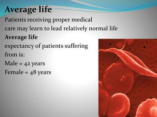
Sickle Cell Anemia
ODAI RJOUB (STUDENT) - HEBRON UNIVERSITY-
BIOLOGY DEPARTMENT-
DEF: is a disorder of the blood caused by an inherited
form of anemia — a condition in which there aren't enough
healthy red blood cells to carry adequate oxygen throughout
your body and presence of a mutated form of hemoglobin,
hemoglobin S (HbS).
HbS disease or sickle cell anemia (the most common form) -
Homozygote for the S globin with usually a severe or
moderately severe phenotype and with the shortest survival
Normaly
your red blood cells are flexible and round, moving easily
through your blood vessels.
In sickle cell anemia,
the red blood cells become rigid and sticky and are shaped
like sickles or crescent moons.
These irregularly shaped cells can get stuck in small blood
vessels, which can slow or block blood flow and oxygen to
parts of the body.
Loss of oxygen.
Polymers or rigid rods
leading to sickled
RBCs
RBCs Stick to blood
vessels
Stasis
Hypoxia
Pain
History :
 The African medical literature reported this condition in the
1870’s where it was locally known as ‘ogbanjes’.
 . 1910 – First Description of Sickle-Shaped Blood Cells by Dr
James Herrick.
 1917 – Genetic basis for SCD were discovered by Dr. V. Emmel.
 1922 – Disease was named “sickle cell anaemia” by Vernon
Mason .
 1927 – Hahn and Gillespie elaborated on Emmel’s work by
demonstrating that the sickling effect was linked to de-
oxygenation.
Causes:
a mutation in the gene that tells your body to make the red, iron-rich
compound that gives blood its red color (hemoglobin).
Produced due to the point mutation in hemoglobin beta gene (HBB gene) found
at chromosome number 11.
It results from the substitution of valine for glutamic acid at position
6 of beta-globin gene
Hemoglobin allows red blood cells to carry oxygen from your lungs to all parts of
your body.
abnormal hemoglobin causes red blood cells to become rigid, sticky and
misshapen.
The sickle cell gene is passed from generation to generation in a pattern of
inheritance called autosomal recessive inheritance.
Inheritance Pattern:
Distrubution:
General Symptoms:
1.Abdominal pain
2.Bone pain
3.Breathlessness
4.Delayed growth
and puberty
5.Fever
6.Jaundice
7.Rapid heart rate
8.Susceptibility to
infections.
Clinical manifestations of sickle cell anemia
1.Vaso-occlusive crisis. 2. Aplastic crisis.
3. Hemolytic crisis 4. Splenic sequestration crisis
1.Vaso-occlusive crisis.
Symptoms:
1.Pain 2.Autosplenectomy
3.Acute chest syndrome
a.Fever
b.Chest pain
c.Hard breathing
d.Pulmonary infiltrate on chest x-ray.
• Management
– Severe: analgesics, Opioid
– Mild: NSAIDs
– New treatment involving
*Adenosine A2a receptor agonists. These medicines may reduce pain-related
complications.
2. Aplastic crisis:
Symptoms:
Pallor
Tachycardia
fatigue
• Paravirus B19
– Divides in RBCs precursors and destroys them
– Stops erythropoiesis for two or three days
– Causes reticulocytopenia
– Disappears within one week with management and
blood transfusions
3.Hemolytic crisis
• Common in patients with G6PD deficiency
4. Splenic sequestration crisis:
Symptoms:
1.Abdominal pain
2.Restlessness
3.Fever
4.Drowsiness
5.Cold extremities
6.Liver and spleen increased in size.
• Acute, painful enlargements of the spleen,
caused by intra splenic trapping of red cells
• Caused by intra splenic trapping of red cells
• Die within 1-2 hours due to circulatory failure
• Auto splenectomy
Complications:
*Hand-Foot syndrome Pain , Fever, Swelling.
*Overwhelming post-splenectomy
infection (OPSI) treated with antibiotics and
supportive care.
*Acute chest Syndrome Chest pain, Shortness of
breath, Fever.
*Stroke Learning problems, Long term disability,
Brain damage,Paralysis, Death.
*cholelithiasis (gall stones) & Cholecytitis
Nausea, Vomiting, Jaundice, Sweating, Clay-
coloured stool.
*Priapism Damge to the Penis and Impotence
*Retinopathy Blindness.
* Sickle cell nephropathy Chronic renal failure.
*Pulmonary hypertension Fatigue, Shortness of breath.
*In pregnancy spontaneous abortion.
*Aseptic bone necrosis.
Diagnosis:
1.Peripheral smear 2.Sickling test
3.Hb electrophoresis 4. Sickle Solubility Test (SST)
5. Newborn screening 6.Dna analysis
1.Peripheral smear
Types of cell in smear:
1.Sickle cells 2.Target cell
2.Sickling test:
METHOD
1. 2drops of 2% sodium
Metabisulphite
2. 1drop of blood Cells with HbS
observed at 15 and 30 minutes.
USE
1.It is a qualitative
test.
2.This test is used to
identify HbS erythrocytes. Cells with HbA
3.Hb electrophoresis:
USE:
1.It is a quantitative test.
2.It not only confirms the presence of HbS but also
quantifies it.
4.Sickle Solubility Test (SST):
•A rapid and inexpensive technique used to screen for the
presence of sickling hemoglobins, can be used at home.
•A positive result must be confirmed by another method
(HPLC or electrophoresis) to confirm the presence of Hb S
and to distinguish Hb AS (carrier state) from Hb SS(sickle
cell disease).
•Disadvantage: Other insoluble hemoglobins, such as Hb C-
Harlem, will also give a positive result.
Method:
Depend on phosphate solubility:
1) Erythrocytes are lysed by saponin.
2) The released hemoglobin is reduced by
sodium hydrosulfite in a phosphate buffer.
3) Reduced HbS is characterized by its very low
solubility and the formation of neumatic liquid crystals
(tactoids).
5.Newborn screening:
In newborns who carry the sickle cell gene, fetal hemoglobin F will predominate,
but a small amount of hemoglobin S will also be present.
• It is performed via the most sensitive Hb isoelectric focusing or HPLC
fractionation and identifies the specific types of hemoglobin present.
6.DNA analysis:
• This test is used to investigate alterations and mutations in the genes that
producehemoglobin components.
•It may be performed to determine whether someone has one or two copies of
the Hb S mutation or has two different gene mutations.
•Genetic testing is most often used for prenatal testing: The usual tests offered
are chorionic villus sampling (CVS) or amniocentesis “14 to 16 weeks”.
•There also may be a small amount of hemoglobin A if they have sickle cell
trait.
Treatment:
1.Pain management 2.Blood transfusion
3.Drugs 4.Bone marrow
transplantation
5. Folic acid daily intake 6. Penicillin
7. Hydroxyurea.
– Reactivates fetal Hb production
– Decreases severity of attacks
– Increases life span
– More effective with Erythropoietin
8. Bone marrow transplant during childhood
9. (5-HMF.) This natural compound binds to red blood cells and
increases their oxygen. This helps prevent the red blood cells
from sickling.
Average life
Patients receiving proper medical
care may learn to lead relatively normal life
Average life
expectancy of patients suffering
from is:
Male = 42 years
Female = 48 years
Prevention:
1.Genetic screening
2.Testing for sickle cells in babies.
a. Chronic vill sampling
B .Amniotic fluid sampling
C . Fetal blood samplin
3. Daily penicillin for newborn babies
with the disease.
sickel cell anemia

sickel cell anemia

  • 1.
    Sickle Cell Anemia ODAIRJOUB (STUDENT) - HEBRON UNIVERSITY- BIOLOGY DEPARTMENT-
  • 2.
    DEF: is adisorder of the blood caused by an inherited form of anemia — a condition in which there aren't enough healthy red blood cells to carry adequate oxygen throughout your body and presence of a mutated form of hemoglobin, hemoglobin S (HbS). HbS disease or sickle cell anemia (the most common form) - Homozygote for the S globin with usually a severe or moderately severe phenotype and with the shortest survival
  • 3.
    Normaly your red bloodcells are flexible and round, moving easily through your blood vessels. In sickle cell anemia, the red blood cells become rigid and sticky and are shaped like sickles or crescent moons. These irregularly shaped cells can get stuck in small blood vessels, which can slow or block blood flow and oxygen to parts of the body.
  • 4.
    Loss of oxygen. Polymersor rigid rods leading to sickled RBCs RBCs Stick to blood vessels Stasis Hypoxia Pain
  • 5.
    History :  TheAfrican medical literature reported this condition in the 1870’s where it was locally known as ‘ogbanjes’.  . 1910 – First Description of Sickle-Shaped Blood Cells by Dr James Herrick.  1917 – Genetic basis for SCD were discovered by Dr. V. Emmel.  1922 – Disease was named “sickle cell anaemia” by Vernon Mason .  1927 – Hahn and Gillespie elaborated on Emmel’s work by demonstrating that the sickling effect was linked to de- oxygenation.
  • 6.
    Causes: a mutation inthe gene that tells your body to make the red, iron-rich compound that gives blood its red color (hemoglobin). Produced due to the point mutation in hemoglobin beta gene (HBB gene) found at chromosome number 11. It results from the substitution of valine for glutamic acid at position 6 of beta-globin gene Hemoglobin allows red blood cells to carry oxygen from your lungs to all parts of your body. abnormal hemoglobin causes red blood cells to become rigid, sticky and misshapen. The sickle cell gene is passed from generation to generation in a pattern of inheritance called autosomal recessive inheritance.
  • 7.
  • 8.
  • 9.
    General Symptoms: 1.Abdominal pain 2.Bonepain 3.Breathlessness 4.Delayed growth and puberty 5.Fever 6.Jaundice 7.Rapid heart rate 8.Susceptibility to infections.
  • 10.
    Clinical manifestations ofsickle cell anemia 1.Vaso-occlusive crisis. 2. Aplastic crisis. 3. Hemolytic crisis 4. Splenic sequestration crisis 1.Vaso-occlusive crisis. Symptoms: 1.Pain 2.Autosplenectomy 3.Acute chest syndrome a.Fever b.Chest pain c.Hard breathing d.Pulmonary infiltrate on chest x-ray. • Management – Severe: analgesics, Opioid – Mild: NSAIDs – New treatment involving *Adenosine A2a receptor agonists. These medicines may reduce pain-related complications.
  • 11.
    2. Aplastic crisis: Symptoms: Pallor Tachycardia fatigue •Paravirus B19 – Divides in RBCs precursors and destroys them – Stops erythropoiesis for two or three days – Causes reticulocytopenia – Disappears within one week with management and blood transfusions 3.Hemolytic crisis • Common in patients with G6PD deficiency
  • 12.
    4. Splenic sequestrationcrisis: Symptoms: 1.Abdominal pain 2.Restlessness 3.Fever 4.Drowsiness 5.Cold extremities 6.Liver and spleen increased in size. • Acute, painful enlargements of the spleen, caused by intra splenic trapping of red cells • Caused by intra splenic trapping of red cells • Die within 1-2 hours due to circulatory failure • Auto splenectomy
  • 13.
    Complications: *Hand-Foot syndrome Pain, Fever, Swelling. *Overwhelming post-splenectomy infection (OPSI) treated with antibiotics and supportive care. *Acute chest Syndrome Chest pain, Shortness of breath, Fever. *Stroke Learning problems, Long term disability, Brain damage,Paralysis, Death. *cholelithiasis (gall stones) & Cholecytitis Nausea, Vomiting, Jaundice, Sweating, Clay- coloured stool.
  • 14.
    *Priapism Damge tothe Penis and Impotence *Retinopathy Blindness. * Sickle cell nephropathy Chronic renal failure. *Pulmonary hypertension Fatigue, Shortness of breath. *In pregnancy spontaneous abortion. *Aseptic bone necrosis.
  • 15.
    Diagnosis: 1.Peripheral smear 2.Sicklingtest 3.Hb electrophoresis 4. Sickle Solubility Test (SST) 5. Newborn screening 6.Dna analysis 1.Peripheral smear Types of cell in smear: 1.Sickle cells 2.Target cell
  • 16.
    2.Sickling test: METHOD 1. 2dropsof 2% sodium Metabisulphite 2. 1drop of blood Cells with HbS observed at 15 and 30 minutes. USE 1.It is a qualitative test. 2.This test is used to identify HbS erythrocytes. Cells with HbA
  • 17.
    3.Hb electrophoresis: USE: 1.It isa quantitative test. 2.It not only confirms the presence of HbS but also quantifies it.
  • 18.
    4.Sickle Solubility Test(SST): •A rapid and inexpensive technique used to screen for the presence of sickling hemoglobins, can be used at home. •A positive result must be confirmed by another method (HPLC or electrophoresis) to confirm the presence of Hb S and to distinguish Hb AS (carrier state) from Hb SS(sickle cell disease). •Disadvantage: Other insoluble hemoglobins, such as Hb C- Harlem, will also give a positive result.
  • 19.
    Method: Depend on phosphatesolubility: 1) Erythrocytes are lysed by saponin. 2) The released hemoglobin is reduced by sodium hydrosulfite in a phosphate buffer. 3) Reduced HbS is characterized by its very low solubility and the formation of neumatic liquid crystals (tactoids).
  • 20.
    5.Newborn screening: In newbornswho carry the sickle cell gene, fetal hemoglobin F will predominate, but a small amount of hemoglobin S will also be present. • It is performed via the most sensitive Hb isoelectric focusing or HPLC fractionation and identifies the specific types of hemoglobin present. 6.DNA analysis: • This test is used to investigate alterations and mutations in the genes that producehemoglobin components. •It may be performed to determine whether someone has one or two copies of the Hb S mutation or has two different gene mutations. •Genetic testing is most often used for prenatal testing: The usual tests offered are chorionic villus sampling (CVS) or amniocentesis “14 to 16 weeks”. •There also may be a small amount of hemoglobin A if they have sickle cell trait.
  • 21.
    Treatment: 1.Pain management 2.Bloodtransfusion 3.Drugs 4.Bone marrow transplantation 5. Folic acid daily intake 6. Penicillin 7. Hydroxyurea. – Reactivates fetal Hb production – Decreases severity of attacks – Increases life span – More effective with Erythropoietin 8. Bone marrow transplant during childhood 9. (5-HMF.) This natural compound binds to red blood cells and increases their oxygen. This helps prevent the red blood cells from sickling.
  • 22.
    Average life Patients receivingproper medical care may learn to lead relatively normal life Average life expectancy of patients suffering from is: Male = 42 years Female = 48 years
  • 23.
    Prevention: 1.Genetic screening 2.Testing forsickle cells in babies. a. Chronic vill sampling B .Amniotic fluid sampling C . Fetal blood samplin 3. Daily penicillin for newborn babies with the disease.