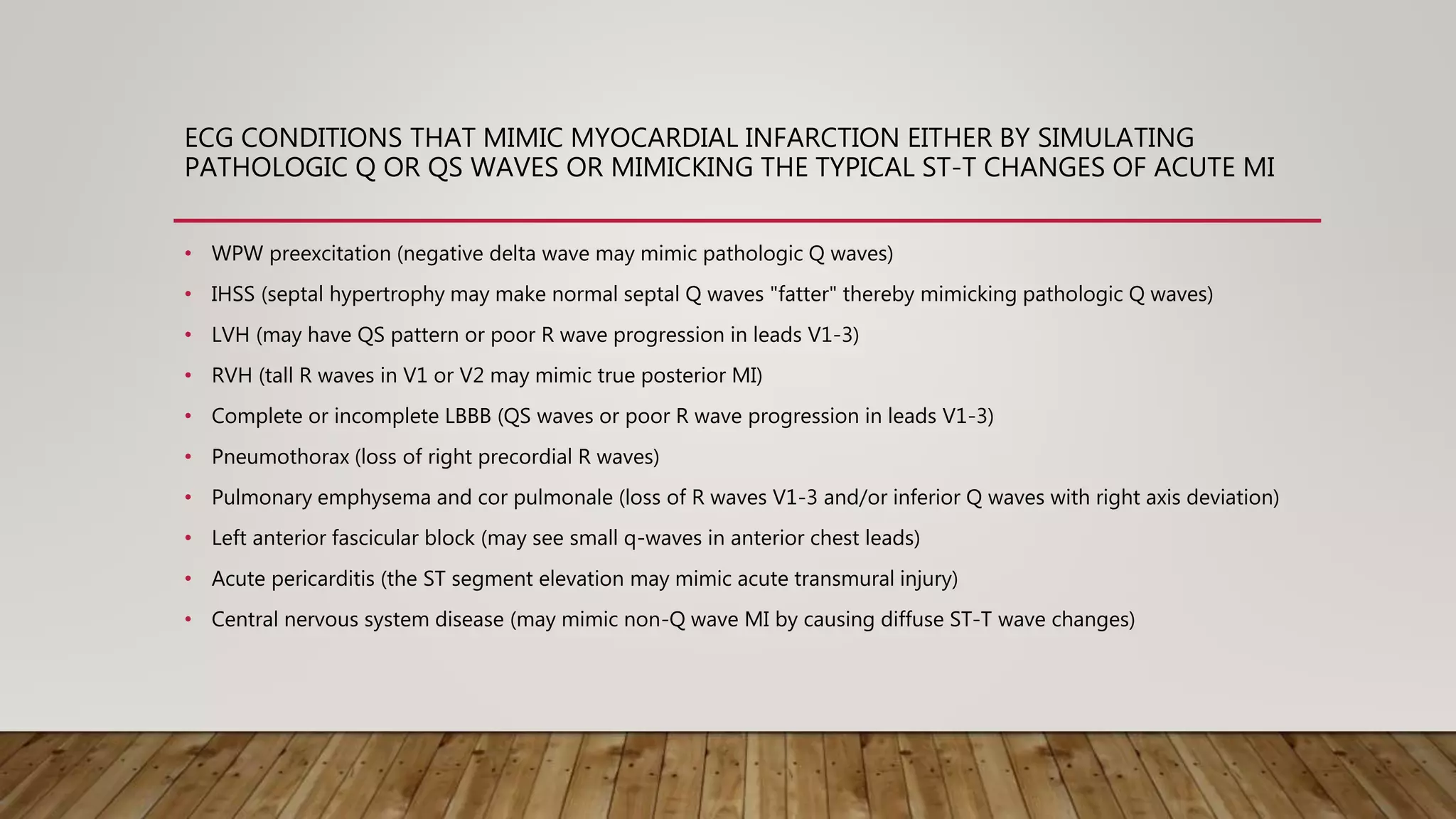
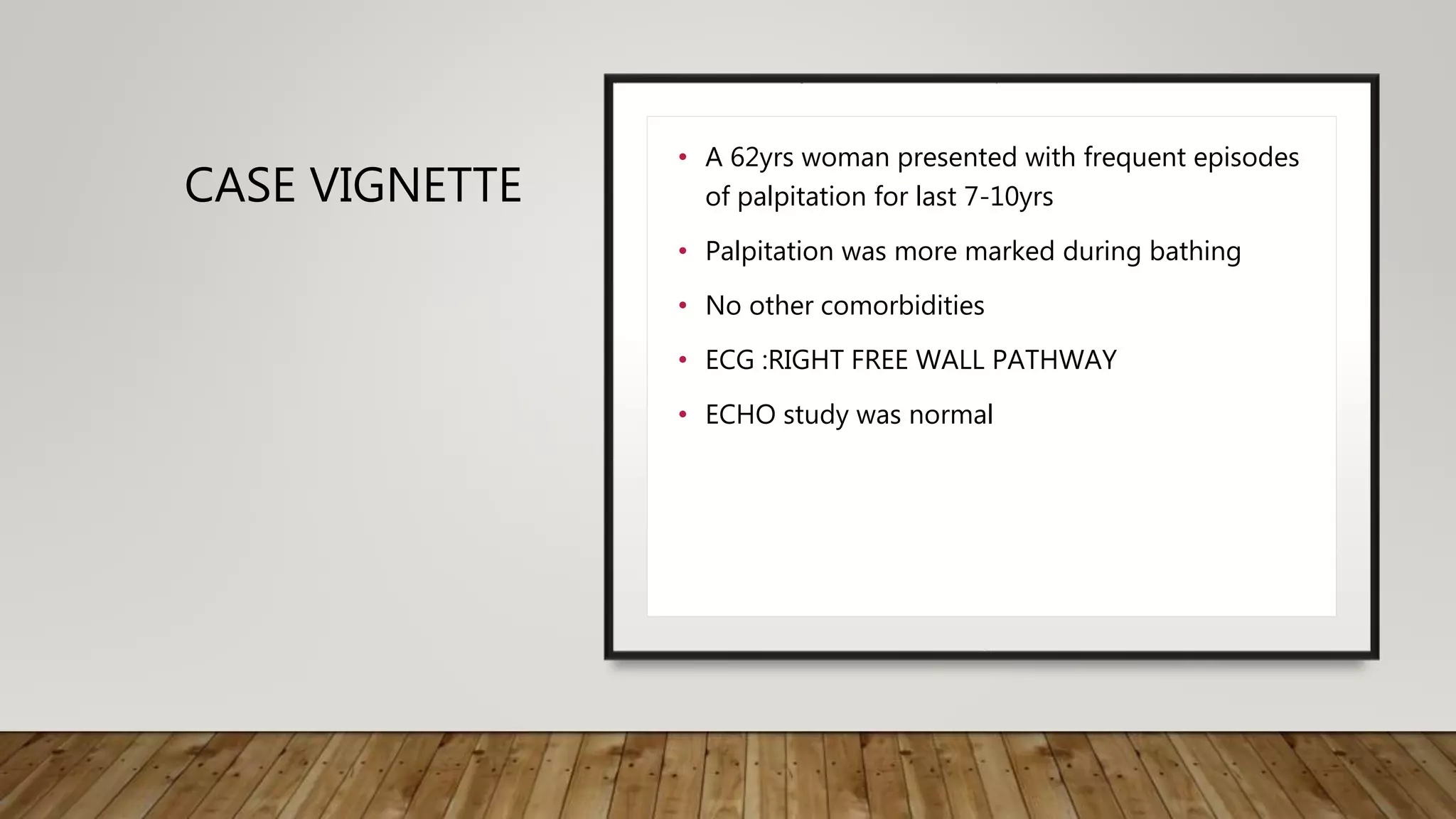
PSEUDO MYOCARDIAL
INFARCTION
AIIMS BHUBANESWAR
ECG CONDITIONS THAT MIMIC MYOCARDIAL INFARCTION EITHER BY SIMULATING
PATHOLOGIC Q OR QS WAVES OR MIMICKING THE TYPICAL ST-T CHANGES OF ACUTE MI
• WPW preexcitation (negative delta wave may mimic pathologic Q waves)
• IHSS (septal hypertrophy may make normal septal Q waves "fatter" thereby mimicking pathologic Q waves)
• LVH (may have QS pattern or poor R wave progression in leads V1-3)
• RVH (tall R waves in V1 or V2 may mimic true posterior MI)
• Complete or incomplete LBBB (QS waves or poor R wave progression in leads V1-3)
• Pneumothorax (loss of right precordial R waves)
• Pulmonary emphysema and cor pulmonale (loss of R waves V1-3 and/or inferior Q waves with right axis deviation)
• Left anterior fascicular block (may see small q-waves in anterior chest leads)
• Acute pericarditis (the ST segment elevation may mimic acute transmural injury)
• Central nervous system disease (may mimic non-Q wave MI by causing diffuse ST-T wave changes)
CASE VIGNETTE
• A 62yrs woman presented with frequent episodes
of palpitation for last 7-10yrs
• Palpitation was more marked during bathing
• No other comorbidities
• ECG :RIGHT FREE WALL PATHWAY
• ECHO study was normal
LOCALISATION KENT BUNDLE
RIGHT LATERAL PATHWAY:THE WOLFF-
PARKINSON-WHITE SYNDROME
• Negative delta and negative QRS in
V1
• QS in inferior leads
• Discordant T in the inferior leads: T is
upright when there is QS in these
leads
• Left axis deviation
IT IS NOT TOO LATE TO LEARN AT ANY AGE

Pseudo infarction

  • 1.
  • 2.
    ECG CONDITIONS THATMIMIC MYOCARDIAL INFARCTION EITHER BY SIMULATING PATHOLOGIC Q OR QS WAVES OR MIMICKING THE TYPICAL ST-T CHANGES OF ACUTE MI • WPW preexcitation (negative delta wave may mimic pathologic Q waves) • IHSS (septal hypertrophy may make normal septal Q waves "fatter" thereby mimicking pathologic Q waves) • LVH (may have QS pattern or poor R wave progression in leads V1-3) • RVH (tall R waves in V1 or V2 may mimic true posterior MI) • Complete or incomplete LBBB (QS waves or poor R wave progression in leads V1-3) • Pneumothorax (loss of right precordial R waves) • Pulmonary emphysema and cor pulmonale (loss of R waves V1-3 and/or inferior Q waves with right axis deviation) • Left anterior fascicular block (may see small q-waves in anterior chest leads) • Acute pericarditis (the ST segment elevation may mimic acute transmural injury) • Central nervous system disease (may mimic non-Q wave MI by causing diffuse ST-T wave changes)
  • 3.
    CASE VIGNETTE • A62yrs woman presented with frequent episodes of palpitation for last 7-10yrs • Palpitation was more marked during bathing • No other comorbidities • ECG :RIGHT FREE WALL PATHWAY • ECHO study was normal
  • 4.
  • 5.
    RIGHT LATERAL PATHWAY:THEWOLFF- PARKINSON-WHITE SYNDROME • Negative delta and negative QRS in V1 • QS in inferior leads • Discordant T in the inferior leads: T is upright when there is QS in these leads • Left axis deviation
  • 6.
    IT IS NOTTOO LATE TO LEARN AT ANY AGE