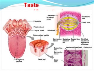
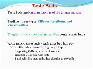
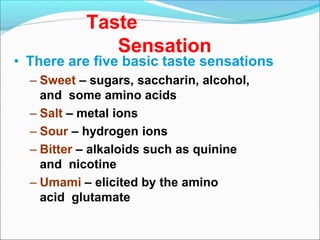
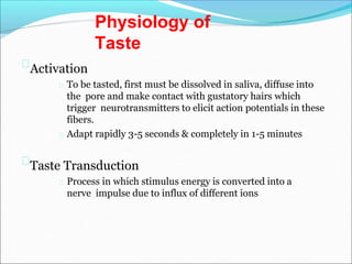
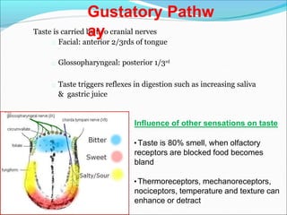
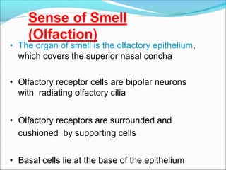
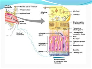
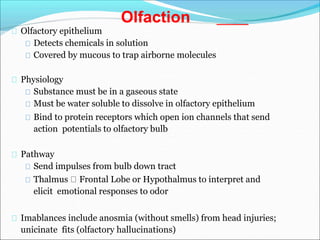
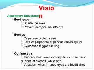
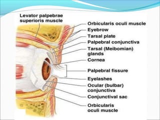
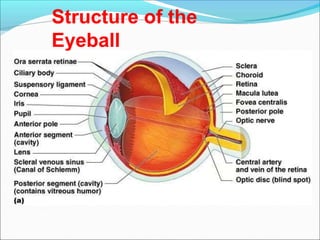
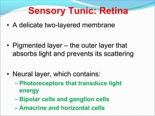
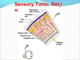
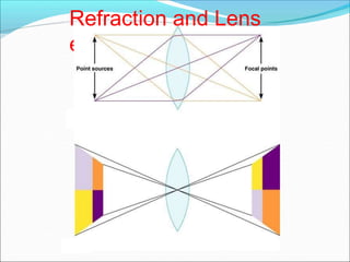
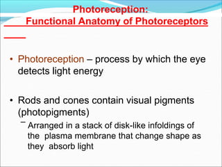
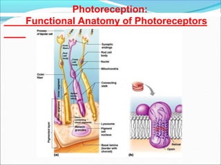
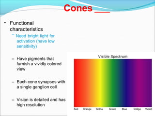
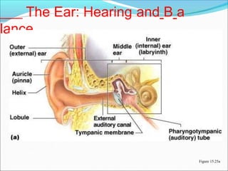
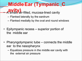
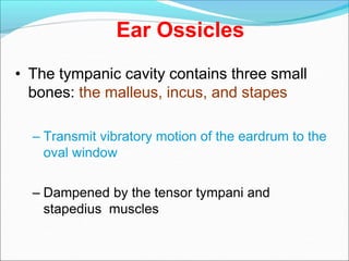
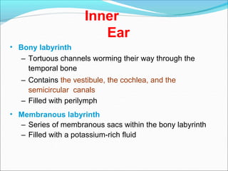
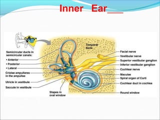
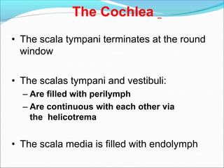
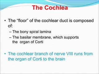
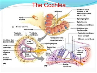
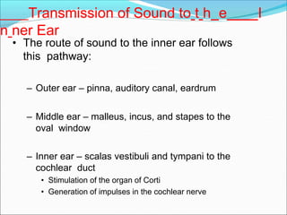
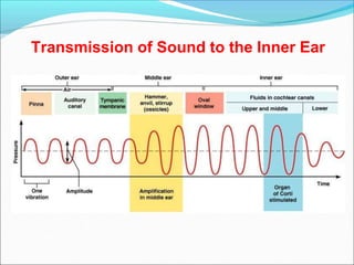
SPECIAL SENSES
Presented by
Ms. Kakade pallavi
Gautam
Moden college of
pharmacy
Five Senses
Vision (Sight)
Smell (olfaction)
Taste
Touch
Hearing
Equilibrium is also considered a special
sense, found in the ear
Taste
(Gustation)
Taste buds are found in papillae of the tongue mucosa
Papillae - three types: filiform, fungiform, and
circumvallate
Fungiform and circumvallate papillae contain taste buds
Appx 10,000 taste buds - each taste bud has 40-
100 epithelial cells made of 3 major types:
Supporting Cells: separate and insulate
Receptor Cells: deal with taste
Basal cells: like stem cells, they give rise to new cells
Taste Buds
Taste
Sensation
• There are five basic taste sensations
– Sweet – sugars, saccharin, alcohol,
and some amino acids
– Salt – metal ions
– Sour – hydrogen ions
– Bitter – alkaloids such as quinine
and nicotine
– Umami – elicited by the amino
acid glutamate
Activation
To be tasted, first must be dissolved in saliva, diffuse into
the pore and make contact with gustatory hairs which
trigger neurotransmitters to elicit action potentials in these
fibers.
Adapt rapidly 3-5 seconds & completely in 1-5 minutes
Taste Transduction
Process in which stimulus energy is converted into a
nerve impulse due to influx of different ions
Physiology of
Taste
Taste is carried by two cranial nerves
Facial: anterior 2/3rds of tongue
Glossopharyngeal: posterior 1/3rd
Taste triggers reflexes in digestion such as increasing saliva
& gastric juice
Gustatory Pathw
ay
Influence of other sensations on taste
•Taste is 80% smell, when olfactory
receptors are blocked food becomes
bland
•Thermoreceptors, mechanoreceptors,
nociceptors, temperature and texture can
enhance or detract
Sense of Smell
(Olfaction)
• The organ of smell is the olfactory epithelium,
which covers the superior nasal concha
• Olfactory receptor cells are bipolar neurons
with radiating olfactory cilia
• Olfactory receptors are surrounded and
cushioned by supporting cells
• Basal cells lie at the base of the epithelium
Olfactory epithelium
Detects chemicals in solution
Covered by mucous to trap airborne molecules
Physiology
Substance must be in a gaseous state
Must be water soluble to dissolve in olfactory epithelium
Bind to protein receptors which open ion channels that send
action potentials to olfactory bulb
Pathway
Send impulses from bulb down tract
Thalmus Frontal Lobe or Hypothalmus to interpret and
elicit emotional responses to odor
Imablances include anosmia (without smells) from head injuries;
unicinate fits (olfactory hallucinations)
Olfaction
Visio
n
Accessory Structures
Eyebrows
Shade the eyes
Prevent perspiration into eye
Eyelids
Palpabrae protects eye
Levator palpebrae superioris raises eyelid
Eyelashes trigger blinking
Conjunctiva
Mucous membrane over eyelids and anterior
surface of eyeball (white part)
Vascular, when irritated eyes are blood shot
Extraocular muscles
Movement is controlled by 6 muscles
Four Rectus muscles:
Superior, Inferior, Lateral,
Medial
Two Oblique muscles:
Superior, Inferior
Nerve Innervation:
abducens, trochlear,
oculomotor
Lens : Divides eye into
anterior and posterior
segments
Transparent, flexible structure
that can change shape to allow
focus of light on retina
Avascular
Extra ocular
muscles
Lacrimal Apparatus
• Consists of the lacrimal gland and
associated ducts
• Lacrimal glands secrete tears
• Tears
– Contain mucus, antibodies, and
lysozyme
– Enter the eye via superolateral
excretory ducts
– Exit the eye medially via the
lacrimal punctum
– Drain into the nasolacrimal duct
Lacrimal Apparatus
Structure of the
Eyeball
Fibrous Tunic
• Forms the outermost coat of the eye and is
composed of:
– Opaque sclera (posteriorly)
– Clear cornea (anteriorly)
• The sclera protects the eye and anchors
extrinsic muscles
• The cornea lets light enter the eye
Vascular Tunic (Uve
a)
• Has three regions: choroid, ciliary
body, and iris
• Choroid region
–A dark brown membrane that
forms the posterior portion of
the uvea
–Supplies blood to all eye tunics
Vascular Tunic
Ciliary Body
• A thickened ring of tissue surrounding the
lens
• Composed of smooth muscle bundles
(ciliary muscles)
• Anchors the suspensory ligament that holds
the lens in place
• The colored part of the eye
• Pupil – central opening of the iris
– Regulates the amount of light entering the eye
during:
• Close vision and bright light – pupils constrict
• Distant vision and dim light – pupils dilate
• Changes in emotional state – pupils dilate when
the subject matter is appealing or requires
problem-solving skills
Iris
Pupil Dilation and Constriction
Sensory Tunic: Retina
• A delicate two-layered membrane
• Pigmented layer – the outer layer that
absorbs light and prevents its scattering
• Neural layer, which contains:
– Photoreceptors that transduce light
energy
– Bipolar cells and ganglion cells
– Amacrine and horizontal cells
Sensory Tunic: Ret i
na
The Retina: Ganglion Cells and
the Optic Disc
• Ganglion cell axons:
– Run along the inner surface of the retina
– Leave the eye as the optic nerve
• The optic disc:
– Is the site where the optic nerve leaves the eye
– Lacks photoreceptors (the blind spot)
The Retina: Ganglion Cells a n d t h e O ptic
Disc
Figure 15.10b
Retinal Photoreceptors
• Rods:
– Respond to dim light
– Are used for peripheral vision
• Cones:
– Respond to bright light
– Have high-acuity color vision
– Are found in the macula lutea
– Are concentrated in the fovea centralis
Blood Supply to the
Retina
• The neural retina receives its blood
supply from two sources
– The outer third receives its blood from
the choroid
– The inner two-thirds is by the central artery
and vein
Inner Chambers and Fluids
• The lens separates the internal eye into anterior
and posterior segments
• The posterior segment is filled with a clear
gel called vitreous humor that:
– Transmits light
– Supports the posterior surface of the lens
– Holds the neural retina firmly against the
pigmented layer
– Contributes to intraocular pressure
Anterior Segment
• Composed of two chambers
– Anterior – between the cornea and the iris
– Posterior – between the iris and the lens
• Aqueous humor
– A plasma like fluid that fills the anterior segment
– Drains via the canal of Schlemm
• Supports, nourishes, and removes wastes
Anterior Segment
Figure 15.12
Refraction and Lenses
• When light passes from one transparent
medium to another its speed changes and it
refracts (bends)
• Light passing through a convex lens (as in
the eye) is bent so that the rays converge to
a focal point
• When a convex lens forms an image, the
image is upside down and reversed right to
left
Refraction and Lens
es
Photoreception:
Functional Anatomy of Photoreceptors
• Photoreception – process by which the eye
detects light energy
• Rods and cones contain visual pigments
(photopigments)
– Arranged in a stack of disk-like infoldings of
the plasma membrane that change shape as
they absorb light
Photoreception:
Functional Anatomy of Photoreceptors
Rods
• Functional characteristics
– Sensitive to dim light and best suited for night vision
– Absorb all wavelengths of visible light
– Perceived input is in gray tones only
– Sum of visual input from many rods feeds into a single
ganglion cell
– Results in fuzzy and indistinct images
Cones
• Functional
characteristics
– Need bright light for
activation (have low
sensitivity)
– Have pigments that
furnish a vividly colored
view
– Each cone synapses with
a single ganglion cell
– Vision is detailed and has
high resolution
The Ear: Hearing and
Balance
• The three parts of the ear are the inner, outer,
and middle ear
• The outer and middle ear are involved
with hearing
• The inner ear functions in both hearing and
equilibrium
The Ear: Hearing and B a
lance
Figure 15.25a
Outer
Ear
Auricle or Pinna
ear composed of elastic cartilage & skin to direct sound
waves to external auditory canal
External auditory meatus
Short curved tube from auricle to eardrum
Lined with skin, sebaceous glands, & ceruminous
glands (secrete earwax)
Tympanic membrane ( ear drum ) boundary
between outer & middle ear
MiddleEar (Tympanic C
avity)
• A small, air-filled, mucosa-lined cavity
– Flanked laterally by the eardrum
– Flanked medially by the oval and round windows
• Epitympanic recess – superior portion of
the middle ear
• Pharyngotympanic tube – connects the middle
ear to the nasopharynx
– Equalizes pressure in the middle ear cavity with
the external air pressure
Ear Ossicles
• The tympanic cavity contains three small
bones: the malleus, incus, and stapes
– Transmit vibratory motion of the eardrum to the
oval window
– Dampened by the tensor tympani and
stapedius muscles
Inner
Ear
• Bony labyrinth
– Tortuous channels worming their way through the
temporal bone
– Contains the vestibule, the cochlea, and the
semicircular canals
– Filled with perilymph
• Membranous labyrinth
– Series of membranous sacs within the bony labyrinth
– Filled with a potassium-rich fluid
Inner Ear
The Cochlea
• The scala tympani terminates at the round
window
• The scalas tympani and vestibuli:
– Are filled with perilymph
– Are continuous with each other via
the helicotrema
• The scala media is filled with endolymph
The Cochlea
• The “floor” of the cochlear duct is composed
of:
– The bony spiral lamina
– The basilar membrane, which supports
the organ of Corti
• The cochlear branch of nerve VIII runs from
the organ of Corti to the brain
The Cochlea
Transmission of Sound to t h e I
n ner Ear
• The route of sound to the inner ear follows
this pathway:
– Outer ear – pinna, auditory canal, eardrum
– Middle ear – malleus, incus, and stapes to the
oval window
– Inner ear – scalas vestibuli and tympani to the
cochlear duct
• Stimulation of the organ of Corti
• Generation of impulses in the cochlear nerve
Transmission of Sound to the Inner Ear
Thank
you

Secial senses (1)

  • 1.
    SPECIAL SENSES Presented by Ms.Kakade pallavi Gautam Moden college of pharmacy
  • 2.
    Five Senses Vision (Sight) Smell(olfaction) Taste Touch Hearing Equilibrium is also considered a special sense, found in the ear
  • 3.
  • 4.
    Taste buds arefound in papillae of the tongue mucosa Papillae - three types: filiform, fungiform, and circumvallate Fungiform and circumvallate papillae contain taste buds Appx 10,000 taste buds - each taste bud has 40- 100 epithelial cells made of 3 major types: Supporting Cells: separate and insulate Receptor Cells: deal with taste Basal cells: like stem cells, they give rise to new cells Taste Buds
  • 5.
    Taste Sensation • There arefive basic taste sensations – Sweet – sugars, saccharin, alcohol, and some amino acids – Salt – metal ions – Sour – hydrogen ions – Bitter – alkaloids such as quinine and nicotine – Umami – elicited by the amino acid glutamate
  • 6.
    Activation To be tasted,first must be dissolved in saliva, diffuse into the pore and make contact with gustatory hairs which trigger neurotransmitters to elicit action potentials in these fibers. Adapt rapidly 3-5 seconds & completely in 1-5 minutes Taste Transduction Process in which stimulus energy is converted into a nerve impulse due to influx of different ions Physiology of Taste
  • 7.
    Taste is carriedby two cranial nerves Facial: anterior 2/3rds of tongue Glossopharyngeal: posterior 1/3rd Taste triggers reflexes in digestion such as increasing saliva & gastric juice Gustatory Pathw ay Influence of other sensations on taste •Taste is 80% smell, when olfactory receptors are blocked food becomes bland •Thermoreceptors, mechanoreceptors, nociceptors, temperature and texture can enhance or detract
  • 8.
    Sense of Smell (Olfaction) •The organ of smell is the olfactory epithelium, which covers the superior nasal concha • Olfactory receptor cells are bipolar neurons with radiating olfactory cilia • Olfactory receptors are surrounded and cushioned by supporting cells • Basal cells lie at the base of the epithelium
  • 10.
    Olfactory epithelium Detects chemicalsin solution Covered by mucous to trap airborne molecules Physiology Substance must be in a gaseous state Must be water soluble to dissolve in olfactory epithelium Bind to protein receptors which open ion channels that send action potentials to olfactory bulb Pathway Send impulses from bulb down tract Thalmus Frontal Lobe or Hypothalmus to interpret and elicit emotional responses to odor Imablances include anosmia (without smells) from head injuries; unicinate fits (olfactory hallucinations) Olfaction
  • 11.
    Visio n Accessory Structures Eyebrows Shade theeyes Prevent perspiration into eye Eyelids Palpabrae protects eye Levator palpebrae superioris raises eyelid Eyelashes trigger blinking Conjunctiva Mucous membrane over eyelids and anterior surface of eyeball (white part) Vascular, when irritated eyes are blood shot
  • 13.
    Extraocular muscles Movement iscontrolled by 6 muscles Four Rectus muscles: Superior, Inferior, Lateral, Medial Two Oblique muscles: Superior, Inferior Nerve Innervation: abducens, trochlear, oculomotor Lens : Divides eye into anterior and posterior segments Transparent, flexible structure that can change shape to allow focus of light on retina Avascular
  • 14.
  • 15.
    Lacrimal Apparatus • Consistsof the lacrimal gland and associated ducts • Lacrimal glands secrete tears • Tears – Contain mucus, antibodies, and lysozyme – Enter the eye via superolateral excretory ducts – Exit the eye medially via the lacrimal punctum – Drain into the nasolacrimal duct
  • 16.
  • 17.
  • 18.
    Fibrous Tunic • Formsthe outermost coat of the eye and is composed of: – Opaque sclera (posteriorly) – Clear cornea (anteriorly) • The sclera protects the eye and anchors extrinsic muscles • The cornea lets light enter the eye
  • 19.
    Vascular Tunic (Uve a) •Has three regions: choroid, ciliary body, and iris • Choroid region –A dark brown membrane that forms the posterior portion of the uvea –Supplies blood to all eye tunics
  • 20.
    Vascular Tunic Ciliary Body •A thickened ring of tissue surrounding the lens • Composed of smooth muscle bundles (ciliary muscles) • Anchors the suspensory ligament that holds the lens in place
  • 21.
    • The coloredpart of the eye • Pupil – central opening of the iris – Regulates the amount of light entering the eye during: • Close vision and bright light – pupils constrict • Distant vision and dim light – pupils dilate • Changes in emotional state – pupils dilate when the subject matter is appealing or requires problem-solving skills Iris
  • 22.
    Pupil Dilation andConstriction
  • 23.
    Sensory Tunic: Retina •A delicate two-layered membrane • Pigmented layer – the outer layer that absorbs light and prevents its scattering • Neural layer, which contains: – Photoreceptors that transduce light energy – Bipolar cells and ganglion cells – Amacrine and horizontal cells
  • 24.
  • 25.
    The Retina: GanglionCells and the Optic Disc • Ganglion cell axons: – Run along the inner surface of the retina – Leave the eye as the optic nerve • The optic disc: – Is the site where the optic nerve leaves the eye – Lacks photoreceptors (the blind spot)
  • 26.
    The Retina: GanglionCells a n d t h e O ptic Disc Figure 15.10b
  • 27.
    Retinal Photoreceptors • Rods: –Respond to dim light – Are used for peripheral vision • Cones: – Respond to bright light – Have high-acuity color vision – Are found in the macula lutea – Are concentrated in the fovea centralis
  • 28.
    Blood Supply tothe Retina • The neural retina receives its blood supply from two sources – The outer third receives its blood from the choroid – The inner two-thirds is by the central artery and vein
  • 29.
    Inner Chambers andFluids • The lens separates the internal eye into anterior and posterior segments • The posterior segment is filled with a clear gel called vitreous humor that: – Transmits light – Supports the posterior surface of the lens – Holds the neural retina firmly against the pigmented layer – Contributes to intraocular pressure
  • 30.
    Anterior Segment • Composedof two chambers – Anterior – between the cornea and the iris – Posterior – between the iris and the lens • Aqueous humor – A plasma like fluid that fills the anterior segment – Drains via the canal of Schlemm • Supports, nourishes, and removes wastes
  • 31.
  • 32.
    Refraction and Lenses •When light passes from one transparent medium to another its speed changes and it refracts (bends) • Light passing through a convex lens (as in the eye) is bent so that the rays converge to a focal point • When a convex lens forms an image, the image is upside down and reversed right to left
  • 33.
  • 34.
    Photoreception: Functional Anatomy ofPhotoreceptors • Photoreception – process by which the eye detects light energy • Rods and cones contain visual pigments (photopigments) – Arranged in a stack of disk-like infoldings of the plasma membrane that change shape as they absorb light
  • 35.
  • 36.
    Rods • Functional characteristics –Sensitive to dim light and best suited for night vision – Absorb all wavelengths of visible light – Perceived input is in gray tones only – Sum of visual input from many rods feeds into a single ganglion cell – Results in fuzzy and indistinct images
  • 37.
    Cones • Functional characteristics – Needbright light for activation (have low sensitivity) – Have pigments that furnish a vividly colored view – Each cone synapses with a single ganglion cell – Vision is detailed and has high resolution
  • 38.
    The Ear: Hearingand Balance • The three parts of the ear are the inner, outer, and middle ear • The outer and middle ear are involved with hearing • The inner ear functions in both hearing and equilibrium
  • 39.
    The Ear: Hearingand B a lance Figure 15.25a
  • 40.
    Outer Ear Auricle or Pinna earcomposed of elastic cartilage & skin to direct sound waves to external auditory canal External auditory meatus Short curved tube from auricle to eardrum Lined with skin, sebaceous glands, & ceruminous glands (secrete earwax) Tympanic membrane ( ear drum ) boundary between outer & middle ear
  • 41.
    MiddleEar (Tympanic C avity) •A small, air-filled, mucosa-lined cavity – Flanked laterally by the eardrum – Flanked medially by the oval and round windows • Epitympanic recess – superior portion of the middle ear • Pharyngotympanic tube – connects the middle ear to the nasopharynx – Equalizes pressure in the middle ear cavity with the external air pressure
  • 42.
    Ear Ossicles • Thetympanic cavity contains three small bones: the malleus, incus, and stapes – Transmit vibratory motion of the eardrum to the oval window – Dampened by the tensor tympani and stapedius muscles
  • 43.
    Inner Ear • Bony labyrinth –Tortuous channels worming their way through the temporal bone – Contains the vestibule, the cochlea, and the semicircular canals – Filled with perilymph • Membranous labyrinth – Series of membranous sacs within the bony labyrinth – Filled with a potassium-rich fluid
  • 44.
  • 45.
    The Cochlea • Thescala tympani terminates at the round window • The scalas tympani and vestibuli: – Are filled with perilymph – Are continuous with each other via the helicotrema • The scala media is filled with endolymph
  • 46.
    The Cochlea • The“floor” of the cochlear duct is composed of: – The bony spiral lamina – The basilar membrane, which supports the organ of Corti • The cochlear branch of nerve VIII runs from the organ of Corti to the brain
  • 47.
  • 48.
    Transmission of Soundto t h e I n ner Ear • The route of sound to the inner ear follows this pathway: – Outer ear – pinna, auditory canal, eardrum – Middle ear – malleus, incus, and stapes to the oval window – Inner ear – scalas vestibuli and tympani to the cochlear duct • Stimulation of the organ of Corti • Generation of impulses in the cochlear nerve
  • 49.
    Transmission of Soundto the Inner Ear
  • 50.