SlideShare a Scribd company logo
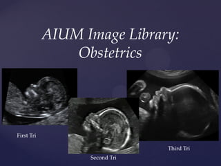
AIUM Image Library:
Obstetrics
First Tri
Third Tri
Second Tri
First Trimester
Ultrasound Examination
{
Gestational sac
Transvaginal (TV) scan showing a sagittal view of a uterus
demonstrating an intrauterine gestational sac. Note the yolk sac
within the gestational sac. The location of the sac should be
reported.
YS
Image of a gestational sac containing an embryo with a
regular fetal heart beat located in the adnexa (ectopic).
Yolk sac
Intrauterine gestational sac containing a yolk sac. No
embryo is identified within the gestational sac.
YS
Prior to the identification of an embryo, the mean sac diameter (MSD)
may be recorded. Note that the measurement does not include the
echogenic rim of tissue.
TV scan demonstrating a uterus containing a
gestational sac with a yolk sac and embryo.
Note the location of the embryo directly adjacent
to the yolk sac.
Crown- Rump Length
“Pseudogestational Sac”
TV scan demonstrating a uterus containing a fluid collection.
This may be associated with an ectopic pregnancy
The presence or absence of CARDIAC ACTIVITY should
be recorded. This may be done by direct visualization,
M-mode or recording a ‘clip’ of the cardiac activity.
Doppler should NOT be used in the first trimester unless
clinically indicated.
FETAL NUMBER should be reported. This
should be specifically stated, even in the case
of a singleton gestation.
FETAL NUMBER should be reported. In the event of a multiple
gestation, amnionicity and chorionicity should be documented
if possible.
1
2
Anterior
PlacentaPosterior
Placenta
Dividing
membrane
Dichorionic-Diamniotic
Embryonic/fetal anatomy appropriate for the
first trimester should be assessed.
Abnormal cranial contour Megacystis at 12 weeks
The uterus and cervix should be evaluated.
Myometrium
Cervix
Myometrium
B
L
The presence of leiomyomata should be recorded. The
measurement of the largest or any potentially clinically
significant leiomyomata may be recorded.
Transvaginal scan in the late first trimester pregnancy showing
a hypoechoic granular collection in the anterior aspect of the
uterus, adjacent to the gestational sac characteristic of
subchorionic hematoma. The calipers outline the hematoma.
Cervix
CDS
Placenta
Septum
3D rendered image of a the same
uterus reveals it to be septate .
Note the gestational sac in the
right side of the abnormal uterus.
Observation of the uterus may lead to the
detection of a uterine anomaly.
TA scan of the transverse uterus
showing splaying of the
endometrium with an eccentrically
located GS on the right.
Bladder
GS
Endo
GS
GS
IUD TV scan of uterus showing
an early gestational sac and
an intensely echogenic 3
dots, consistent with a
foreign body.
3D rendering reveals an early
gestational sac with an
echogenic choriodecidual
ring. There is an unusual IUD
within the uterus.
TV scan demonstrating a uterus containing a
gestational sac with a yolk sac. The appearance of the
cervix and the fluid in the cul-de-sac is noted.
Gestational Sac
Yolk Sac
CDS
The presence, location and size of adnexal
masses should be recorded.
Thick walled cyst with
circumferential color flow
characteristic of a normal corpus
luteum
Irregular cyst with mural
nodule and color flow.
Pathology confirmed an ovarian
malignancy. Note the adjacent
intrauterine pregnancy.
Embryo
The appearance of the nuchal region should be
assessed as part of the first trimester scan where a
live fetus is present.
For those patients desiring risk assessment for
aneuploidy, a specific measurement of the nuchal
translucency may be performed in accordance with
the stated guidelines.
Second and Third Trimester
Ultrasound Examinations
FETAL CARDIAC ACTIVITY should be reported,
including any abnormalities of rate or rhythm.
FETAL NUMBER should be reported.
Multiple gestations require chorionicity, amnionicity, comparison of
fetal sizes, estimation of amniotic fluid on each side of the membrane
and fetal genitalia when visualized.
Dichorionic-Diamniotic Twins
Placentae
Membrane
Lambda Sign
Multiple gestations require chorionicity, amnionicity, comparison of fetal
sizes, estimation of amniotic fluid on each side of the membrane and fetal
genitalia when visualized.
Monochorionic- Diamniotic Twins
Membrane
Placenta
FETAL PRESENTATION should be reported.
Cephalic Breech
Head
Cervix
Buttock
Cervix
A qualitative or semi-quantitative assessment of
AMNIOTIC FLUID VOLUME should be reported.
Subjective Assessment
Single deepest
vertical pocket
2-diameter pocket
Amniotic fluid index
Placental location, appearance, and relationship to the
internal cervical os should be recorded.
TA scan showing an anterior
placenta completely covering
the cervical os
TA scan showing a posterior
placenta, with the edge of the
placenta well away from the
cervix.
Internal Os
Placental edge
If the cervix appears short or is not adequately seen during
transabdominal evaluation, at transvaginal or transperineal
approach can be considered.
TV scan showing a short cervix with
funneling at 18 weeks gestation.
TV scan showing a vasa previa
which was not detected by
transabdominal imaging.
The UMBILICAL CORD should be imaged and the
number of vessels in the cord evaluated when possible.
Transverse view through a free
floating loop of cord demonstrating
2 arteries and 1 vein.
Transverse view of the lower fetal
pelvis using color Doppler to
identify the umbilical arteries as
they course around the bladder
Measured at the level of the thalami and cavum septi
pellucidi. The cerebellar hemispheres should not be visible
in this imaging plane. Measurement is from the outer
edge of the proximal skull to the inner edge of the distal
skull.
Biparietal Diameter
Head circumference is measured around the outer
perimeter of the skull at the same level as the BPD.
FEMORAL DIAPHYSIS LENGTH should not include
the distal femoral epiphysis. The beam of insonation
should be perpendicular to the shaft of the bone.
ABDOMINAL CIRCUMFERENCE is measured on a
transverse view of the fetal abdomen The stomach
and the junction of the umbilical vein and portal
sinus should be seen. A single rib should be seen.
The ellipse is at the skin edge.
ABDOMINAL DIAMETER is measured on a
transverse view of the fetal abdomen The stomach
and the junction of the umbilical vein and portal
sinus should be seen. A single rib should be seen.
The cursers are placed on the outer skin edge.
Evaluation of the uterus
and adnexa at 18 weeks
gestation reveals a left
pedunculated fibroid and
the adjacent normal ovary.
Pedunculated
leiomyoma
Amniotic Fluid
Ovary
Uterus and
Adnexa should
be evaluated
when
appropriate.
Fetal Anatomic Survey
The following areas of assessment
represent the minimal elements of a
standard examination of fetal anatomy.
Cerebellum
Choroid plexus
Cisterna magna
Lateral cerebral ventricles
Midline falx
Cavum septi pellucidi
In an transverse axial view of the fetal head, the CSP appears as a fluid
filled rectangular structure in the anterior midline; situated between the
frontal horns of the lateral ventricle.
PITFALL: Do not confuse the CSP with the
Columns of the Fornix (CF)
Note the parallel line
traveling through the CF.
PITFALL: Do not confuse the CSP with the
Columns of the Fornix (CF)
“Columns of the fornix, not to be mistaken for the cavum
septi pellucidi on prenatal ultrasound"
(http://www.jultrasoundmed.org/content/27/1/25.full?sid=bbf
a6200-abc0-4398-9ce0-a53f8039a007)
"The cavum septum septi pellucidi: why is it important“
(http://www.jultrasoundmed.org/content/29/3/427.full?sid=bb
fa6200-abc0-4398-9ce0-a53f8039a007)
Nuchal fold measurement may be helpful during a
certain gestational age interval in modifying the risk
for aneuploidy.
Axial scan through the fetal head which includes the thalami and the
cerebellum. The measurement is made from the occipital bone to the
outer skin edge.
Normal NF Thick NF
View of the feta lower face showing the surface of the
lips and the the two nares.
4-chamber view
Apical Axial View
Cardiac Outflow Tracts
If technically feasible, views of the cardiac
outflow tracts should be attempted.
RVOT LVOT
Sagittal view of cardiac outflow tracts.
Left Ventricular Outflow Tract. Note the continuity in
the interventricular septum (yellow arrows). LV= Left
Ventricle; Ao = Aorta; RV= Right Ventricle.
Right ventricular outflow tract. RV= Right Ventricle,
PA= Pulmonary Artery, Ao= Aorta
STOMACH : Presence, Size and Situs
Evaluation of situs demonstrated on split image of the
abdomen and thorax. Fetal position within the uterus
must be evaluated,. In this case, the fetus is vertex and
the left side is down.
Kidneys
Coronal view demonstrating both fetal kidneys.
Bladder
Umbilical cord insertion site
into the fetal abdomen
Umbilical Cord Vessel Number
Can be demonstrated by color Doppler as the umbilical
arteries course around the bladder, or by
transverse/longitudinal imaging of a free loop of cord
with color or gray scale.
Cervical, Thoracic, Lumbar and Sacral spine
Transverse view of sacral spine showing the centrum
(C) and lamina (L) .
L
C
Legs
Arms
Male genitalia in second trimester.
Note the stream of urine (arrow).
Female
Second Trimester Third Trimester
ALARA: As Low As Reasonably Achievable

More Related Content

What's hot

SIÊU ÂM DOPPLER TRONG SẢN KHOA
SIÊU ÂM DOPPLER TRONG SẢN KHOASIÊU ÂM DOPPLER TRONG SẢN KHOA
SIÊU ÂM DOPPLER TRONG SẢN KHOA
SoM
 
Kênh nhĩ thất - Atrioventricular septal defect - Siêu âm tim thai
Kênh nhĩ thất - Atrioventricular septal defect - Siêu âm tim thaiKênh nhĩ thất - Atrioventricular septal defect - Siêu âm tim thai
Kênh nhĩ thất - Atrioventricular septal defect - Siêu âm tim thai
Võ Tá Sơn
 
SIÊU ÂM CHẨN ĐOÁN TRONG PHỤ KHOA
SIÊU ÂM CHẨN ĐOÁN TRONG PHỤ KHOASIÊU ÂM CHẨN ĐOÁN TRONG PHỤ KHOA
SIÊU ÂM CHẨN ĐOÁN TRONG PHỤ KHOA
SoM
 
SIÊU ÂM QUÝ HAI THAI KỲ
SIÊU ÂM QUÝ HAI THAI KỲSIÊU ÂM QUÝ HAI THAI KỲ
SIÊU ÂM QUÝ HAI THAI KỲ
SoM
 
Bai 13 siêu âm trong đa thai
Bai 13 siêu âm trong đa thaiBai 13 siêu âm trong đa thai
Bai 13 siêu âm trong đa thai
Lan Đặng
 
CÁC KHẢI NIỆM CHÍNH YẾU VỀ VAI TRÒ CỦA SIÊU ÂM TRONG NỬA ĐẦU THAI KỲ
CÁC KHẢI NIỆM CHÍNH YẾU VỀ VAI TRÒ CỦA SIÊU ÂM TRONG NỬA ĐẦU THAI KỲCÁC KHẢI NIỆM CHÍNH YẾU VỀ VAI TRÒ CỦA SIÊU ÂM TRONG NỬA ĐẦU THAI KỲ
CÁC KHẢI NIỆM CHÍNH YẾU VỀ VAI TRÒ CỦA SIÊU ÂM TRONG NỬA ĐẦU THAI KỲ
SoM
 
SONG THAI VÀ CÁC VẤN ĐỀ CÓ LIÊN QUAN
SONG THAI VÀ CÁC VẤN ĐỀ CÓ LIÊN QUANSONG THAI VÀ CÁC VẤN ĐỀ CÓ LIÊN QUAN
SONG THAI VÀ CÁC VẤN ĐỀ CÓ LIÊN QUAN
SoM
 
Các mặt cắt cơ bản trong siêu âm ths nguyễn cảnh chương
Các mặt cắt cơ bản trong siêu âm   ths nguyễn cảnh chươngCác mặt cắt cơ bản trong siêu âm   ths nguyễn cảnh chương
Các mặt cắt cơ bản trong siêu âm ths nguyễn cảnh chương
Lan Đặng
 
1. Cac chi so sinh hoc thai nhi, GS Michel Collet
1. Cac chi so sinh hoc thai nhi, GS Michel Collet1. Cac chi so sinh hoc thai nhi, GS Michel Collet
1. Cac chi so sinh hoc thai nhi, GS Michel Collet
Nguyen Lam
 
Hình ảnh phân loại siêu âm trước sinh Holoprosencephaly
Hình ảnh phân loại siêu âm trước sinh HoloprosencephalyHình ảnh phân loại siêu âm trước sinh Holoprosencephaly
Hình ảnh phân loại siêu âm trước sinh Holoprosencephaly
Võ Tá Sơn
 
17. Sieu am banh nhau, GS Michel Collet
17. Sieu am banh nhau, GS Michel Collet17. Sieu am banh nhau, GS Michel Collet
17. Sieu am banh nhau, GS Michel Collet
Nguyen Lam
 
5. Sieu am tim thai binh thuong, GS Michel Collet
5. Sieu am tim thai binh thuong, GS Michel Collet5. Sieu am tim thai binh thuong, GS Michel Collet
5. Sieu am tim thai binh thuong, GS Michel Collet
Nguyen Lam
 
KHÂU CẦM MÁU TRONG MỔ SINH NHAU TIỀN ĐẠO TRUNG TÂM
KHÂU CẦM MÁU TRONG MỔ SINH NHAU TIỀN ĐẠO TRUNG TÂMKHÂU CẦM MÁU TRONG MỔ SINH NHAU TIỀN ĐẠO TRUNG TÂM
KHÂU CẦM MÁU TRONG MỔ SINH NHAU TIỀN ĐẠO TRUNG TÂM
SoM
 
Thai đoạn kẽ vòi tử cung, interstitial ectopic pregnancy, siêu âm, võ tá sơn
Thai đoạn kẽ vòi tử cung, interstitial ectopic pregnancy, siêu âm, võ tá sơnThai đoạn kẽ vòi tử cung, interstitial ectopic pregnancy, siêu âm, võ tá sơn
Thai đoạn kẽ vòi tử cung, interstitial ectopic pregnancy, siêu âm, võ tá sơn
Võ Tá Sơn
 
KHÂU EO CỔ TỬ CUNG
KHÂU EO CỔ TỬ CUNGKHÂU EO CỔ TỬ CUNG
KHÂU EO CỔ TỬ CUNG
SoM
 
8. Sieu am benh ly buong trung, GS Michel Collet
8. Sieu am benh ly buong trung, GS Michel Collet8. Sieu am benh ly buong trung, GS Michel Collet
8. Sieu am benh ly buong trung, GS Michel Collet
Nguyen Lam
 
SIÊU ÂM 3 THÁNG CUỐI THAI KỲ
SIÊU ÂM 3 THÁNG CUỐI THAI KỲSIÊU ÂM 3 THÁNG CUỐI THAI KỲ
SIÊU ÂM 3 THÁNG CUỐI THAI KỲ
SoM
 
KHẢO SÁT SIÊU ÂM SẢN KHOA
KHẢO SÁT SIÊU ÂM SẢN KHOAKHẢO SÁT SIÊU ÂM SẢN KHOA
KHẢO SÁT SIÊU ÂM SẢN KHOA
SoM
 
10. Sieu am khao sat mat thai, GS Michel Collet
10. Sieu am khao sat mat thai, GS Michel Collet10. Sieu am khao sat mat thai, GS Michel Collet
10. Sieu am khao sat mat thai, GS Michel Collet
Nguyen Lam
 
4. Sieu am bat thuong hinh thai quy I thai ky, GS Michel Collet
4. Sieu am bat thuong hinh thai quy I thai ky, GS Michel Collet4. Sieu am bat thuong hinh thai quy I thai ky, GS Michel Collet
4. Sieu am bat thuong hinh thai quy I thai ky, GS Michel Collet
Nguyen Lam
 

What's hot (20)

SIÊU ÂM DOPPLER TRONG SẢN KHOA
SIÊU ÂM DOPPLER TRONG SẢN KHOASIÊU ÂM DOPPLER TRONG SẢN KHOA
SIÊU ÂM DOPPLER TRONG SẢN KHOA
 
Kênh nhĩ thất - Atrioventricular septal defect - Siêu âm tim thai
Kênh nhĩ thất - Atrioventricular septal defect - Siêu âm tim thaiKênh nhĩ thất - Atrioventricular septal defect - Siêu âm tim thai
Kênh nhĩ thất - Atrioventricular septal defect - Siêu âm tim thai
 
SIÊU ÂM CHẨN ĐOÁN TRONG PHỤ KHOA
SIÊU ÂM CHẨN ĐOÁN TRONG PHỤ KHOASIÊU ÂM CHẨN ĐOÁN TRONG PHỤ KHOA
SIÊU ÂM CHẨN ĐOÁN TRONG PHỤ KHOA
 
SIÊU ÂM QUÝ HAI THAI KỲ
SIÊU ÂM QUÝ HAI THAI KỲSIÊU ÂM QUÝ HAI THAI KỲ
SIÊU ÂM QUÝ HAI THAI KỲ
 
Bai 13 siêu âm trong đa thai
Bai 13 siêu âm trong đa thaiBai 13 siêu âm trong đa thai
Bai 13 siêu âm trong đa thai
 
CÁC KHẢI NIỆM CHÍNH YẾU VỀ VAI TRÒ CỦA SIÊU ÂM TRONG NỬA ĐẦU THAI KỲ
CÁC KHẢI NIỆM CHÍNH YẾU VỀ VAI TRÒ CỦA SIÊU ÂM TRONG NỬA ĐẦU THAI KỲCÁC KHẢI NIỆM CHÍNH YẾU VỀ VAI TRÒ CỦA SIÊU ÂM TRONG NỬA ĐẦU THAI KỲ
CÁC KHẢI NIỆM CHÍNH YẾU VỀ VAI TRÒ CỦA SIÊU ÂM TRONG NỬA ĐẦU THAI KỲ
 
SONG THAI VÀ CÁC VẤN ĐỀ CÓ LIÊN QUAN
SONG THAI VÀ CÁC VẤN ĐỀ CÓ LIÊN QUANSONG THAI VÀ CÁC VẤN ĐỀ CÓ LIÊN QUAN
SONG THAI VÀ CÁC VẤN ĐỀ CÓ LIÊN QUAN
 
Các mặt cắt cơ bản trong siêu âm ths nguyễn cảnh chương
Các mặt cắt cơ bản trong siêu âm   ths nguyễn cảnh chươngCác mặt cắt cơ bản trong siêu âm   ths nguyễn cảnh chương
Các mặt cắt cơ bản trong siêu âm ths nguyễn cảnh chương
 
1. Cac chi so sinh hoc thai nhi, GS Michel Collet
1. Cac chi so sinh hoc thai nhi, GS Michel Collet1. Cac chi so sinh hoc thai nhi, GS Michel Collet
1. Cac chi so sinh hoc thai nhi, GS Michel Collet
 
Hình ảnh phân loại siêu âm trước sinh Holoprosencephaly
Hình ảnh phân loại siêu âm trước sinh HoloprosencephalyHình ảnh phân loại siêu âm trước sinh Holoprosencephaly
Hình ảnh phân loại siêu âm trước sinh Holoprosencephaly
 
17. Sieu am banh nhau, GS Michel Collet
17. Sieu am banh nhau, GS Michel Collet17. Sieu am banh nhau, GS Michel Collet
17. Sieu am banh nhau, GS Michel Collet
 
5. Sieu am tim thai binh thuong, GS Michel Collet
5. Sieu am tim thai binh thuong, GS Michel Collet5. Sieu am tim thai binh thuong, GS Michel Collet
5. Sieu am tim thai binh thuong, GS Michel Collet
 
KHÂU CẦM MÁU TRONG MỔ SINH NHAU TIỀN ĐẠO TRUNG TÂM
KHÂU CẦM MÁU TRONG MỔ SINH NHAU TIỀN ĐẠO TRUNG TÂMKHÂU CẦM MÁU TRONG MỔ SINH NHAU TIỀN ĐẠO TRUNG TÂM
KHÂU CẦM MÁU TRONG MỔ SINH NHAU TIỀN ĐẠO TRUNG TÂM
 
Thai đoạn kẽ vòi tử cung, interstitial ectopic pregnancy, siêu âm, võ tá sơn
Thai đoạn kẽ vòi tử cung, interstitial ectopic pregnancy, siêu âm, võ tá sơnThai đoạn kẽ vòi tử cung, interstitial ectopic pregnancy, siêu âm, võ tá sơn
Thai đoạn kẽ vòi tử cung, interstitial ectopic pregnancy, siêu âm, võ tá sơn
 
KHÂU EO CỔ TỬ CUNG
KHÂU EO CỔ TỬ CUNGKHÂU EO CỔ TỬ CUNG
KHÂU EO CỔ TỬ CUNG
 
8. Sieu am benh ly buong trung, GS Michel Collet
8. Sieu am benh ly buong trung, GS Michel Collet8. Sieu am benh ly buong trung, GS Michel Collet
8. Sieu am benh ly buong trung, GS Michel Collet
 
SIÊU ÂM 3 THÁNG CUỐI THAI KỲ
SIÊU ÂM 3 THÁNG CUỐI THAI KỲSIÊU ÂM 3 THÁNG CUỐI THAI KỲ
SIÊU ÂM 3 THÁNG CUỐI THAI KỲ
 
KHẢO SÁT SIÊU ÂM SẢN KHOA
KHẢO SÁT SIÊU ÂM SẢN KHOAKHẢO SÁT SIÊU ÂM SẢN KHOA
KHẢO SÁT SIÊU ÂM SẢN KHOA
 
10. Sieu am khao sat mat thai, GS Michel Collet
10. Sieu am khao sat mat thai, GS Michel Collet10. Sieu am khao sat mat thai, GS Michel Collet
10. Sieu am khao sat mat thai, GS Michel Collet
 
4. Sieu am bat thuong hinh thai quy I thai ky, GS Michel Collet
4. Sieu am bat thuong hinh thai quy I thai ky, GS Michel Collet4. Sieu am bat thuong hinh thai quy I thai ky, GS Michel Collet
4. Sieu am bat thuong hinh thai quy I thai ky, GS Michel Collet
 

Similar to Hình ảnh siêu âm cơ bản thai nhi quý 1 2 3 AIUM 2019

Ovarian ectopic pregnancy
Ovarian ectopic pregnancyOvarian ectopic pregnancy
Ovarian ectopic pregnancy
Ritesh Mahajan
 
first trimester ultrasound and overview of obs imaging
first trimester ultrasound and overview of obs imagingfirst trimester ultrasound and overview of obs imaging
first trimester ultrasound and overview of obs imaging
Bharat Jain
 
first trimester ultrasound and overview of obs imaging
first trimester ultrasound and overview of obs imagingfirst trimester ultrasound and overview of obs imaging
first trimester ultrasound and overview of obs imaging
Bharat Jain
 
2nd trimester ultrasound..
2nd trimester ultrasound..2nd trimester ultrasound..
2nd trimester ultrasound..
Soumitra Halder
 
Evaluation of fetal anatomy
Evaluation of fetal anatomyEvaluation of fetal anatomy
Evaluation of fetal anatomy
dypradio
 
Prenatal diagnosis of congenital anomalies 3
Prenatal diagnosis of congenital anomalies 3Prenatal diagnosis of congenital anomalies 3
Prenatal diagnosis of congenital anomalies 3
DrAhmed Badr
 
Placenta ultrasound
Placenta ultrasoundPlacenta ultrasound
Placenta ultrasoundDoaa Gadalla
 
Presentation1.pptx, ultrasound examination of the 2nd & 3rd trimester pregnancy.
Presentation1.pptx, ultrasound examination of the 2nd & 3rd trimester pregnancy.Presentation1.pptx, ultrasound examination of the 2nd & 3rd trimester pregnancy.
Presentation1.pptx, ultrasound examination of the 2nd & 3rd trimester pregnancy.Abdellah Nazeer
 
utrasound in Early pregnancy
utrasound in Early pregnancyutrasound in Early pregnancy
utrasound in Early pregnancy
vinothmezoss
 
Ectopic pregnancy
Ectopic pregnancyEctopic pregnancy
Ectopic pregnancy
Nikita Sharma
 
Ultrasound and usg doppler in obstetrics
Ultrasound and usg doppler in obstetricsUltrasound and usg doppler in obstetrics
Ultrasound and usg doppler in obstetrics
dr. gokul reshmi mariappan
 
Hysterosalpingography
HysterosalpingographyHysterosalpingography
Hysterosalpingographyairwave12
 
ULTRASOUND & DOPPLER DURING PREGNANCY.pptx
ULTRASOUND & DOPPLER DURING PREGNANCY.pptxULTRASOUND & DOPPLER DURING PREGNANCY.pptx
ULTRASOUND & DOPPLER DURING PREGNANCY.pptx
Gullu Agarwal
 
anorectal malformation
anorectal malformationanorectal malformation
anorectal malformation
Pushpa Latha
 
The dilemma of eccentrically located gestational sac
The dilemma of eccentrically located gestational sacThe dilemma of eccentrically located gestational sac
The dilemma of eccentrically located gestational sac
Ahmed Al Amely
 
OBSTETRICS EMERGENCIES
OBSTETRICS EMERGENCIESOBSTETRICS EMERGENCIES
OBSTETRICS EMERGENCIES
Lohith Varma
 
placenta-140315062308-phpapp01.pdf
placenta-140315062308-phpapp01.pdfplacenta-140315062308-phpapp01.pdf
placenta-140315062308-phpapp01.pdf
MuhamadAznorAqwaAzma
 
Ectopic Pregnancy: New ESHRE terminology
Ectopic Pregnancy: New ESHRE terminologyEctopic Pregnancy: New ESHRE terminology
Ectopic Pregnancy: New ESHRE terminology
Ahmed Al Amely
 
Fetal Neurosonogram
Fetal Neurosonogram Fetal Neurosonogram
Fetal Neurosonogram
nasrat1949
 

Similar to Hình ảnh siêu âm cơ bản thai nhi quý 1 2 3 AIUM 2019 (20)

Ovarian ectopic pregnancy
Ovarian ectopic pregnancyOvarian ectopic pregnancy
Ovarian ectopic pregnancy
 
Level II usg
Level II usgLevel II usg
Level II usg
 
first trimester ultrasound and overview of obs imaging
first trimester ultrasound and overview of obs imagingfirst trimester ultrasound and overview of obs imaging
first trimester ultrasound and overview of obs imaging
 
first trimester ultrasound and overview of obs imaging
first trimester ultrasound and overview of obs imagingfirst trimester ultrasound and overview of obs imaging
first trimester ultrasound and overview of obs imaging
 
2nd trimester ultrasound..
2nd trimester ultrasound..2nd trimester ultrasound..
2nd trimester ultrasound..
 
Evaluation of fetal anatomy
Evaluation of fetal anatomyEvaluation of fetal anatomy
Evaluation of fetal anatomy
 
Prenatal diagnosis of congenital anomalies 3
Prenatal diagnosis of congenital anomalies 3Prenatal diagnosis of congenital anomalies 3
Prenatal diagnosis of congenital anomalies 3
 
Placenta ultrasound
Placenta ultrasoundPlacenta ultrasound
Placenta ultrasound
 
Presentation1.pptx, ultrasound examination of the 2nd & 3rd trimester pregnancy.
Presentation1.pptx, ultrasound examination of the 2nd & 3rd trimester pregnancy.Presentation1.pptx, ultrasound examination of the 2nd & 3rd trimester pregnancy.
Presentation1.pptx, ultrasound examination of the 2nd & 3rd trimester pregnancy.
 
utrasound in Early pregnancy
utrasound in Early pregnancyutrasound in Early pregnancy
utrasound in Early pregnancy
 
Ectopic pregnancy
Ectopic pregnancyEctopic pregnancy
Ectopic pregnancy
 
Ultrasound and usg doppler in obstetrics
Ultrasound and usg doppler in obstetricsUltrasound and usg doppler in obstetrics
Ultrasound and usg doppler in obstetrics
 
Hysterosalpingography
HysterosalpingographyHysterosalpingography
Hysterosalpingography
 
ULTRASOUND & DOPPLER DURING PREGNANCY.pptx
ULTRASOUND & DOPPLER DURING PREGNANCY.pptxULTRASOUND & DOPPLER DURING PREGNANCY.pptx
ULTRASOUND & DOPPLER DURING PREGNANCY.pptx
 
anorectal malformation
anorectal malformationanorectal malformation
anorectal malformation
 
The dilemma of eccentrically located gestational sac
The dilemma of eccentrically located gestational sacThe dilemma of eccentrically located gestational sac
The dilemma of eccentrically located gestational sac
 
OBSTETRICS EMERGENCIES
OBSTETRICS EMERGENCIESOBSTETRICS EMERGENCIES
OBSTETRICS EMERGENCIES
 
placenta-140315062308-phpapp01.pdf
placenta-140315062308-phpapp01.pdfplacenta-140315062308-phpapp01.pdf
placenta-140315062308-phpapp01.pdf
 
Ectopic Pregnancy: New ESHRE terminology
Ectopic Pregnancy: New ESHRE terminologyEctopic Pregnancy: New ESHRE terminology
Ectopic Pregnancy: New ESHRE terminology
 
Fetal Neurosonogram
Fetal Neurosonogram Fetal Neurosonogram
Fetal Neurosonogram
 

More from Võ Tá Sơn

Thai bam vet mo cu RMT - VOTASON 2023.pdf
Thai bam vet mo cu RMT - VOTASON 2023.pdfThai bam vet mo cu RMT - VOTASON 2023.pdf
Thai bam vet mo cu RMT - VOTASON 2023.pdf
Võ Tá Sơn
 
YHSS 55 (13_10 S)-3 NGUYEN HAI DANG.pdf
YHSS 55 (13_10 S)-3 NGUYEN HAI DANG.pdfYHSS 55 (13_10 S)-3 NGUYEN HAI DANG.pdf
YHSS 55 (13_10 S)-3 NGUYEN HAI DANG.pdf
Võ Tá Sơn
 
Sinh thiết gai rau CVS những điều mẹ bầu nên biết
Sinh thiết gai rau CVS những điều mẹ bầu nên biếtSinh thiết gai rau CVS những điều mẹ bầu nên biết
Sinh thiết gai rau CVS những điều mẹ bầu nên biết
Võ Tá Sơn
 
Chọc ối amniocentesis những điều mẹ bầu cần biết
Chọc ối amniocentesis những điều mẹ bầu cần biếtChọc ối amniocentesis những điều mẹ bầu cần biết
Chọc ối amniocentesis những điều mẹ bầu cần biết
Võ Tá Sơn
 
Mang thai ở từ tuổi 35 - Pregnancy at 35 years or older - ACOG SMFM 2022.pdf
Mang thai ở từ tuổi 35 - Pregnancy at 35 years or older - ACOG SMFM 2022.pdfMang thai ở từ tuổi 35 - Pregnancy at 35 years or older - ACOG SMFM 2022.pdf
Mang thai ở từ tuổi 35 - Pregnancy at 35 years or older - ACOG SMFM 2022.pdf
Võ Tá Sơn
 
wigby2016 Expanding the phenotype of Triple X syndrome- A comparison of prena...
wigby2016 Expanding the phenotype of Triple X syndrome- A comparison of prena...wigby2016 Expanding the phenotype of Triple X syndrome- A comparison of prena...
wigby2016 Expanding the phenotype of Triple X syndrome- A comparison of prena...
Võ Tá Sơn
 
Prenatal Diagnosis - 2022 - Wu - Prenatal diagnosis of Cornelia de Lange synd...
Prenatal Diagnosis - 2022 - Wu - Prenatal diagnosis of Cornelia de Lange synd...Prenatal Diagnosis - 2022 - Wu - Prenatal diagnosis of Cornelia de Lange synd...
Prenatal Diagnosis - 2022 - Wu - Prenatal diagnosis of Cornelia de Lange synd...
Võ Tá Sơn
 
Day ron bam mang - mach mau tien dao - 2021 bv tu du
Day ron bam mang -  mach mau tien dao - 2021 bv tu duDay ron bam mang -  mach mau tien dao - 2021 bv tu du
Day ron bam mang - mach mau tien dao - 2021 bv tu du
Võ Tá Sơn
 
Prediction and prevention of spontaneous preterm birth 2021 [votason.net]
Prediction and prevention of spontaneous preterm birth 2021 [votason.net]Prediction and prevention of spontaneous preterm birth 2021 [votason.net]
Prediction and prevention of spontaneous preterm birth 2021 [votason.net]
Võ Tá Sơn
 
VISUOG fetal goitre images- HÌNH ẢNH BƯỚU GIÁP THAI NHI
VISUOG fetal goitre images- HÌNH ẢNH BƯỚU GIÁP THAI NHIVISUOG fetal goitre images- HÌNH ẢNH BƯỚU GIÁP THAI NHI
VISUOG fetal goitre images- HÌNH ẢNH BƯỚU GIÁP THAI NHI
Võ Tá Sơn
 
Oligohydramnios - etiology, diagnosis, and management - uptodate 7/2021
Oligohydramnios - etiology, diagnosis, and management  - uptodate 7/2021Oligohydramnios - etiology, diagnosis, and management  - uptodate 7/2021
Oligohydramnios - etiology, diagnosis, and management - uptodate 7/2021
Võ Tá Sơn
 
Amnioinfusion - uptodate 7 2021
Amnioinfusion  - uptodate 7 2021Amnioinfusion  - uptodate 7 2021
Amnioinfusion - uptodate 7 2021
Võ Tá Sơn
 
2020 HƯỚNG DẪN VỀ CHUYÊN MÔN KỸ THUẬT TRONG SÀNG LỌC, CHẨN ĐOÁN, ĐIỀU TRỊ TRƯ...
2020 HƯỚNG DẪN VỀ CHUYÊN MÔN KỸ THUẬT TRONG SÀNG LỌC, CHẨN ĐOÁN, ĐIỀU TRỊ TRƯ...2020 HƯỚNG DẪN VỀ CHUYÊN MÔN KỸ THUẬT TRONG SÀNG LỌC, CHẨN ĐOÁN, ĐIỀU TRỊ TRƯ...
2020 HƯỚNG DẪN VỀ CHUYÊN MÔN KỸ THUẬT TRONG SÀNG LỌC, CHẨN ĐOÁN, ĐIỀU TRỊ TRƯ...
Võ Tá Sơn
 
Toxoplasma và thai kỳ, Toxoplasmosis and pregnancy, uptodate 6 2021
Toxoplasma và thai kỳ, Toxoplasmosis and pregnancy, uptodate 6 2021Toxoplasma và thai kỳ, Toxoplasmosis and pregnancy, uptodate 6 2021
Toxoplasma và thai kỳ, Toxoplasmosis and pregnancy, uptodate 6 2021
Võ Tá Sơn
 
ACOG screening for fetal chromosomal abnormalities 2020
ACOG screening for fetal chromosomal abnormalities 2020ACOG screening for fetal chromosomal abnormalities 2020
ACOG screening for fetal chromosomal abnormalities 2020
Võ Tá Sơn
 
Hướng dẫn sàng lọc và dự phòng tiền sản giật 2021 Bộ Y Tế
Hướng dẫn sàng lọc và dự phòng tiền sản giật 2021 Bộ Y TếHướng dẫn sàng lọc và dự phòng tiền sản giật 2021 Bộ Y Tế
Hướng dẫn sàng lọc và dự phòng tiền sản giật 2021 Bộ Y Tế
Võ Tá Sơn
 
Posttest isuog mid trimester ultrasound course 2021
Posttest isuog mid trimester ultrasound course 2021Posttest isuog mid trimester ultrasound course 2021
Posttest isuog mid trimester ultrasound course 2021
Võ Tá Sơn
 
Visuog miscarriage puv say thai bs vo ta son 2020
Visuog miscarriage puv say thai bs vo ta son 2020Visuog miscarriage puv say thai bs vo ta son 2020
Visuog miscarriage puv say thai bs vo ta son 2020
Võ Tá Sơn
 
Đặc điểm điện di huyết sắc tố và kiểu gene hội chứng thai tích dịch do Hb Bart's
Đặc điểm điện di huyết sắc tố và kiểu gene hội chứng thai tích dịch do Hb Bart'sĐặc điểm điện di huyết sắc tố và kiểu gene hội chứng thai tích dịch do Hb Bart's
Đặc điểm điện di huyết sắc tố và kiểu gene hội chứng thai tích dịch do Hb Bart's
Võ Tá Sơn
 
Liao2011 phân tích máu cuống rốn để khẳng định chẩn đoán nhanh trước sinh bện...
Liao2011 phân tích máu cuống rốn để khẳng định chẩn đoán nhanh trước sinh bện...Liao2011 phân tích máu cuống rốn để khẳng định chẩn đoán nhanh trước sinh bện...
Liao2011 phân tích máu cuống rốn để khẳng định chẩn đoán nhanh trước sinh bện...
Võ Tá Sơn
 

More from Võ Tá Sơn (20)

Thai bam vet mo cu RMT - VOTASON 2023.pdf
Thai bam vet mo cu RMT - VOTASON 2023.pdfThai bam vet mo cu RMT - VOTASON 2023.pdf
Thai bam vet mo cu RMT - VOTASON 2023.pdf
 
YHSS 55 (13_10 S)-3 NGUYEN HAI DANG.pdf
YHSS 55 (13_10 S)-3 NGUYEN HAI DANG.pdfYHSS 55 (13_10 S)-3 NGUYEN HAI DANG.pdf
YHSS 55 (13_10 S)-3 NGUYEN HAI DANG.pdf
 
Sinh thiết gai rau CVS những điều mẹ bầu nên biết
Sinh thiết gai rau CVS những điều mẹ bầu nên biếtSinh thiết gai rau CVS những điều mẹ bầu nên biết
Sinh thiết gai rau CVS những điều mẹ bầu nên biết
 
Chọc ối amniocentesis những điều mẹ bầu cần biết
Chọc ối amniocentesis những điều mẹ bầu cần biếtChọc ối amniocentesis những điều mẹ bầu cần biết
Chọc ối amniocentesis những điều mẹ bầu cần biết
 
Mang thai ở từ tuổi 35 - Pregnancy at 35 years or older - ACOG SMFM 2022.pdf
Mang thai ở từ tuổi 35 - Pregnancy at 35 years or older - ACOG SMFM 2022.pdfMang thai ở từ tuổi 35 - Pregnancy at 35 years or older - ACOG SMFM 2022.pdf
Mang thai ở từ tuổi 35 - Pregnancy at 35 years or older - ACOG SMFM 2022.pdf
 
wigby2016 Expanding the phenotype of Triple X syndrome- A comparison of prena...
wigby2016 Expanding the phenotype of Triple X syndrome- A comparison of prena...wigby2016 Expanding the phenotype of Triple X syndrome- A comparison of prena...
wigby2016 Expanding the phenotype of Triple X syndrome- A comparison of prena...
 
Prenatal Diagnosis - 2022 - Wu - Prenatal diagnosis of Cornelia de Lange synd...
Prenatal Diagnosis - 2022 - Wu - Prenatal diagnosis of Cornelia de Lange synd...Prenatal Diagnosis - 2022 - Wu - Prenatal diagnosis of Cornelia de Lange synd...
Prenatal Diagnosis - 2022 - Wu - Prenatal diagnosis of Cornelia de Lange synd...
 
Day ron bam mang - mach mau tien dao - 2021 bv tu du
Day ron bam mang -  mach mau tien dao - 2021 bv tu duDay ron bam mang -  mach mau tien dao - 2021 bv tu du
Day ron bam mang - mach mau tien dao - 2021 bv tu du
 
Prediction and prevention of spontaneous preterm birth 2021 [votason.net]
Prediction and prevention of spontaneous preterm birth 2021 [votason.net]Prediction and prevention of spontaneous preterm birth 2021 [votason.net]
Prediction and prevention of spontaneous preterm birth 2021 [votason.net]
 
VISUOG fetal goitre images- HÌNH ẢNH BƯỚU GIÁP THAI NHI
VISUOG fetal goitre images- HÌNH ẢNH BƯỚU GIÁP THAI NHIVISUOG fetal goitre images- HÌNH ẢNH BƯỚU GIÁP THAI NHI
VISUOG fetal goitre images- HÌNH ẢNH BƯỚU GIÁP THAI NHI
 
Oligohydramnios - etiology, diagnosis, and management - uptodate 7/2021
Oligohydramnios - etiology, diagnosis, and management  - uptodate 7/2021Oligohydramnios - etiology, diagnosis, and management  - uptodate 7/2021
Oligohydramnios - etiology, diagnosis, and management - uptodate 7/2021
 
Amnioinfusion - uptodate 7 2021
Amnioinfusion  - uptodate 7 2021Amnioinfusion  - uptodate 7 2021
Amnioinfusion - uptodate 7 2021
 
2020 HƯỚNG DẪN VỀ CHUYÊN MÔN KỸ THUẬT TRONG SÀNG LỌC, CHẨN ĐOÁN, ĐIỀU TRỊ TRƯ...
2020 HƯỚNG DẪN VỀ CHUYÊN MÔN KỸ THUẬT TRONG SÀNG LỌC, CHẨN ĐOÁN, ĐIỀU TRỊ TRƯ...2020 HƯỚNG DẪN VỀ CHUYÊN MÔN KỸ THUẬT TRONG SÀNG LỌC, CHẨN ĐOÁN, ĐIỀU TRỊ TRƯ...
2020 HƯỚNG DẪN VỀ CHUYÊN MÔN KỸ THUẬT TRONG SÀNG LỌC, CHẨN ĐOÁN, ĐIỀU TRỊ TRƯ...
 
Toxoplasma và thai kỳ, Toxoplasmosis and pregnancy, uptodate 6 2021
Toxoplasma và thai kỳ, Toxoplasmosis and pregnancy, uptodate 6 2021Toxoplasma và thai kỳ, Toxoplasmosis and pregnancy, uptodate 6 2021
Toxoplasma và thai kỳ, Toxoplasmosis and pregnancy, uptodate 6 2021
 
ACOG screening for fetal chromosomal abnormalities 2020
ACOG screening for fetal chromosomal abnormalities 2020ACOG screening for fetal chromosomal abnormalities 2020
ACOG screening for fetal chromosomal abnormalities 2020
 
Hướng dẫn sàng lọc và dự phòng tiền sản giật 2021 Bộ Y Tế
Hướng dẫn sàng lọc và dự phòng tiền sản giật 2021 Bộ Y TếHướng dẫn sàng lọc và dự phòng tiền sản giật 2021 Bộ Y Tế
Hướng dẫn sàng lọc và dự phòng tiền sản giật 2021 Bộ Y Tế
 
Posttest isuog mid trimester ultrasound course 2021
Posttest isuog mid trimester ultrasound course 2021Posttest isuog mid trimester ultrasound course 2021
Posttest isuog mid trimester ultrasound course 2021
 
Visuog miscarriage puv say thai bs vo ta son 2020
Visuog miscarriage puv say thai bs vo ta son 2020Visuog miscarriage puv say thai bs vo ta son 2020
Visuog miscarriage puv say thai bs vo ta son 2020
 
Đặc điểm điện di huyết sắc tố và kiểu gene hội chứng thai tích dịch do Hb Bart's
Đặc điểm điện di huyết sắc tố và kiểu gene hội chứng thai tích dịch do Hb Bart'sĐặc điểm điện di huyết sắc tố và kiểu gene hội chứng thai tích dịch do Hb Bart's
Đặc điểm điện di huyết sắc tố và kiểu gene hội chứng thai tích dịch do Hb Bart's
 
Liao2011 phân tích máu cuống rốn để khẳng định chẩn đoán nhanh trước sinh bện...
Liao2011 phân tích máu cuống rốn để khẳng định chẩn đoán nhanh trước sinh bện...Liao2011 phân tích máu cuống rốn để khẳng định chẩn đoán nhanh trước sinh bện...
Liao2011 phân tích máu cuống rốn để khẳng định chẩn đoán nhanh trước sinh bện...
 

Recently uploaded

micro teaching on communication m.sc nursing.pdf
micro teaching on communication m.sc nursing.pdfmicro teaching on communication m.sc nursing.pdf
micro teaching on communication m.sc nursing.pdf
Anurag Sharma
 
Hemodialysis: Chapter 3, Dialysis Water Unit - Dr.Gawad
Hemodialysis: Chapter 3, Dialysis Water Unit - Dr.GawadHemodialysis: Chapter 3, Dialysis Water Unit - Dr.Gawad
Hemodialysis: Chapter 3, Dialysis Water Unit - Dr.Gawad
NephroTube - Dr.Gawad
 
Knee anatomy and clinical tests 2024.pdf
Knee anatomy and clinical tests 2024.pdfKnee anatomy and clinical tests 2024.pdf
Knee anatomy and clinical tests 2024.pdf
vimalpl1234
 
HOT NEW PRODUCT! BIG SALES FAST SHIPPING NOW FROM CHINA!! EU KU DB BK substit...
HOT NEW PRODUCT! BIG SALES FAST SHIPPING NOW FROM CHINA!! EU KU DB BK substit...HOT NEW PRODUCT! BIG SALES FAST SHIPPING NOW FROM CHINA!! EU KU DB BK substit...
HOT NEW PRODUCT! BIG SALES FAST SHIPPING NOW FROM CHINA!! EU KU DB BK substit...
GL Anaacs
 
ANATOMY AND PHYSIOLOGY OF URINARY SYSTEM.pptx
ANATOMY AND PHYSIOLOGY OF URINARY SYSTEM.pptxANATOMY AND PHYSIOLOGY OF URINARY SYSTEM.pptx
ANATOMY AND PHYSIOLOGY OF URINARY SYSTEM.pptx
Swetaba Besh
 
NVBDCP.pptx Nation vector borne disease control program
NVBDCP.pptx Nation vector borne disease control programNVBDCP.pptx Nation vector borne disease control program
NVBDCP.pptx Nation vector borne disease control program
Sapna Thakur
 
Lung Cancer: Artificial Intelligence, Synergetics, Complex System Analysis, S...
Lung Cancer: Artificial Intelligence, Synergetics, Complex System Analysis, S...Lung Cancer: Artificial Intelligence, Synergetics, Complex System Analysis, S...
Lung Cancer: Artificial Intelligence, Synergetics, Complex System Analysis, S...
Oleg Kshivets
 
Flu Vaccine Alert in Bangalore Karnataka
Flu Vaccine Alert in Bangalore KarnatakaFlu Vaccine Alert in Bangalore Karnataka
Flu Vaccine Alert in Bangalore Karnataka
addon Scans
 
Surat @ℂall @Girls ꧁❤8527049040❤꧂@ℂall @Girls Service Vip Top Model Safe
Surat @ℂall @Girls ꧁❤8527049040❤꧂@ℂall @Girls Service Vip Top Model SafeSurat @ℂall @Girls ꧁❤8527049040❤꧂@ℂall @Girls Service Vip Top Model Safe
Surat @ℂall @Girls ꧁❤8527049040❤꧂@ℂall @Girls Service Vip Top Model Safe
Savita Shen $i11
 
BRACHYTHERAPY OVERVIEW AND APPLICATORS
BRACHYTHERAPY OVERVIEW  AND  APPLICATORSBRACHYTHERAPY OVERVIEW  AND  APPLICATORS
BRACHYTHERAPY OVERVIEW AND APPLICATORS
Krishan Murari
 
ACUTE SCROTUM.....pdf. ACUTE SCROTAL CONDITIOND
ACUTE SCROTUM.....pdf. ACUTE SCROTAL CONDITIONDACUTE SCROTUM.....pdf. ACUTE SCROTAL CONDITIOND
ACUTE SCROTUM.....pdf. ACUTE SCROTAL CONDITIOND
DR SETH JOTHAM
 
The Normal Electrocardiogram - Part I of II
The Normal Electrocardiogram - Part I of IIThe Normal Electrocardiogram - Part I of II
The Normal Electrocardiogram - Part I of II
MedicoseAcademics
 
Novas diretrizes da OMS para os cuidados perinatais de mais qualidade
Novas diretrizes da OMS para os cuidados perinatais de mais qualidadeNovas diretrizes da OMS para os cuidados perinatais de mais qualidade
Novas diretrizes da OMS para os cuidados perinatais de mais qualidade
Prof. Marcus Renato de Carvalho
 
Pharynx and Clinical Correlations BY Dr.Rabia Inam Gandapore.pptx
Pharynx and Clinical Correlations BY Dr.Rabia Inam Gandapore.pptxPharynx and Clinical Correlations BY Dr.Rabia Inam Gandapore.pptx
Pharynx and Clinical Correlations BY Dr.Rabia Inam Gandapore.pptx
Dr. Rabia Inam Gandapore
 
Are There Any Natural Remedies To Treat Syphilis.pdf
Are There Any Natural Remedies To Treat Syphilis.pdfAre There Any Natural Remedies To Treat Syphilis.pdf
Are There Any Natural Remedies To Treat Syphilis.pdf
Little Cross Family Clinic
 
Alcohol_Dr. Jeenal Mistry MD Pharmacology.pdf
Alcohol_Dr. Jeenal Mistry MD Pharmacology.pdfAlcohol_Dr. Jeenal Mistry MD Pharmacology.pdf
Alcohol_Dr. Jeenal Mistry MD Pharmacology.pdf
Dr Jeenal Mistry
 
Pulmonary Thromboembolism - etilogy, types, medical- Surgical and nursing man...
Pulmonary Thromboembolism - etilogy, types, medical- Surgical and nursing man...Pulmonary Thromboembolism - etilogy, types, medical- Surgical and nursing man...
Pulmonary Thromboembolism - etilogy, types, medical- Surgical and nursing man...
VarunMahajani
 
Triangles of Neck and Clinical Correlation by Dr. RIG.pptx
Triangles of Neck and Clinical Correlation by Dr. RIG.pptxTriangles of Neck and Clinical Correlation by Dr. RIG.pptx
Triangles of Neck and Clinical Correlation by Dr. RIG.pptx
Dr. Rabia Inam Gandapore
 
TEST BANK for Operations Management, 14th Edition by William J. Stevenson, Ve...
TEST BANK for Operations Management, 14th Edition by William J. Stevenson, Ve...TEST BANK for Operations Management, 14th Edition by William J. Stevenson, Ve...
TEST BANK for Operations Management, 14th Edition by William J. Stevenson, Ve...
kevinkariuki227
 
How STIs Influence the Development of Pelvic Inflammatory Disease.pptx
How STIs Influence the Development of Pelvic Inflammatory Disease.pptxHow STIs Influence the Development of Pelvic Inflammatory Disease.pptx
How STIs Influence the Development of Pelvic Inflammatory Disease.pptx
FFragrant
 

Recently uploaded (20)

micro teaching on communication m.sc nursing.pdf
micro teaching on communication m.sc nursing.pdfmicro teaching on communication m.sc nursing.pdf
micro teaching on communication m.sc nursing.pdf
 
Hemodialysis: Chapter 3, Dialysis Water Unit - Dr.Gawad
Hemodialysis: Chapter 3, Dialysis Water Unit - Dr.GawadHemodialysis: Chapter 3, Dialysis Water Unit - Dr.Gawad
Hemodialysis: Chapter 3, Dialysis Water Unit - Dr.Gawad
 
Knee anatomy and clinical tests 2024.pdf
Knee anatomy and clinical tests 2024.pdfKnee anatomy and clinical tests 2024.pdf
Knee anatomy and clinical tests 2024.pdf
 
HOT NEW PRODUCT! BIG SALES FAST SHIPPING NOW FROM CHINA!! EU KU DB BK substit...
HOT NEW PRODUCT! BIG SALES FAST SHIPPING NOW FROM CHINA!! EU KU DB BK substit...HOT NEW PRODUCT! BIG SALES FAST SHIPPING NOW FROM CHINA!! EU KU DB BK substit...
HOT NEW PRODUCT! BIG SALES FAST SHIPPING NOW FROM CHINA!! EU KU DB BK substit...
 
ANATOMY AND PHYSIOLOGY OF URINARY SYSTEM.pptx
ANATOMY AND PHYSIOLOGY OF URINARY SYSTEM.pptxANATOMY AND PHYSIOLOGY OF URINARY SYSTEM.pptx
ANATOMY AND PHYSIOLOGY OF URINARY SYSTEM.pptx
 
NVBDCP.pptx Nation vector borne disease control program
NVBDCP.pptx Nation vector borne disease control programNVBDCP.pptx Nation vector borne disease control program
NVBDCP.pptx Nation vector borne disease control program
 
Lung Cancer: Artificial Intelligence, Synergetics, Complex System Analysis, S...
Lung Cancer: Artificial Intelligence, Synergetics, Complex System Analysis, S...Lung Cancer: Artificial Intelligence, Synergetics, Complex System Analysis, S...
Lung Cancer: Artificial Intelligence, Synergetics, Complex System Analysis, S...
 
Flu Vaccine Alert in Bangalore Karnataka
Flu Vaccine Alert in Bangalore KarnatakaFlu Vaccine Alert in Bangalore Karnataka
Flu Vaccine Alert in Bangalore Karnataka
 
Surat @ℂall @Girls ꧁❤8527049040❤꧂@ℂall @Girls Service Vip Top Model Safe
Surat @ℂall @Girls ꧁❤8527049040❤꧂@ℂall @Girls Service Vip Top Model SafeSurat @ℂall @Girls ꧁❤8527049040❤꧂@ℂall @Girls Service Vip Top Model Safe
Surat @ℂall @Girls ꧁❤8527049040❤꧂@ℂall @Girls Service Vip Top Model Safe
 
BRACHYTHERAPY OVERVIEW AND APPLICATORS
BRACHYTHERAPY OVERVIEW  AND  APPLICATORSBRACHYTHERAPY OVERVIEW  AND  APPLICATORS
BRACHYTHERAPY OVERVIEW AND APPLICATORS
 
ACUTE SCROTUM.....pdf. ACUTE SCROTAL CONDITIOND
ACUTE SCROTUM.....pdf. ACUTE SCROTAL CONDITIONDACUTE SCROTUM.....pdf. ACUTE SCROTAL CONDITIOND
ACUTE SCROTUM.....pdf. ACUTE SCROTAL CONDITIOND
 
The Normal Electrocardiogram - Part I of II
The Normal Electrocardiogram - Part I of IIThe Normal Electrocardiogram - Part I of II
The Normal Electrocardiogram - Part I of II
 
Novas diretrizes da OMS para os cuidados perinatais de mais qualidade
Novas diretrizes da OMS para os cuidados perinatais de mais qualidadeNovas diretrizes da OMS para os cuidados perinatais de mais qualidade
Novas diretrizes da OMS para os cuidados perinatais de mais qualidade
 
Pharynx and Clinical Correlations BY Dr.Rabia Inam Gandapore.pptx
Pharynx and Clinical Correlations BY Dr.Rabia Inam Gandapore.pptxPharynx and Clinical Correlations BY Dr.Rabia Inam Gandapore.pptx
Pharynx and Clinical Correlations BY Dr.Rabia Inam Gandapore.pptx
 
Are There Any Natural Remedies To Treat Syphilis.pdf
Are There Any Natural Remedies To Treat Syphilis.pdfAre There Any Natural Remedies To Treat Syphilis.pdf
Are There Any Natural Remedies To Treat Syphilis.pdf
 
Alcohol_Dr. Jeenal Mistry MD Pharmacology.pdf
Alcohol_Dr. Jeenal Mistry MD Pharmacology.pdfAlcohol_Dr. Jeenal Mistry MD Pharmacology.pdf
Alcohol_Dr. Jeenal Mistry MD Pharmacology.pdf
 
Pulmonary Thromboembolism - etilogy, types, medical- Surgical and nursing man...
Pulmonary Thromboembolism - etilogy, types, medical- Surgical and nursing man...Pulmonary Thromboembolism - etilogy, types, medical- Surgical and nursing man...
Pulmonary Thromboembolism - etilogy, types, medical- Surgical and nursing man...
 
Triangles of Neck and Clinical Correlation by Dr. RIG.pptx
Triangles of Neck and Clinical Correlation by Dr. RIG.pptxTriangles of Neck and Clinical Correlation by Dr. RIG.pptx
Triangles of Neck and Clinical Correlation by Dr. RIG.pptx
 
TEST BANK for Operations Management, 14th Edition by William J. Stevenson, Ve...
TEST BANK for Operations Management, 14th Edition by William J. Stevenson, Ve...TEST BANK for Operations Management, 14th Edition by William J. Stevenson, Ve...
TEST BANK for Operations Management, 14th Edition by William J. Stevenson, Ve...
 
How STIs Influence the Development of Pelvic Inflammatory Disease.pptx
How STIs Influence the Development of Pelvic Inflammatory Disease.pptxHow STIs Influence the Development of Pelvic Inflammatory Disease.pptx
How STIs Influence the Development of Pelvic Inflammatory Disease.pptx
 

Hình ảnh siêu âm cơ bản thai nhi quý 1 2 3 AIUM 2019

  • 1. AIUM Image Library: Obstetrics First Tri Third Tri Second Tri
  • 3. { Gestational sac Transvaginal (TV) scan showing a sagittal view of a uterus demonstrating an intrauterine gestational sac. Note the yolk sac within the gestational sac. The location of the sac should be reported. YS
  • 4. Image of a gestational sac containing an embryo with a regular fetal heart beat located in the adnexa (ectopic).
  • 5. Yolk sac Intrauterine gestational sac containing a yolk sac. No embryo is identified within the gestational sac. YS
  • 6. Prior to the identification of an embryo, the mean sac diameter (MSD) may be recorded. Note that the measurement does not include the echogenic rim of tissue.
  • 7. TV scan demonstrating a uterus containing a gestational sac with a yolk sac and embryo. Note the location of the embryo directly adjacent to the yolk sac.
  • 9. “Pseudogestational Sac” TV scan demonstrating a uterus containing a fluid collection. This may be associated with an ectopic pregnancy
  • 10. The presence or absence of CARDIAC ACTIVITY should be recorded. This may be done by direct visualization, M-mode or recording a ‘clip’ of the cardiac activity. Doppler should NOT be used in the first trimester unless clinically indicated.
  • 11. FETAL NUMBER should be reported. This should be specifically stated, even in the case of a singleton gestation.
  • 12. FETAL NUMBER should be reported. In the event of a multiple gestation, amnionicity and chorionicity should be documented if possible. 1 2
  • 14. Embryonic/fetal anatomy appropriate for the first trimester should be assessed. Abnormal cranial contour Megacystis at 12 weeks
  • 15. The uterus and cervix should be evaluated. Myometrium Cervix Myometrium B L
  • 16. The presence of leiomyomata should be recorded. The measurement of the largest or any potentially clinically significant leiomyomata may be recorded.
  • 17. Transvaginal scan in the late first trimester pregnancy showing a hypoechoic granular collection in the anterior aspect of the uterus, adjacent to the gestational sac characteristic of subchorionic hematoma. The calipers outline the hematoma. Cervix CDS Placenta
  • 18. Septum 3D rendered image of a the same uterus reveals it to be septate . Note the gestational sac in the right side of the abnormal uterus. Observation of the uterus may lead to the detection of a uterine anomaly. TA scan of the transverse uterus showing splaying of the endometrium with an eccentrically located GS on the right. Bladder GS Endo GS
  • 19. GS IUD TV scan of uterus showing an early gestational sac and an intensely echogenic 3 dots, consistent with a foreign body. 3D rendering reveals an early gestational sac with an echogenic choriodecidual ring. There is an unusual IUD within the uterus.
  • 20. TV scan demonstrating a uterus containing a gestational sac with a yolk sac. The appearance of the cervix and the fluid in the cul-de-sac is noted. Gestational Sac Yolk Sac CDS
  • 21. The presence, location and size of adnexal masses should be recorded. Thick walled cyst with circumferential color flow characteristic of a normal corpus luteum Irregular cyst with mural nodule and color flow. Pathology confirmed an ovarian malignancy. Note the adjacent intrauterine pregnancy. Embryo
  • 22. The appearance of the nuchal region should be assessed as part of the first trimester scan where a live fetus is present.
  • 23. For those patients desiring risk assessment for aneuploidy, a specific measurement of the nuchal translucency may be performed in accordance with the stated guidelines.
  • 24. Second and Third Trimester Ultrasound Examinations
  • 25. FETAL CARDIAC ACTIVITY should be reported, including any abnormalities of rate or rhythm.
  • 26. FETAL NUMBER should be reported.
  • 27. Multiple gestations require chorionicity, amnionicity, comparison of fetal sizes, estimation of amniotic fluid on each side of the membrane and fetal genitalia when visualized. Dichorionic-Diamniotic Twins Placentae Membrane Lambda Sign
  • 28. Multiple gestations require chorionicity, amnionicity, comparison of fetal sizes, estimation of amniotic fluid on each side of the membrane and fetal genitalia when visualized. Monochorionic- Diamniotic Twins Membrane Placenta
  • 29. FETAL PRESENTATION should be reported. Cephalic Breech Head Cervix Buttock Cervix
  • 30. A qualitative or semi-quantitative assessment of AMNIOTIC FLUID VOLUME should be reported. Subjective Assessment Single deepest vertical pocket 2-diameter pocket
  • 32. Placental location, appearance, and relationship to the internal cervical os should be recorded. TA scan showing an anterior placenta completely covering the cervical os TA scan showing a posterior placenta, with the edge of the placenta well away from the cervix. Internal Os Placental edge
  • 33. If the cervix appears short or is not adequately seen during transabdominal evaluation, at transvaginal or transperineal approach can be considered. TV scan showing a short cervix with funneling at 18 weeks gestation. TV scan showing a vasa previa which was not detected by transabdominal imaging.
  • 34. The UMBILICAL CORD should be imaged and the number of vessels in the cord evaluated when possible. Transverse view through a free floating loop of cord demonstrating 2 arteries and 1 vein. Transverse view of the lower fetal pelvis using color Doppler to identify the umbilical arteries as they course around the bladder
  • 35. Measured at the level of the thalami and cavum septi pellucidi. The cerebellar hemispheres should not be visible in this imaging plane. Measurement is from the outer edge of the proximal skull to the inner edge of the distal skull. Biparietal Diameter
  • 36. Head circumference is measured around the outer perimeter of the skull at the same level as the BPD.
  • 37. FEMORAL DIAPHYSIS LENGTH should not include the distal femoral epiphysis. The beam of insonation should be perpendicular to the shaft of the bone.
  • 38. ABDOMINAL CIRCUMFERENCE is measured on a transverse view of the fetal abdomen The stomach and the junction of the umbilical vein and portal sinus should be seen. A single rib should be seen. The ellipse is at the skin edge.
  • 39. ABDOMINAL DIAMETER is measured on a transverse view of the fetal abdomen The stomach and the junction of the umbilical vein and portal sinus should be seen. A single rib should be seen. The cursers are placed on the outer skin edge.
  • 40. Evaluation of the uterus and adnexa at 18 weeks gestation reveals a left pedunculated fibroid and the adjacent normal ovary. Pedunculated leiomyoma Amniotic Fluid Ovary Uterus and Adnexa should be evaluated when appropriate.
  • 41. Fetal Anatomic Survey The following areas of assessment represent the minimal elements of a standard examination of fetal anatomy.
  • 48. In an transverse axial view of the fetal head, the CSP appears as a fluid filled rectangular structure in the anterior midline; situated between the frontal horns of the lateral ventricle.
  • 49. PITFALL: Do not confuse the CSP with the Columns of the Fornix (CF) Note the parallel line traveling through the CF.
  • 50. PITFALL: Do not confuse the CSP with the Columns of the Fornix (CF) “Columns of the fornix, not to be mistaken for the cavum septi pellucidi on prenatal ultrasound" (http://www.jultrasoundmed.org/content/27/1/25.full?sid=bbf a6200-abc0-4398-9ce0-a53f8039a007) "The cavum septum septi pellucidi: why is it important“ (http://www.jultrasoundmed.org/content/29/3/427.full?sid=bb fa6200-abc0-4398-9ce0-a53f8039a007)
  • 51. Nuchal fold measurement may be helpful during a certain gestational age interval in modifying the risk for aneuploidy. Axial scan through the fetal head which includes the thalami and the cerebellum. The measurement is made from the occipital bone to the outer skin edge. Normal NF Thick NF
  • 52. View of the feta lower face showing the surface of the lips and the the two nares.
  • 54. Cardiac Outflow Tracts If technically feasible, views of the cardiac outflow tracts should be attempted. RVOT LVOT Sagittal view of cardiac outflow tracts.
  • 55. Left Ventricular Outflow Tract. Note the continuity in the interventricular septum (yellow arrows). LV= Left Ventricle; Ao = Aorta; RV= Right Ventricle.
  • 56. Right ventricular outflow tract. RV= Right Ventricle, PA= Pulmonary Artery, Ao= Aorta
  • 57. STOMACH : Presence, Size and Situs
  • 58. Evaluation of situs demonstrated on split image of the abdomen and thorax. Fetal position within the uterus must be evaluated,. In this case, the fetus is vertex and the left side is down.
  • 59. Kidneys Coronal view demonstrating both fetal kidneys.
  • 61. Umbilical cord insertion site into the fetal abdomen
  • 62. Umbilical Cord Vessel Number Can be demonstrated by color Doppler as the umbilical arteries course around the bladder, or by transverse/longitudinal imaging of a free loop of cord with color or gray scale.
  • 63. Cervical, Thoracic, Lumbar and Sacral spine
  • 64. Transverse view of sacral spine showing the centrum (C) and lamina (L) . L C
  • 65. Legs
  • 66. Arms
  • 67. Male genitalia in second trimester. Note the stream of urine (arrow).
  • 69. ALARA: As Low As Reasonably Achievable