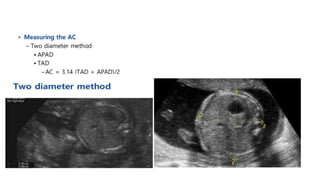
Fetal Biometry parameters
Stepwise Approach
1. Fetal lie and presentation
2. Fetal cardiac activity
3. Number of fetuses in the uterus
4. Adequacy of amniotic fluid
5. Localization of the placenta
6. Fetal biometry
2.Fetal cardiac activity
•
5.Amniotic fluid Estimation
Fetal biometry
Fetal biometric parameters are antenatal
ultrasound measurements that are used to
indirectly assess the growth and well being
of the fetus.
Standard parameters
Biparietal diameter
• The BPD is the maximum diameter of a transverse
section of the fetal skull at the level of the parietal
eminences.
• Measured from the outer edge of the proximal skull to
the inner edge of the distal skull at the level of thalami &
cavum septum pellucidum.
• Easy to obtain.
• More accurate than CRL .
• More accurate in predicting EDD than LMP.
Problems
• Incorrect angle
• Incorrect rotation
• Incorrect level
• Midline not
horizontal
Head Circumference:
Abdominal Circumference
• The single most important measurement to make in late pregnancy. It reflects more of fetal size and weight
rather than age. Serial measurements are useful in monitoring growth of the fetus.
Femoral Length
FL
Assigning GA in second and third trimester
• In the second and third trimesters, gestational age can be assigned
based on a single measurement, such as the BPD,corrected BPD,HC,
or FL.
• Head measurements that take into account the shape of the fetal head
namely, the corrected BPD and the HC are more accurate than the BPD
alone or FL alone in the second trimester.
• Accuracy of the corrected BPD and the HC before 20 weeks is
approximately ±1.2 weeks.
• By the end of the third trimester, the accuracy of gestational age
estimation by head measurements is about ±3.5 weeks
• The accuracy of the FL is similar to that of head
measurements by third trimester.
• The AC is a poor predictor of gestational age, particularly later in
pregnancy, and should not be used on its own to assign gestational
age
• Composite age formulas estimate gestational age via two or more
fetal measurements such as the BPD, HC, FL, and AC ,the accuracy of gestational
age estimation using these composite age formulas is similar to the
accuracy of the corrected BPD and HC and is more accurate than age estimation
using the FL.
• One drawback is the potential to miss an abnormal measurement or anomaly, for
example,if the fetal head is abnormally small and the FL and AC are normal for
gestational age, the composite age formula that incorporates measurements of
the BPD,HC,FL,and AC will be an underestimation of the true age.
• Formulas that use measurements of the fetal head, abdomen, and
femur have a mean error of 15% (±2 standard deviations [SDs]).
• Formulas that use fewer than three measurements of fetal body
parts perform less well(i.e.,have larger standard deviations).
• Adding other measurements to the head, abdomen, and femur, such
as the thigh circumference or thickness of thigh soft tissue or
three-dimensional volume calculations, does not improve accuracy of weight estimation.
• Despite considerable improvements in sonographic equipment, the
accuracy of estimating fetal weight has not changed since the development of formulas 3 decades
ago.
The fetal biometry necessary for measuring the fetal weight was as follows:
• Sheppard (BPD, AC),
• Campbell (AC),
• Hadlockl-I(AC, FL),
• Hadlock II (BPD, AC, FL),
• Hadlock III (HC, AC, FLx HC),
• Hadlock IV (BPD, HC, AC, FL)}
IUGR
• The “normal” neonate is one whose birth weight is between the 10th
and 90th percentile as per the gestational age, gender and race with
no feature of malnutrition and growth retardation.
• IUGR is a pathology involving reduced fetal growth potential of a
specific infant as per the race and gender of the fetus
Definition:
• Most commonly defined as a birth weight below the 10th percentile
considering gender and gestational age; a birth weight lower than
2500 grams in a pregnancy of 37 weeks or more; the birth weight is
more than 2 SD below the average mean.
Types:
Two different types of IUGR:
 Symmetrical(primary):
• Symmetric IUGR is characterized by all internal organs being reduced in
size.
• indicates that the fetus has developed slowly throughout the duration of
the pregnancy and was thus affected from a very early stage
• 20–30% of all cases of IUGR
• Causes: early intrauterine infections cytomegalovirus, rubella or
toxoplasmosis, chromosomal abnormalities,, anemia and maternal
substance abuse
 Asymmetric (secondary):
• characterized by the head and brain being normal in size, but the
abdomen is smaller
• accounts for 70–80% of all cases of IUGR
• this is not evident until the third trimester.
• The most common causes are placental insufficiency and pre-
eclampsia.
Small for gestational age (SGA):
• SGA definition has been used for those neonates whose birth weight is less
than the 10th percentile for that particular gestational age or two standard
deviations below the population norms on the growth charts
• This definition considers only the birth weight without any consideration of
the impaired development.
• 70% of the newborns with a birth weight below the 10th percentile are
small in their constitutional factors including maternal height, weight,
ethnicity, and parity, although they are proportionate, healthy, well
developed and well nourished (e.g. born to parents who are small and/or
into an ethnic population that is smaller than the reference population)
• Intrauterine fetal weight is usually determined according to the
relevant formulas (most often M. J. Shepard's and F. P. Hadlock's)
which include BPD, HC, FL and AC measurements.
• The parameter classically affected is AC, so the highest diagnostic
accuracy of IUGR is achieved when this dimension is used.
• The sensitivity of the latter examination is as high as 95% if the AC
value during the measurement is below the 2.5th percentile
• Thus, ultrasound fetal biometry remains the “golden standard” for
assessing IUGR in case of singleton and multiple pregnancies. The
EFW, for example, has fairly high sensitivity of 89% for FGR,but its
positive predictive value is only 45%.
• Supplementary ultrasound examination methods, e.g., Doppler test
and measurements of the quantity of amniotic fluid, are helpful in
providing additional information about fetal growth and development
during the prenatal period.
References:
• Ultrasound in Obstetrics and gynecology: A Practical Approach
• Fetal biometry: Relevance in obstetrical practice
• Callen's Ultrasonography in Obstetrics and Gynecology
Thank You!!!

Fetal biometry parameters lk

  • 1.
  • 3.
    Stepwise Approach 1. Fetallie and presentation 2. Fetal cardiac activity 3. Number of fetuses in the uterus 4. Adequacy of amniotic fluid 5. Localization of the placenta 6. Fetal biometry
  • 6.
  • 9.
  • 10.
    Fetal biometry Fetal biometricparameters are antenatal ultrasound measurements that are used to indirectly assess the growth and well being of the fetus.
  • 11.
  • 12.
    Biparietal diameter • TheBPD is the maximum diameter of a transverse section of the fetal skull at the level of the parietal eminences. • Measured from the outer edge of the proximal skull to the inner edge of the distal skull at the level of thalami & cavum septum pellucidum. • Easy to obtain. • More accurate than CRL . • More accurate in predicting EDD than LMP.
  • 17.
    Problems • Incorrect angle •Incorrect rotation • Incorrect level • Midline not horizontal
  • 18.
  • 22.
    Abdominal Circumference • Thesingle most important measurement to make in late pregnancy. It reflects more of fetal size and weight rather than age. Serial measurements are useful in monitoring growth of the fetus.
  • 31.
  • 32.
  • 37.
    Assigning GA insecond and third trimester • In the second and third trimesters, gestational age can be assigned based on a single measurement, such as the BPD,corrected BPD,HC, or FL. • Head measurements that take into account the shape of the fetal head namely, the corrected BPD and the HC are more accurate than the BPD alone or FL alone in the second trimester. • Accuracy of the corrected BPD and the HC before 20 weeks is approximately ±1.2 weeks.
  • 38.
    • By theend of the third trimester, the accuracy of gestational age estimation by head measurements is about ±3.5 weeks • The accuracy of the FL is similar to that of head measurements by third trimester. • The AC is a poor predictor of gestational age, particularly later in pregnancy, and should not be used on its own to assign gestational age
  • 39.
    • Composite ageformulas estimate gestational age via two or more fetal measurements such as the BPD, HC, FL, and AC ,the accuracy of gestational age estimation using these composite age formulas is similar to the accuracy of the corrected BPD and HC and is more accurate than age estimation using the FL. • One drawback is the potential to miss an abnormal measurement or anomaly, for example,if the fetal head is abnormally small and the FL and AC are normal for gestational age, the composite age formula that incorporates measurements of the BPD,HC,FL,and AC will be an underestimation of the true age.
  • 43.
    • Formulas thatuse measurements of the fetal head, abdomen, and femur have a mean error of 15% (±2 standard deviations [SDs]). • Formulas that use fewer than three measurements of fetal body parts perform less well(i.e.,have larger standard deviations). • Adding other measurements to the head, abdomen, and femur, such as the thigh circumference or thickness of thigh soft tissue or three-dimensional volume calculations, does not improve accuracy of weight estimation. • Despite considerable improvements in sonographic equipment, the accuracy of estimating fetal weight has not changed since the development of formulas 3 decades ago.
  • 45.
    The fetal biometrynecessary for measuring the fetal weight was as follows: • Sheppard (BPD, AC), • Campbell (AC), • Hadlockl-I(AC, FL), • Hadlock II (BPD, AC, FL), • Hadlock III (HC, AC, FLx HC), • Hadlock IV (BPD, HC, AC, FL)}
  • 47.
    IUGR • The “normal”neonate is one whose birth weight is between the 10th and 90th percentile as per the gestational age, gender and race with no feature of malnutrition and growth retardation. • IUGR is a pathology involving reduced fetal growth potential of a specific infant as per the race and gender of the fetus
  • 48.
    Definition: • Most commonlydefined as a birth weight below the 10th percentile considering gender and gestational age; a birth weight lower than 2500 grams in a pregnancy of 37 weeks or more; the birth weight is more than 2 SD below the average mean.
  • 49.
    Types: Two different typesof IUGR:  Symmetrical(primary): • Symmetric IUGR is characterized by all internal organs being reduced in size. • indicates that the fetus has developed slowly throughout the duration of the pregnancy and was thus affected from a very early stage • 20–30% of all cases of IUGR • Causes: early intrauterine infections cytomegalovirus, rubella or toxoplasmosis, chromosomal abnormalities,, anemia and maternal substance abuse
  • 50.
     Asymmetric (secondary): •characterized by the head and brain being normal in size, but the abdomen is smaller • accounts for 70–80% of all cases of IUGR • this is not evident until the third trimester. • The most common causes are placental insufficiency and pre- eclampsia.
  • 51.
    Small for gestationalage (SGA): • SGA definition has been used for those neonates whose birth weight is less than the 10th percentile for that particular gestational age or two standard deviations below the population norms on the growth charts • This definition considers only the birth weight without any consideration of the impaired development. • 70% of the newborns with a birth weight below the 10th percentile are small in their constitutional factors including maternal height, weight, ethnicity, and parity, although they are proportionate, healthy, well developed and well nourished (e.g. born to parents who are small and/or into an ethnic population that is smaller than the reference population)
  • 52.
    • Intrauterine fetalweight is usually determined according to the relevant formulas (most often M. J. Shepard's and F. P. Hadlock's) which include BPD, HC, FL and AC measurements. • The parameter classically affected is AC, so the highest diagnostic accuracy of IUGR is achieved when this dimension is used. • The sensitivity of the latter examination is as high as 95% if the AC value during the measurement is below the 2.5th percentile
  • 53.
    • Thus, ultrasoundfetal biometry remains the “golden standard” for assessing IUGR in case of singleton and multiple pregnancies. The EFW, for example, has fairly high sensitivity of 89% for FGR,but its positive predictive value is only 45%. • Supplementary ultrasound examination methods, e.g., Doppler test and measurements of the quantity of amniotic fluid, are helpful in providing additional information about fetal growth and development during the prenatal period.
  • 54.
    References: • Ultrasound inObstetrics and gynecology: A Practical Approach • Fetal biometry: Relevance in obstetrical practice • Callen's Ultrasonography in Obstetrics and Gynecology
  • 55.