MRI is a valuable tool for assessing endometrial cancer by depicting tumor size, extension into the myometrium or parametrium, cervical invasion, and lymphadenopathy. It plays an important role in pre-operative planning by identifying high-risk features that may require lymph node dissection or adjuvant therapy. While endometrial cancer is surgically staged, MRI can accurately assess key features to guide treatment. It can also differentiate endometrial cancer from benign conditions like hyperplasia, adenomyosis, or fibroids.
Description of various ultrasound features of benign and suspicious thyroid nodules with multiple ultrasound systems for risk stratification of malignancy.
New Directions in Targeted Therapeutic Approaches for Older Adults With Mantl...i3 Health
i3 Health is pleased to make the speaker slides from this activity available for use as a non-accredited self-study or teaching resource.
This slide deck presented by Dr. Kami Maddocks, Professor-Clinical in the Division of Hematology and
Associate Division Director for Ambulatory Operations
The Ohio State University Comprehensive Cancer Center, will provide insight into new directions in targeted therapeutic approaches for older adults with mantle cell lymphoma.
STATEMENT OF NEED
Mantle cell lymphoma (MCL) is a rare, aggressive B-cell non-Hodgkin lymphoma (NHL) accounting for 5% to 7% of all lymphomas. Its prognosis ranges from indolent disease that does not require treatment for years to very aggressive disease, which is associated with poor survival (Silkenstedt et al, 2021). Typically, MCL is diagnosed at advanced stage and in older patients who cannot tolerate intensive therapy (NCCN, 2022). Although recent advances have slightly increased remission rates, recurrence and relapse remain very common, leading to a median overall survival between 3 and 6 years (LLS, 2021). Though there are several effective options, progress is still needed towards establishing an accepted frontline approach for MCL (Castellino et al, 2022). Treatment selection and management of MCL are complicated by the heterogeneity of prognosis, advanced age and comorbidities of patients, and lack of an established standard approach for treatment, making it vital that clinicians be familiar with the latest research and advances in this area. In this activity chaired by Michael Wang, MD, Professor in the Department of Lymphoma & Myeloma at MD Anderson Cancer Center, expert faculty will discuss prognostic factors informing treatment, the promising results of recent trials in new therapeutic approaches, and the implications of treatment resistance in therapeutic selection for MCL.
Target Audience
Hematology/oncology fellows, attending faculty, and other health care professionals involved in the treatment of patients with mantle cell lymphoma (MCL).
Learning Objectives
1.) Identify clinical and biological prognostic factors that can guide treatment decision making for older adults with MCL
2.) Evaluate emerging data on targeted therapeutic approaches for treatment-naive and relapsed/refractory MCL and their applicability to older adults
3.) Assess mechanisms of resistance to targeted therapies for MCL and their implications for treatment selection
Title: Sense of Taste
Presenter: Dr. Faiza, Assistant Professor of Physiology
Qualifications:
MBBS (Best Graduate, AIMC Lahore)
FCPS Physiology
ICMT, CHPE, DHPE (STMU)
MPH (GC University, Faisalabad)
MBA (Virtual University of Pakistan)
Learning Objectives:
Describe the structure and function of taste buds.
Describe the relationship between the taste threshold and taste index of common substances.
Explain the chemical basis and signal transduction of taste perception for each type of primary taste sensation.
Recognize different abnormalities of taste perception and their causes.
Key Topics:
Significance of Taste Sensation:
Differentiation between pleasant and harmful food
Influence on behavior
Selection of food based on metabolic needs
Receptors of Taste:
Taste buds on the tongue
Influence of sense of smell, texture of food, and pain stimulation (e.g., by pepper)
Primary and Secondary Taste Sensations:
Primary taste sensations: Sweet, Sour, Salty, Bitter, Umami
Chemical basis and signal transduction mechanisms for each taste
Taste Threshold and Index:
Taste threshold values for Sweet (sucrose), Salty (NaCl), Sour (HCl), and Bitter (Quinine)
Taste index relationship: Inversely proportional to taste threshold
Taste Blindness:
Inability to taste certain substances, particularly thiourea compounds
Example: Phenylthiocarbamide
Structure and Function of Taste Buds:
Composition: Epithelial cells, Sustentacular/Supporting cells, Taste cells, Basal cells
Features: Taste pores, Taste hairs/microvilli, and Taste nerve fibers
Location of Taste Buds:
Found in papillae of the tongue (Fungiform, Circumvallate, Foliate)
Also present on the palate, tonsillar pillars, epiglottis, and proximal esophagus
Mechanism of Taste Stimulation:
Interaction of taste substances with receptors on microvilli
Signal transduction pathways for Umami, Sweet, Bitter, Sour, and Salty tastes
Taste Sensitivity and Adaptation:
Decrease in sensitivity with age
Rapid adaptation of taste sensation
Role of Saliva in Taste:
Dissolution of tastants to reach receptors
Washing away the stimulus
Taste Preferences and Aversions:
Mechanisms behind taste preference and aversion
Influence of receptors and neural pathways
Impact of Sensory Nerve Damage:
Degeneration of taste buds if the sensory nerve fiber is cut
Abnormalities of Taste Detection:
Conditions: Ageusia, Hypogeusia, Dysgeusia (parageusia)
Causes: Nerve damage, neurological disorders, infections, poor oral hygiene, adverse drug effects, deficiencies, aging, tobacco use, altered neurotransmitter levels
Neurotransmitters and Taste Threshold:
Effects of serotonin (5-HT) and norepinephrine (NE) on taste sensitivity
Supertasters:
25% of the population with heightened sensitivity to taste, especially bitterness
Increased number of fungiform papillae
Acute scrotum is a general term referring to an emergency condition affecting the contents or the wall of the scrotum.
There are a number of conditions that present acutely, predominantly with pain and/or swelling
A careful and detailed history and examination, and in some cases, investigations allow differentiation between these diagnoses. A prompt diagnosis is essential as the patient may require urgent surgical intervention
Testicular torsion refers to twisting of the spermatic cord, causing ischaemia of the testicle.
Testicular torsion results from inadequate fixation of the testis to the tunica vaginalis producing ischemia from reduced arterial inflow and venous outflow obstruction.
The prevalence of testicular torsion in adult patients hospitalized with acute scrotal pain is approximately 25 to 50 percent
These simplified slides by Dr. Sidra Arshad present an overview of the non-respiratory functions of the respiratory tract.
Learning objectives:
1. Enlist the non-respiratory functions of the respiratory tract
2. Briefly explain how these functions are carried out
3. Discuss the significance of dead space
4. Differentiate between minute ventilation and alveolar ventilation
5. Describe the cough and sneeze reflexes
Study Resources:
1. Chapter 39, Guyton and Hall Textbook of Medical Physiology, 14th edition
2. Chapter 34, Ganong’s Review of Medical Physiology, 26th edition
3. Chapter 17, Human Physiology by Lauralee Sherwood, 9th edition
4. Non-respiratory functions of the lungs https://academic.oup.com/bjaed/article/13/3/98/278874
Prix Galien International 2024 Forum ProgramLevi Shapiro
June 20, 2024, Prix Galien International and Jerusalem Ethics Forum in ROME. Detailed agenda including panels:
- ADVANCES IN CARDIOLOGY: A NEW PARADIGM IS COMING
- WOMEN’S HEALTH: FERTILITY PRESERVATION
- WHAT’S NEW IN THE TREATMENT OF INFECTIOUS,
ONCOLOGICAL AND INFLAMMATORY SKIN DISEASES?
- ARTIFICIAL INTELLIGENCE AND ETHICS
- GENE THERAPY
- BEYOND BORDERS: GLOBAL INITIATIVES FOR DEMOCRATIZING LIFE SCIENCE TECHNOLOGIES AND PROMOTING ACCESS TO HEALTHCARE
- ETHICAL CHALLENGES IN LIFE SCIENCES
- Prix Galien International Awards Ceremony
Recomendações da OMS sobre cuidados maternos e neonatais para uma experiência pós-natal positiva.
Em consonância com os ODS – Objetivos do Desenvolvimento Sustentável e a Estratégia Global para a Saúde das Mulheres, Crianças e Adolescentes, e aplicando uma abordagem baseada nos direitos humanos, os esforços de cuidados pós-natais devem expandir-se para além da cobertura e da simples sobrevivência, de modo a incluir cuidados de qualidade.
Estas diretrizes visam melhorar a qualidade dos cuidados pós-natais essenciais e de rotina prestados às mulheres e aos recém-nascidos, com o objetivo final de melhorar a saúde e o bem-estar materno e neonatal.
Uma “experiência pós-natal positiva” é um resultado importante para todas as mulheres que dão à luz e para os seus recém-nascidos, estabelecendo as bases para a melhoria da saúde e do bem-estar a curto e longo prazo. Uma experiência pós-natal positiva é definida como aquela em que as mulheres, pessoas que gestam, os recém-nascidos, os casais, os pais, os cuidadores e as famílias recebem informação consistente, garantia e apoio de profissionais de saúde motivados; e onde um sistema de saúde flexível e com recursos reconheça as necessidades das mulheres e dos bebês e respeite o seu contexto cultural.
Estas diretrizes consolidadas apresentam algumas recomendações novas e já bem fundamentadas sobre cuidados pós-natais de rotina para mulheres e neonatos que recebem cuidados no pós-parto em unidades de saúde ou na comunidade, independentemente dos recursos disponíveis.
É fornecido um conjunto abrangente de recomendações para cuidados durante o período puerperal, com ênfase nos cuidados essenciais que todas as mulheres e recém-nascidos devem receber, e com a devida atenção à qualidade dos cuidados; isto é, a entrega e a experiência do cuidado recebido. Estas diretrizes atualizam e ampliam as recomendações da OMS de 2014 sobre cuidados pós-natais da mãe e do recém-nascido e complementam as atuais diretrizes da OMS sobre a gestão de complicações pós-natais.
O estabelecimento da amamentação e o manejo das principais intercorrências é contemplada.
Recomendamos muito.
Vamos discutir essas recomendações no nosso curso de pós-graduação em Aleitamento no Instituto Ciclos.
Esta publicação só está disponível em inglês até o momento.
Prof. Marcus Renato de Carvalho
www.agostodourado.com
Pulmonary Thromboembolism - etilogy, types, medical- Surgical and nursing man...VarunMahajani
Disruption of blood supply to lung alveoli due to blockage of one or more pulmonary blood vessels is called as Pulmonary thromboembolism. In this presentation we will discuss its causes, types and its management in depth.
Pulmonary Thromboembolism - etilogy, types, medical- Surgical and nursing man...
Presentation1, radiological imaging of endometrial carcinoma.
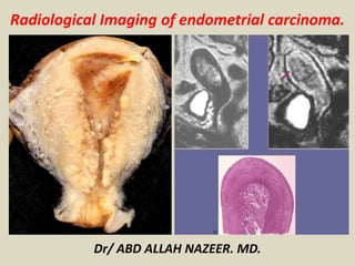
1. Dr/ ABD ALLAH NAZEER. MD.
Radiological Imaging of endometrial carcinoma.
2. Endometrial cancer
-4th most common cancer in women
-Most common gynecologic malignancy
-Peak incidence: 55 and 65 years
-Risk factors:
-Obesity.
-Nulliparity.
-Late menopause.
-Unopposed estrogen exposure.
-Tamoxifen.
3. -Over 80% of patients present with early stage
disease due to symptoms of abnormal uterine
bleeding, with an overall excellent prognosis.
-Endometrial cancer is traditionally surgically staged
and the treatment varies by stage, grade, regional
guidelines, and expertise.
-MRI is a valuable adjunct to pre-operative planning
as it can accurately depict endometrial cancer . The
goal of MRI is to identify patients preoperatively who
would benefit from abdominopelvic lymph node
dissection and adjuvant therapy while avoiding
overtreating early stage patients with unnecessary
lymphadenectomies.
4.
5.
6.
7.
8.
9.
10. Imaging techniques
Ultrasound
TVUS is often used for the initial evaluation in women with history
of postmenopausal bleeding because it is quick, inexpensive, and
does not expose the patient to ionizing radiation. ECs typically
present as thickening of the endometrium and TVUS diagnosis of
endometrial cancer is based on endometrial thickness that is
measured in the anteroposterior dimension. The sensitivity and
specificity of TVUS for detecting EC approach 96% and 61%,
respectively, when an endometrial thickness threshold of 5 mm, in
postmenopausal women, is used to define abnormal endometrial
thickening. A meta-analysis suggested a sensitivity of 68-100% and a
specificity of 71-90% for subjective assessment of deep myometrial
invasion. Furthermore, the negative predictive value of a thin
endometrium is very high. Also, cancer is more likely when the
endometrium has a heterogeneous echotexture and irregular or
poorly defined margins.
11. A 67-year-old female with endometrial cancer. (A) Longitudinal transabdominal scan. (B) Transvaginal
scan and a 3-D reconstructed ultrasonography image (C) through the uterus demonstrate a thickened and
heterogeneous endometrium measuring 2.0 cm (arrows). Note regular endometrial–myometrial border
with no signs of invasion (arrowheads). (D) Note increased vascularity in the color Doppler US (black arrow).
15. Ultrasound images of a female patient, aged 55 years with malignant thickened
endometrium (confirmed to be endometrial adenocarcinoma by pathological examination).
B-mode ultrasound image (A), Power Doppler image (B) and TV elastogram (C) show
endometrial thickness = 18 mm with increase vascularity, dark blue color and SR = 11.54.
16.
17. Image planes for high-resolution T2-weighted imaging of the uterus. Initial high resolution
T2-weighted sagittal images of the pelvis are obtained. Axial oblique images are obtained
by scanning perpendicular (dotted blue lines) to the long axis of the endometrial cavity
(solid red line). These sequences can then be used for accurate assessment of myometrial
invasion by tumour. If there is suspected cervical invasion, further imaging is recommended
on a plane perpendicular to the long axis of the endocervical canal.
44. Grade I endometrial carcinoma
on top of endometrial
hyperplasia. A and B Sagittal and
axial T2-weighted images of the
uterus show endometrial mass
with bright signal intensity filling
and distending the endometrial
cavity with thinning of the
myometrium. C Axial T1-
weighted. D Axial Gd- T1-
weighted image showing the
endometrial mass enhancing less
than the myometrium. E and F
DW-MRI and corresponding ADC
map. There is heterogeneous iso-
intense signal with foci of high
signal intensity and
corresponding low signal on ADC
map (consistent with endometrial
hyperplasia with foci of
malignant transformation). ADC
value was 1.4 × 10⁻³ mm2/s in
areas of endometrial hyperplasia
and 0.74 × 10⁻³ mm2/s in areas
of endometrial carcinoma.
45.
46.
47.
48.
49. Stage 11A (A) Sagittal T2W image show a hyperintense signal intensity tumor distending the endometrial cavity (arrow). (B) On T1W
post-contrast image, the tumor (arrow) is low in signal compared to the enhancing adjacent myometrium. It presents restricted diffusion
with high signal on DW images (C) and low signal on ADC map (D) (arrows). (E) It presents with high FDG uptake on FDG-PET/CT (arrow)
50.
51.
52.
53.
54.
55. (A,B) cervical
cancer and (C,D)
endometrial
cancer on
sagittal T2-
weighted (left
column) and
contrast-
enhanced T1-
weighted (right
column) MRI
images.
Abbreviations:
T, tumor; M,
myometrium.
56. 77-year-old female patient with
pathologically diagnosed G2
endometrioid endometrial carcinoma. A
and B: Sagittal and axial T2 W images
showing a hyper-intense endometrial
mass relative to the myometrium
occupying the fundus and anterior wall
of the uterine cavity. C and D: Sagittal
and Axial Delayed Gd-T1 W images
showing the endometrial mass to be
more conspicuous and contrasting
against the enhanced myometrium with
deep myometrial invasion >50%. E, F:
Axial ADC map and corresponding high b
value DW image showing diffusion
restriction of the mass in the form of low
signal on ADC map and high signal on
DWI. The ADC value 0.75 × 10⁻³ mm2/s.
On DWI, there is well delineation of
depth of myometrial invasion >50%.
Note the associated Right ovarian
endometrioid carcinoma (∗).
64. A) Axial T2-weighted image shows a 6 cm sized endometrial mass with multiple
hyperintense cystic lesions (arrowhead) and hypointense fibrous stroma (arrow) .
69. Summary.
- MRI is excellent at depicting uterine anatomy and
assessing endometrial cancer.
-Tumor size and extension
-Myometrial invasion
-Parametrial/cervical invasion
-Lymphadenopathy
-Although currently endometrial cancer is staged
surgically, MRI does play a critical role in pre-operative
planning.
-Differentiating endometrial cancer from co-existing
endometrial hyperplasia, adenomyosis, fibroids,
and polyps is important in the accurate assessment
of endometrial cancer.