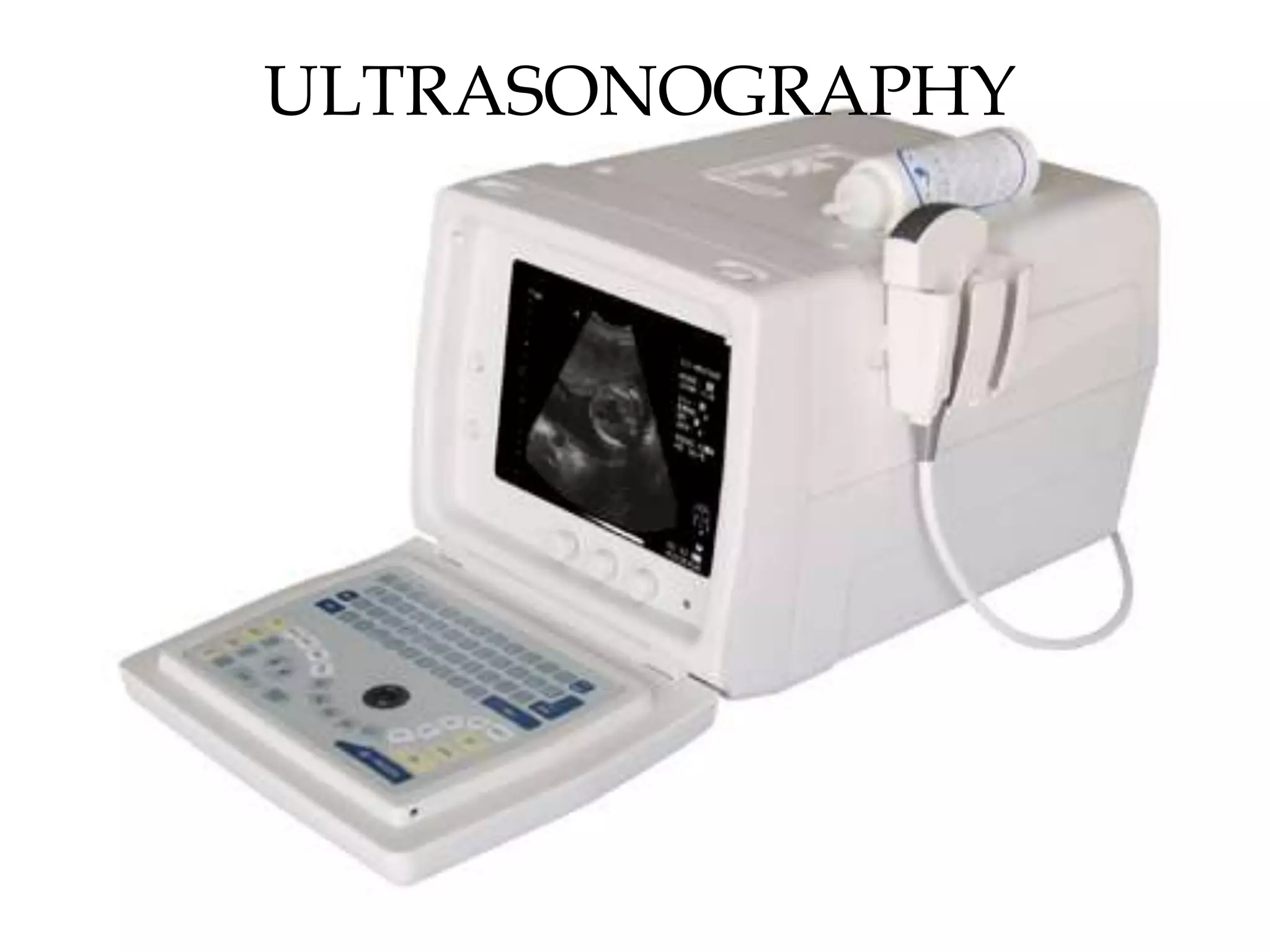
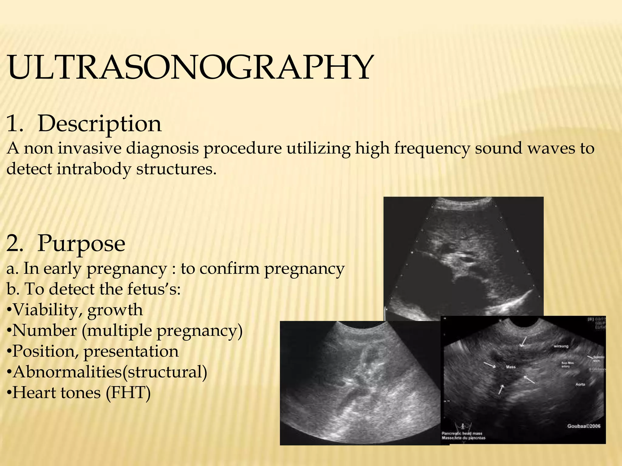
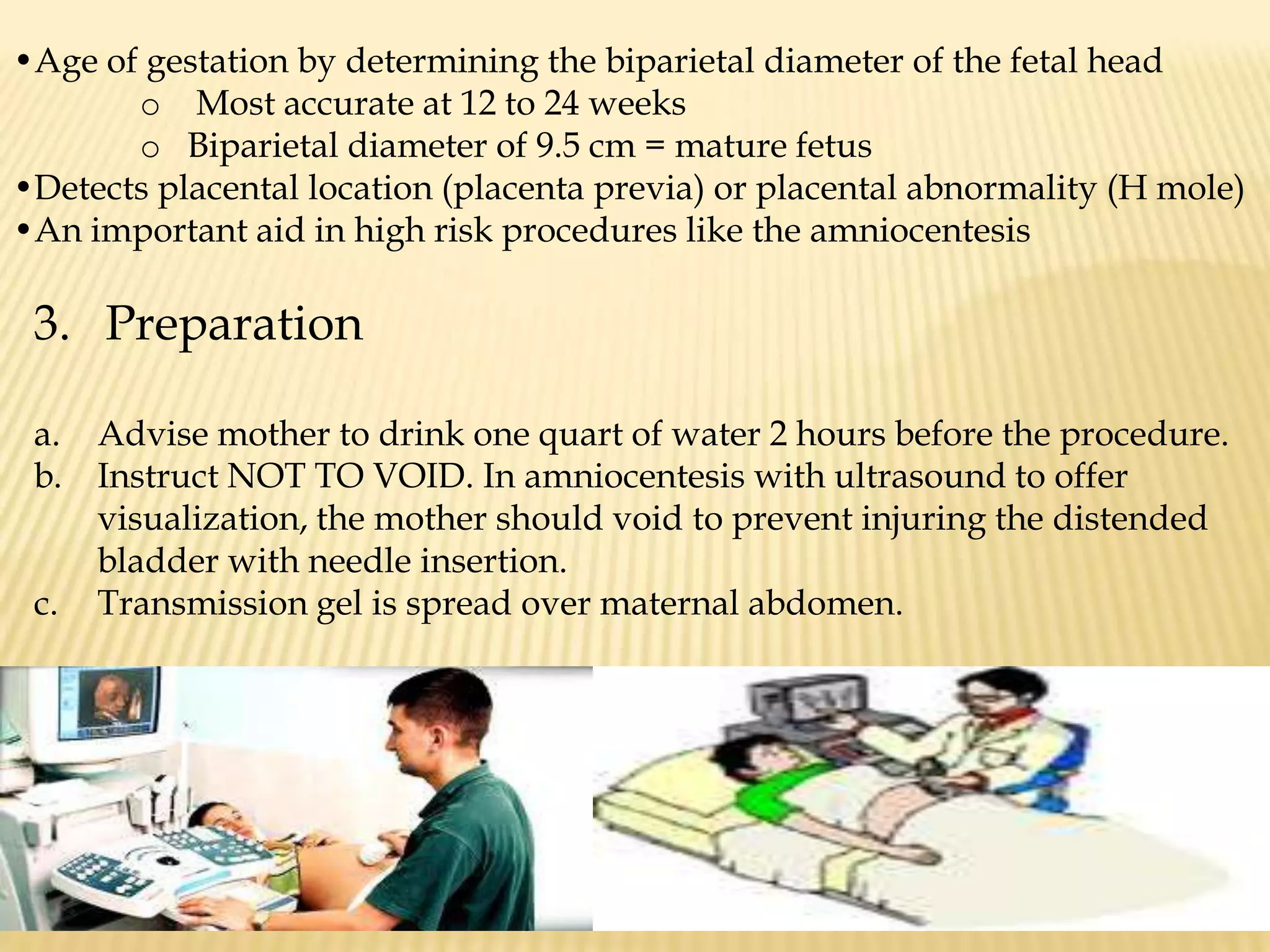
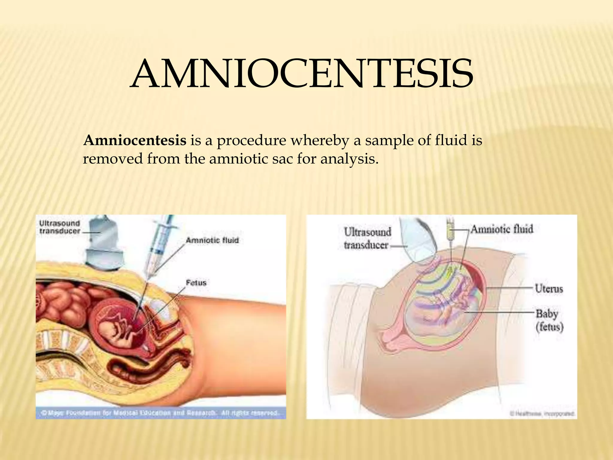
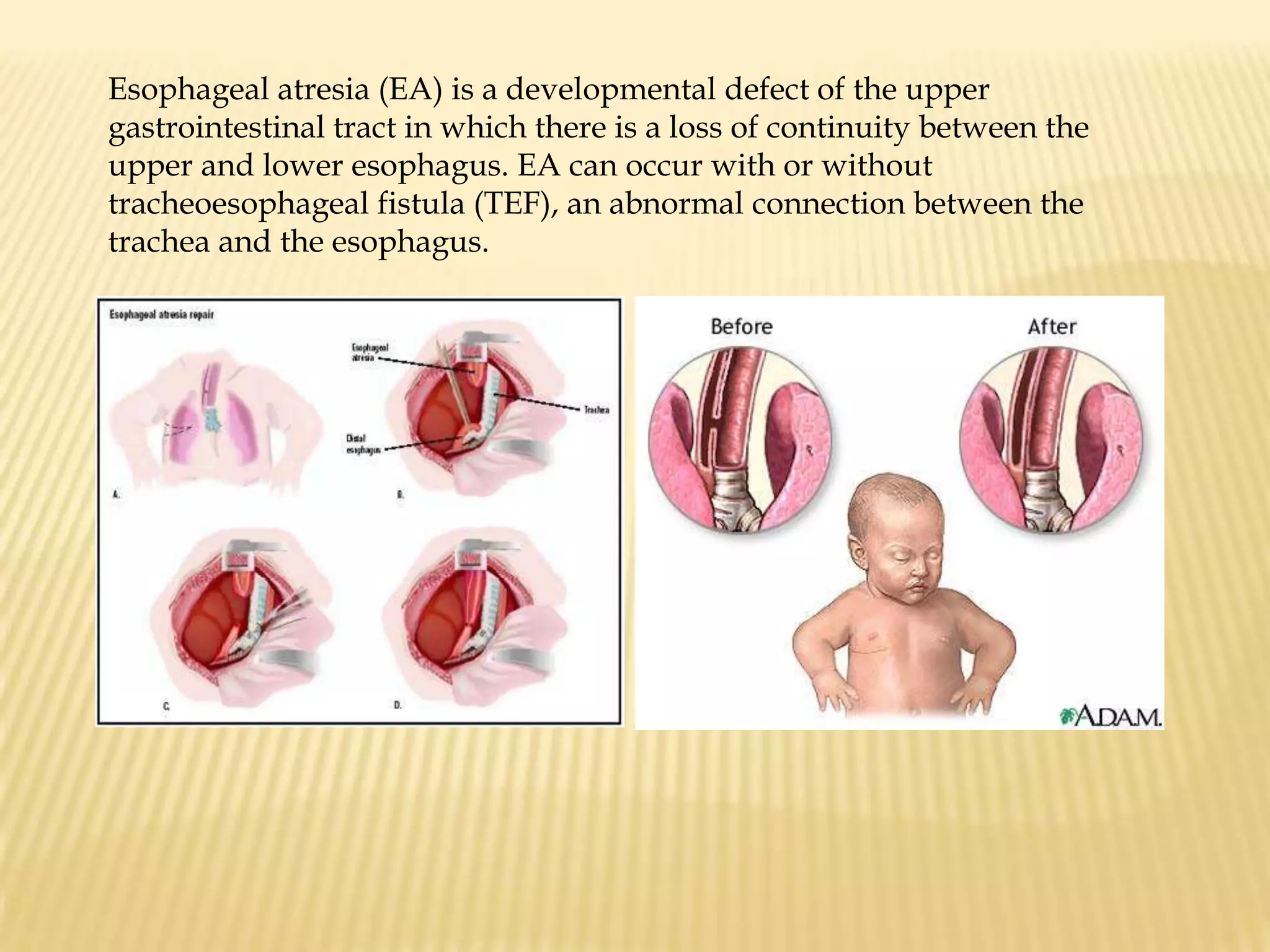
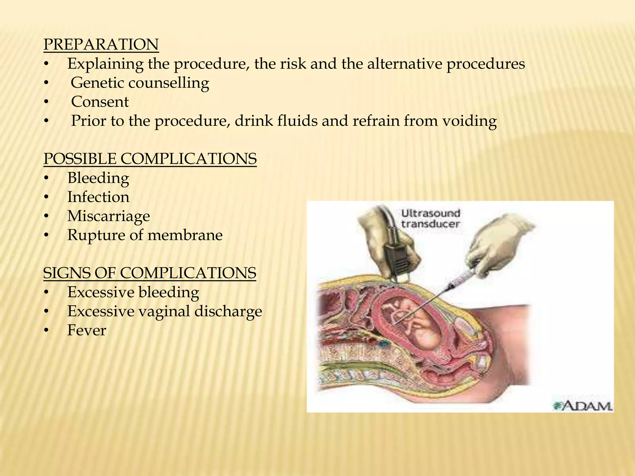
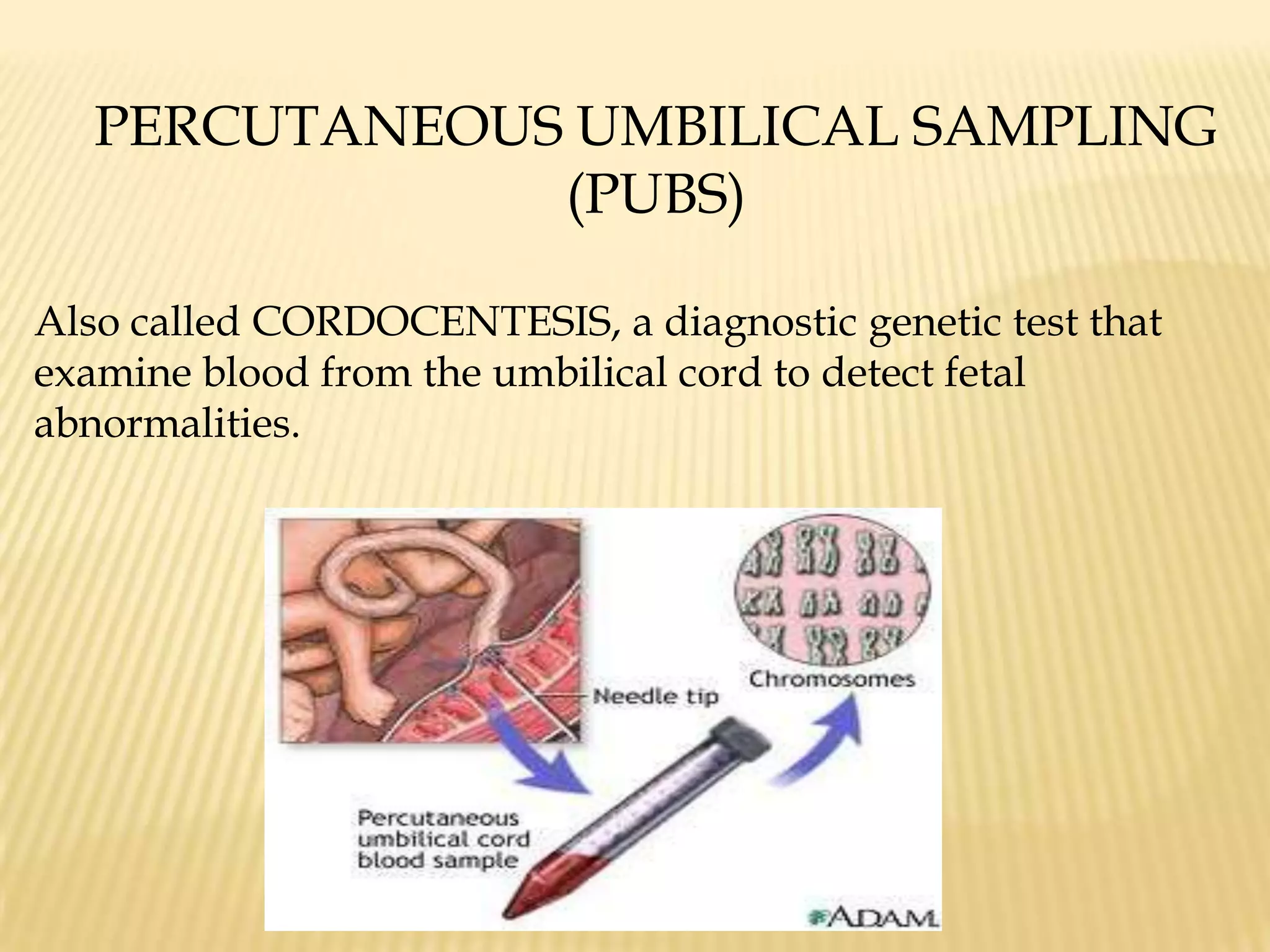
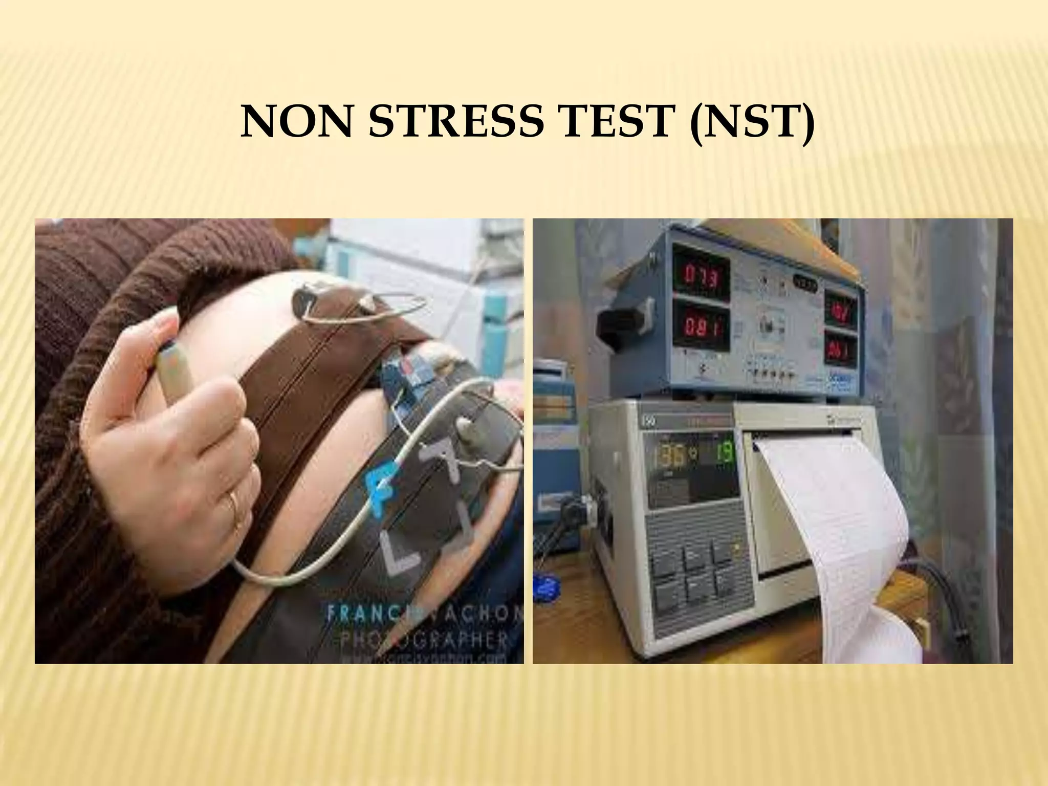
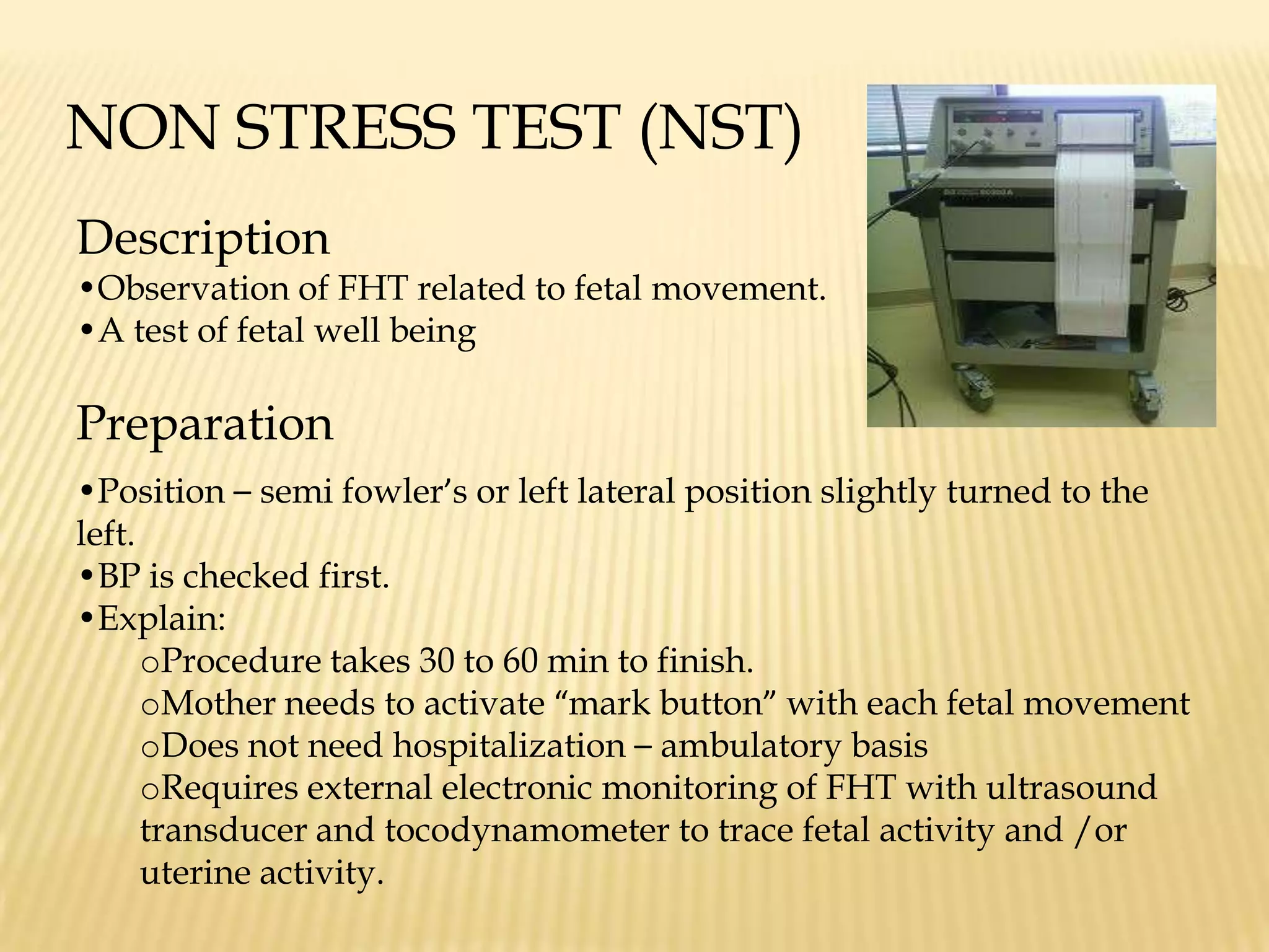
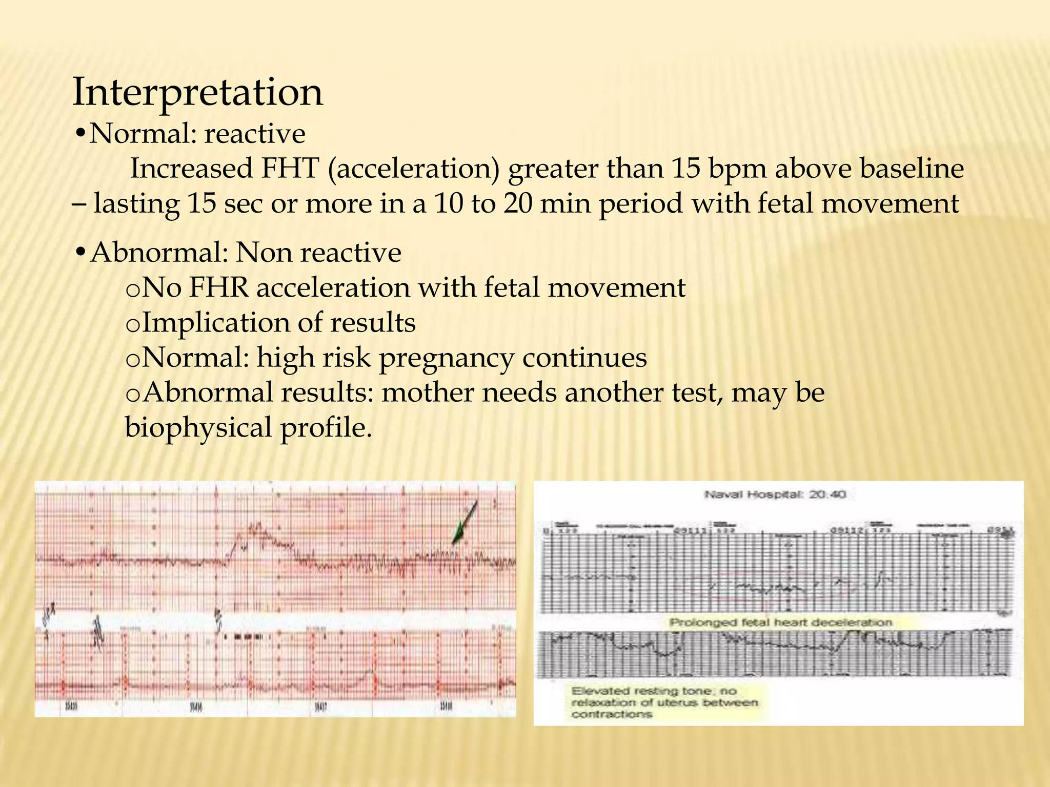
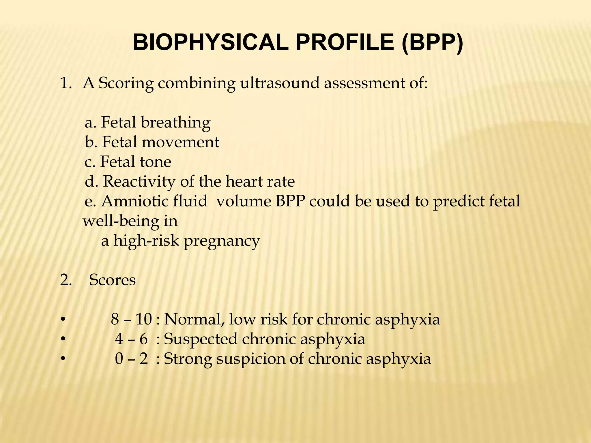
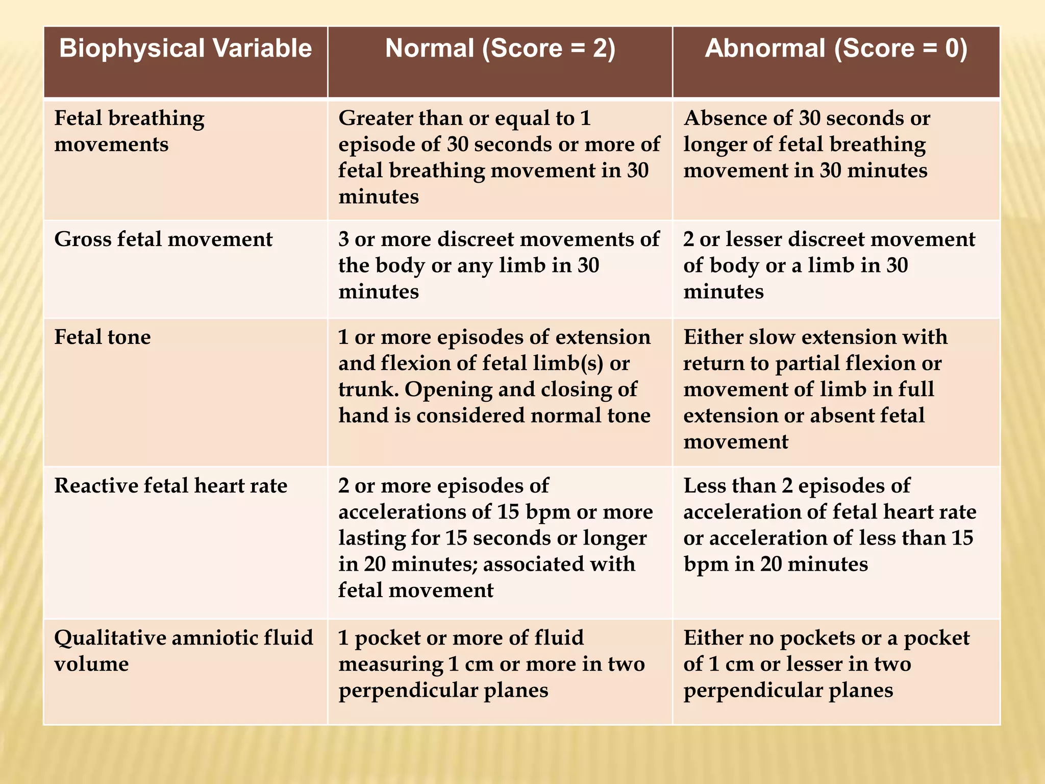
Diagnostic test in high risk pregnancy/prenatal determination of fetal status
ULTRASONOGRAPHY
ULTRASONOGRAPHY Description A non invasive diagnosis procedure utilizing high frequency sound waves to detect intrabody structures.Purposea. In early pregnancy : to confirm pregnancyb. To detect the fetus’s:Viability, growth
Number (multiple pregnancy)
Position, presentation
Abnormalities(structural)
Heart tones (FHT)Age of gestation by determining the biparietal diameter of the fetal head
    Most accurate at 12 to 24 weeks
Biparietal diameter of 9.5 cm = mature fetus
Detects placental location (placenta previa) or placental abnormality (H mole)
An important aid in high risk procedures like the amniocentesis PreparationAdvise mother to drink one quart of water 2 hours before the procedure.Instruct NOT TO VOID. In amniocentesis with ultrasound to offer visualization, the mother should void to prevent injuring the distended bladder with needle insertion.Transmission gel is spread over maternal abdomen.
d.   Psychological  support is given to the mother/father (couple):Explain the reasons for the procedure together with its benefits and the preparations.
Explain that there is no known risk with infrequent and brief exposure to high frequency sound waves.
    Encourage verbalization of fears and concerns. Explain further that:
Procedure is non invasive and safe for mother and fetus.
Confinement is not needed
No need for dye and here is no X ray irradiation.
Procedures take a short time (about 30 min) to accomplish.FETOSCOPYFetoscopy is a procedure to allow the visualization of the baby while it is still in the uterus with the aide of fetoscope, a thin flexible instrument.  Fetoscopy is usually done around the 18th week of pregnancy because the placenta and the baby is well developed. PURPOSES OF FETOSCOPY To evaluate fetal birth defects, such as spina bifida
 To collect blood samples from the umbilical cord
 To collect skin tissue samples from the babyPREPARATION Do not eat or drink for 8 hours before the procedure.
 The doctor will check the fetal heart rate.
 You may be asked to take medicine like Demerol to prevent the baby  from       moving around a lot. You are required to remove your clothes around your waist down – leaving   your stomach exposed. You need to lie down but your upper body will be slightly raised. Your stomach will be cleaned and a local anesthetic will be given to the   area where the incision will be done.Doctor will determine the position of the fetus and the placenta using ultrasound.
 Your health care provider will determine the best place to make an incision and then examine the baby.
 After the procedure, fetoscope is removed and the incision will be closed.
 An ultrasound will be done again, to check the status of the baby.RISKS INVOLVED Miscarriage
 Excessive bleeding, infection or excessive leakage  of the amniotic fluid
 Preterm rupture which can cause early delivery of the baby

NCM Skills 3/3/2011

  • 1.
    Diagnostic test inhigh risk pregnancy/prenatal determination of fetal status
  • 2.
  • 3.
    ULTRASONOGRAPHY Description Anon invasive diagnosis procedure utilizing high frequency sound waves to detect intrabody structures.Purposea. In early pregnancy : to confirm pregnancyb. To detect the fetus’s:Viability, growth
  • 4.
  • 5.
  • 6.
  • 7.
    Heart tones (FHT)Ageof gestation by determining the biparietal diameter of the fetal head
  • 8.
    Most accurate at 12 to 24 weeks
  • 9.
    Biparietal diameter of9.5 cm = mature fetus
  • 10.
    Detects placental location(placenta previa) or placental abnormality (H mole)
  • 11.
    An important aidin high risk procedures like the amniocentesis PreparationAdvise mother to drink one quart of water 2 hours before the procedure.Instruct NOT TO VOID. In amniocentesis with ultrasound to offer visualization, the mother should void to prevent injuring the distended bladder with needle insertion.Transmission gel is spread over maternal abdomen.
  • 12.
    d. Psychological support is given to the mother/father (couple):Explain the reasons for the procedure together with its benefits and the preparations.
  • 13.
    Explain that thereis no known risk with infrequent and brief exposure to high frequency sound waves.
  • 14.
    Encourage verbalization of fears and concerns. Explain further that:
  • 15.
    Procedure is noninvasive and safe for mother and fetus.
  • 16.
  • 17.
    No need fordye and here is no X ray irradiation.
  • 18.
    Procedures take ashort time (about 30 min) to accomplish.FETOSCOPYFetoscopy is a procedure to allow the visualization of the baby while it is still in the uterus with the aide of fetoscope, a thin flexible instrument. Fetoscopy is usually done around the 18th week of pregnancy because the placenta and the baby is well developed. PURPOSES OF FETOSCOPY To evaluate fetal birth defects, such as spina bifida
  • 19.
    To collectblood samples from the umbilical cord
  • 20.
    To collectskin tissue samples from the babyPREPARATION Do not eat or drink for 8 hours before the procedure.
  • 21.
    The doctorwill check the fetal heart rate.
  • 22.
    You maybe asked to take medicine like Demerol to prevent the baby from moving around a lot. You are required to remove your clothes around your waist down – leaving your stomach exposed. You need to lie down but your upper body will be slightly raised. Your stomach will be cleaned and a local anesthetic will be given to the area where the incision will be done.Doctor will determine the position of the fetus and the placenta using ultrasound.
  • 23.
    Your healthcare provider will determine the best place to make an incision and then examine the baby.
  • 24.
    After theprocedure, fetoscope is removed and the incision will be closed.
  • 25.
    An ultrasoundwill be done again, to check the status of the baby.RISKS INVOLVED Miscarriage
  • 26.
    Excessive bleeding,infection or excessive leakage of the amniotic fluid
  • 27.
    Preterm rupturewhich can cause early delivery of the baby
  • 28.
    Mixing themother and the baby’s bloodCOMPLICATIONS Moderate or severe abdominal pain or cramping
  • 29.
  • 30.
  • 31.
    Fluid orblood leaking from the vagina or from the cut in the belly AMNIOCENTESISAmniocentesis is a procedure whereby a sample of fluid is removed from the amniotic sac for analysis.
  • 32.
    AMNIOCENTESISEntering amniotic sacto aspirate amniotic fluid for a variety of diagnostic exams to detect fetal well being.
  • 33.
  • 34.
    Trauma: fetus, placenta, umbilical cord and maternal surface
  • 35.
  • 36.
  • 37.
  • 38.
    Prepare for ultrasonography:to locate placenta and to provide visualization to a blind procedure.
  • 39.
    Ultrasound in amniocentesis:client needs to void.
  • 40.
    Pelvic ultrasound only:client should not void Increase oral fluid: take 1 quart water 2 hours before.
  • 41.
    Prepare needle: G20-22; 3” -6”.
  • 42.
    Prepare for administrationof local anesthesia of abdomen.
  • 43.
    Provide psychological support.Amnioticfluid to be aspirated up to 30 ml at 15 – 18 weeks of gestation.Aftercare Monitor for 30-60 min
  • 44.
  • 45.
  • 46.
  • 47.
    Fever and chillsPurposesof amniotic fluid analysisTo determine the maturity of the lungs of the fetus Result – L/S ratio of 2:1 means mature lungs, done between the 32 to 36 AOG, evaluation of any abnormal AFP test done between the 15th and 20th weeks of pregnancy For genetic testing, chromosome analysis and evaluation of abnormal AFP testAlpha- fetoprotein (AFP) levels: increasing- high levels may indicate the presence of neural defects such as spina bifida or tracheoesophagealatresia.Low AFP level may indicate the potential force down syndrome
  • 48.
    Genetic disorders: forchromosomal studies, testing like hemophilia, sickle cell anemia
  • 49.
    Fetal distress: passageof meconium in cephalic presentation( not significant in breech presentation)
  • 50.
    spina bifidaA birthdefect (a congenital malformation) in which there is a bonydefect in the vertebral column so that part of the spinal cord, which isnormally protected within the vertebral column, is exposed. People withspina bifida can suffer from bladder and bowel incontinence, cognitive(learning) problems and limited mobility. Spina bifida is caused by the failure ofthe neural tube to close during embryonic development. The neural tube is theembryonal structure that gives rise to the brain and spinal cord. The risk ofspina bifida varies according to country, ethnic group and socioeconomicstatus. In the US as a whole, spina bifida occurs in about one in every 1-2,000births. The risk of spina bifida and other neural tube defects such asanencephaly can be decreased by women taking folic acid supplements beforethey conceive and during the first months of their pregnancy.
  • 51.
    Esophageal atresia (EA)is a developmental defect of the upper gastrointestinal tract in which there is a loss of continuity between the upper and lower esophagus. EA can occur with or without tracheoesophageal fistula (TEF), an abnormal connection between the trachea and the esophagus.
  • 52.
    X-RAY: LATERAL PELVIMETRY Indication for radiography to determine pelvic size and shape: suspected cephalopelvic disproportion (CPD)history of injury/disease of the pelvic and spineprevious difficult deliverycases of maternal deformity or limp
  • 53.
  • 54.
    CHORIONIC VILLI SAMPLINGChorionicVilli Sampling (CVS) is removal of a small piece of chorionic villi from the uterus to screen the baby for genetics defects. CVS needs abdominal ultrasound to determine the position of the uterus, the size of the gestational sac, and the position of the placenta within the uterus.TWO METHODS OF CVSTranscervical procedure - performed by inserting a thin plastic tube through the vagina and cervix to reach the placenta.Transabdominal procedure - performed by inserting a needle through the abdomen and the uterus and into the placenta. PURPOSEDetecting genetic disorders, used to study DNA, chromosomes and certainsigns of disease in the developing baby. It is usually done 10-12 weeksAOG. Test results take about 2 weeks. It does not detect neural tubedefects or Rh incompatibility.
  • 55.
    PREPARATIONExplaining the procedure,the risk and the alternative procedures
  • 56.
  • 57.
  • 58.
    Prior tothe procedure, drink fluids and refrain from voidingPOSSIBLE COMPLICATIONSBleeding
  • 59.
  • 60.
  • 61.
    Rupture of membraneSIGNSOF COMPLICATIONSExcessive bleeding
  • 62.
  • 63.
    Fever PERCUTANEOUS UMBILICALSAMPLING (PUBS)Also called CORDOCENTESIS, a diagnostic genetic test that examine blood from the umbilical cord to detect fetal abnormalities.
  • 64.
    PUBS is usuallydone when diagnostic information can not be obtained through amniocentesis ,CVS and ultrasound. It is usually done at 18 weeks AOG. It does not help identify neural tube defects. PURPOSE Detects chromosone abnormalities (i.e. Down Syndrome) and blood disorder such as fetal hemolytic disease.
  • 65.
    Helps todiagnose any of the following concerns:
  • 66.
  • 67.
  • 68.
    Fetal anemiaRISKSBlood lossfrom the puncture site
  • 69.
  • 70.
    Drop in fetalheart rate
  • 71.
    Premature rupture ofmembranesSIGNS OF COMPLICATIONSFever
  • 72.
  • 73.
    Leaking of amnioticfluidNON STRESS TEST (NST)
  • 74.
    NON STRESS TEST(NST)DescriptionObservation of FHT related to fetal movement.
  • 75.
    A test offetal well beingPreparation Position – semi fowler’s or left lateral position slightly turned to the left.
  • 76.
  • 77.
  • 78.
    Procedure takes 30to 60 min to finish.
  • 79.
    Mother needs toactivate “mark button” with each fetal movement
  • 80.
    Does not needhospitalization – ambulatory basis
  • 81.
    Requires external electronicmonitoring of FHT with ultrasound transducer and tocodynamometer to trace fetal activity and /or uterine activity.Interpretation Normal: reactive Increased FHT (acceleration) greater than 15 bpm above baseline – lasting 15 sec or more in a 10 to 20 min period with fetal movement Abnormal: Non reactive
  • 82.
    No FHR accelerationwith fetal movement
  • 83.
  • 84.
    Normal: high riskpregnancy continues
  • 85.
    Abnormal results: motherneeds another test, may be biophysical profile.OXYTOCIN CHALLENGE TEST (OCT) or CONTRACTION STRESS TEST (CST)PURPOSEObservation of response of the fetus to induced uterine contractions
  • 86.
    A test offeto-placental well-beingPREPARATIONSemi-Fowler’s or left lateral position
  • 87.
    BP is checkedpriorly and q 15 minutes during the test
  • 88.
  • 89.
    Procedure takes 1 to 3 hours to finish
  • 90.
    Mother receives oxytocin of increasing dosage “piggybacked” to the mainline and aimed to cause 3 uterine contractions in 10 minutes May be done on outpatient basis
  • 91.
    Requires external electronicFHT monitoring with ultrasound transducer and tocodynamometer to detect uterine activity.INTERPRETATIONNormal : Negative – no late decelerations of FHR with each of three contractions during a 10-minute interval
  • 92.
    Abnormal : Positive– with late decelerations of FHR with three contractions in 10 minutes IMPLICATIONS OF RESULTSNormal result – pregnancy continues; normal result of OCT may require weekly tests
  • 93.
    Abnormal result –may indicate a need to terminate pregnancyBIOPHYSICAL PROFILE (BPP)A Scoring combining ultrasound assessment of: a. Fetal breathing b. Fetal movement c. Fetal tone d. Reactivity of the heart rate e. Amniotic fluid volume BPP could be used to predict fetal well-being in a high-risk pregnancyScores 8 – 10 : Normal, low risk for chronic asphyxia
  • 94.
    4 – 6 : Suspected chronic asphyxia
  • 95.
    0 – 2 : Strong suspicion of chronic asphyxia