Download as PDF, PPTX


![NERVE supply of the SCALP
Sensory innervation of the scalp is from two major sources, cranial nerves or
cervical nerves, depending on whether it is anterior or posterior to the ears
and the vertex of the head .
The occipitofrontalis muscle is innervated by branches of the facial nerve [VII].
Anterior to the ears and the vertex
Branches of the trigeminal nerve [V] supply the scalp anterior to the ears and the vertex of the head .
These branches are:
1. Supratrochlear, (Ophthalmic )
2. Supra-orbital, (Ophthalmic )
3. Zygomaticotemporal, ( Maxillary )
4. Auriculotemporal ( Mandibular)
Mohamed el fiky](https://image.slidesharecdn.com/anatomyofscalpandface-190206052019/75/Anatomy-of-scalp-and-face-12-2048.jpg)
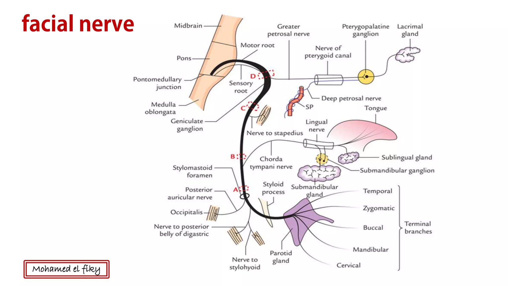
![Nerve supply of the Face
During development a cranial nerve becomes associated with each of the pharyngeal arches. Because the face is
primarily derived from the first and second pharyngeal arches, innervation of neighboring facial structures is as
follows:
• The trigeminal nerve [V] innervates facial structures derived from the first arch.
• The facial nerve [VII] innervates facial structures derived from the second arch.
Sensory innervation
Because the face is derived developmentally from a number of structures originating from the first pharyngeal
arch, cutaneous innervation of the face is by branches of the trigeminal nerve [V]. The trigeminal nerve [V] divides
into three major divisions-the ophthalmic [V1], maxillary [V2], and mandibular [V 3] nerves-before leaving the
middle cranial fossa (Fig. 8.61). Each of these divisions passes out of the cranial cavity to innervate a part of the
face, so most of the skin covering the face is innervated solely by branches of the trigeminal nerve [V]. The
exception is a small area covering the angle and lower border of the ramus of the mandible and parts of the ear,
where the facial [VII], vagus [X], and cervical nerves contribute to the innervation. Mohamed el fiky](https://image.slidesharecdn.com/anatomyofscalpandface-190206052019/75/Anatomy-of-scalp-and-face-13-2048.jpg)
![Nerve supply of the Face
Sensory innervation
The trigeminal nerve [V] divides into three major divisions-the ophthalmic [V1], maxillary [V2], and
mandibular [V 3] to innervate the face, so most of the skin covering the face is innervated solely by branches
of the trigeminal nerve [V]. The exception is a small area covering the angle and lower border of the ramus
of the mandible and parts of the ear, where the facial [VII], vagus [X], and cervical nerves contribute to the
innervation.
Mohamed el fiky](https://image.slidesharecdn.com/anatomyofscalpandface-190206052019/75/Anatomy-of-scalp-and-face-14-2048.jpg)

![The ophthalmic nerve [V 1] exits the skull through the
superior orbital fissure and enters the orbit. Its branches
(Fig. 8.61) that innervate the face include:
• the supra-orbital and supratrochlear nerves, which leave
the orbit superiorly and innervate the upper eyelid,
forehead, and scalp
• the infratrochlear nerve, which exits the orbit in the
medial angle to innervate the medial half of the upper
eyelid, the skin in the area of the medial angle, and the side
of the nose;
• the lacrimal nerve, which exits the orbit in the lateral
angle to innervate the lateral half of the upper eyelid and
the skin in the area of the lateral angle; and
• the external nasal nerve, which supplies the anterior part
of the nose .
Ophthalmic nerve [V1,]
Mohamed el fiky](https://image.slidesharecdn.com/anatomyofscalpandface-190206052019/75/Anatomy-of-scalp-and-face-16-2048.jpg)
![The maxillary nerve [V 2] exits the skull through the
foramen rotundum. Branches that innervate the face
include:
• Zygomaticotemporal branch, which exits the
zygomatic bone and supplies a small area of the anterior
temple above the zygomatic arch;
• Zygomaticofacial branch, which exits the zygomatic
bone and supplies a small area of skin over the
zygomatic bone; and
• infra-orbital nerve, which exits the maxilla through the
infra-orbital foramen and immediately divides into
multiple branches to supply the lower eyelid, cheek, side
of the nose, and upper lip .
Maxillary nerve [V2]
Mohamed el fiky](https://image.slidesharecdn.com/anatomyofscalpandface-190206052019/75/Anatomy-of-scalp-and-face-17-2048.jpg)
![The mandibular nerve [V 3] exits the skull through the
foramen ovale. Branches (Fig. 8.62) innervating the face
include:
• the auriculotemporal nerve, which enters the face just
posterior to the temporomandibular joint, passes through
the parotid gland, and ascends just anterior to the ear to
supply the external acoustic meatus, the surface of the
tympanic membrane (eardrum), and a large area of the
temple;
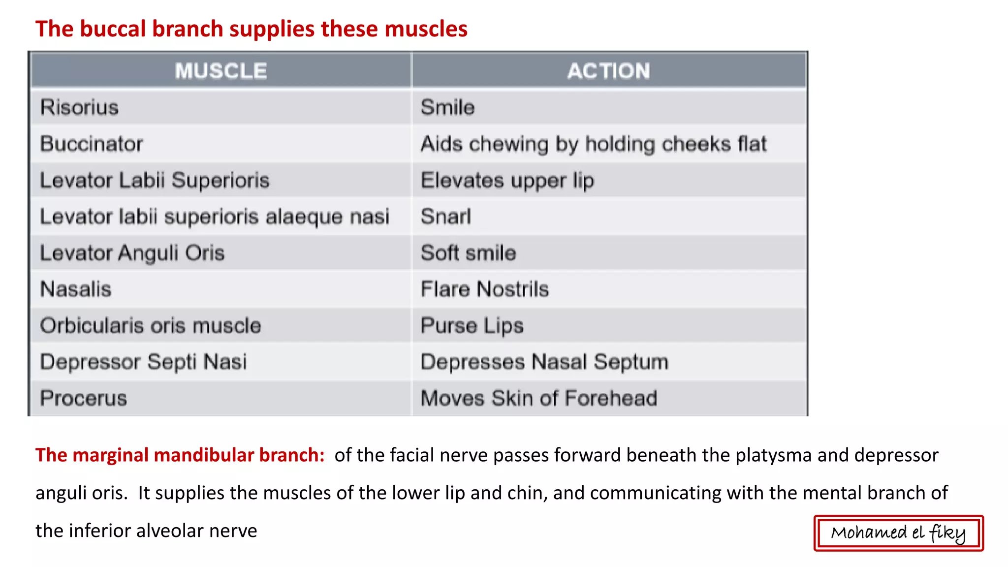
• the buccal nerve, which is on the surface of the
buccinator muscle supplying the cheek; and
• the mental nerve, which exits the mandible through the
mental foramen and immediately divides into multiple
branches to supply the skin and mucous membrane of the
lower lip and skin of the chin
Mandibular nerve [V3]
Mohamed el fiky](https://image.slidesharecdn.com/anatomyofscalpandface-190206052019/75/Anatomy-of-scalp-and-face-18-2048.jpg)


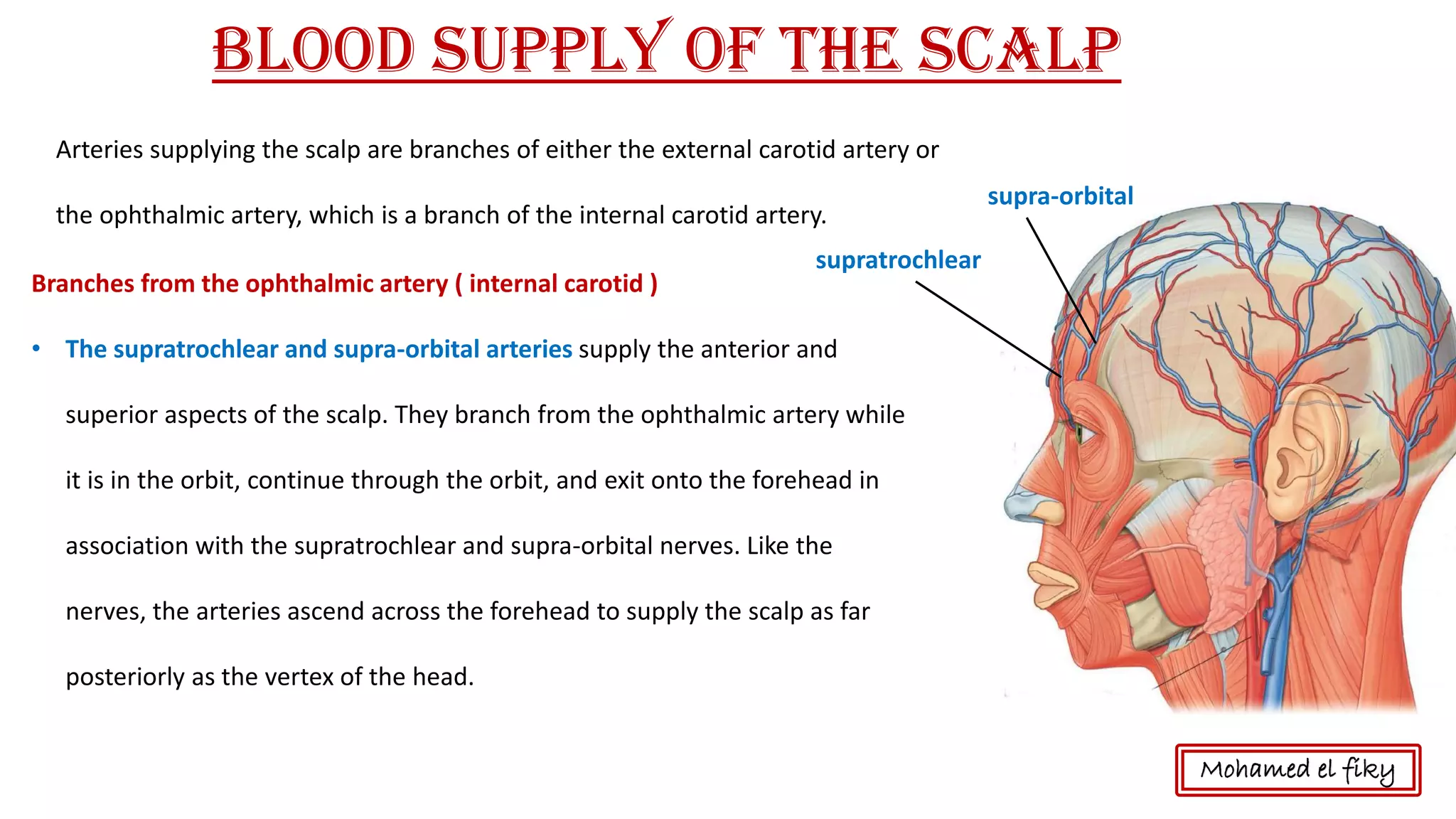
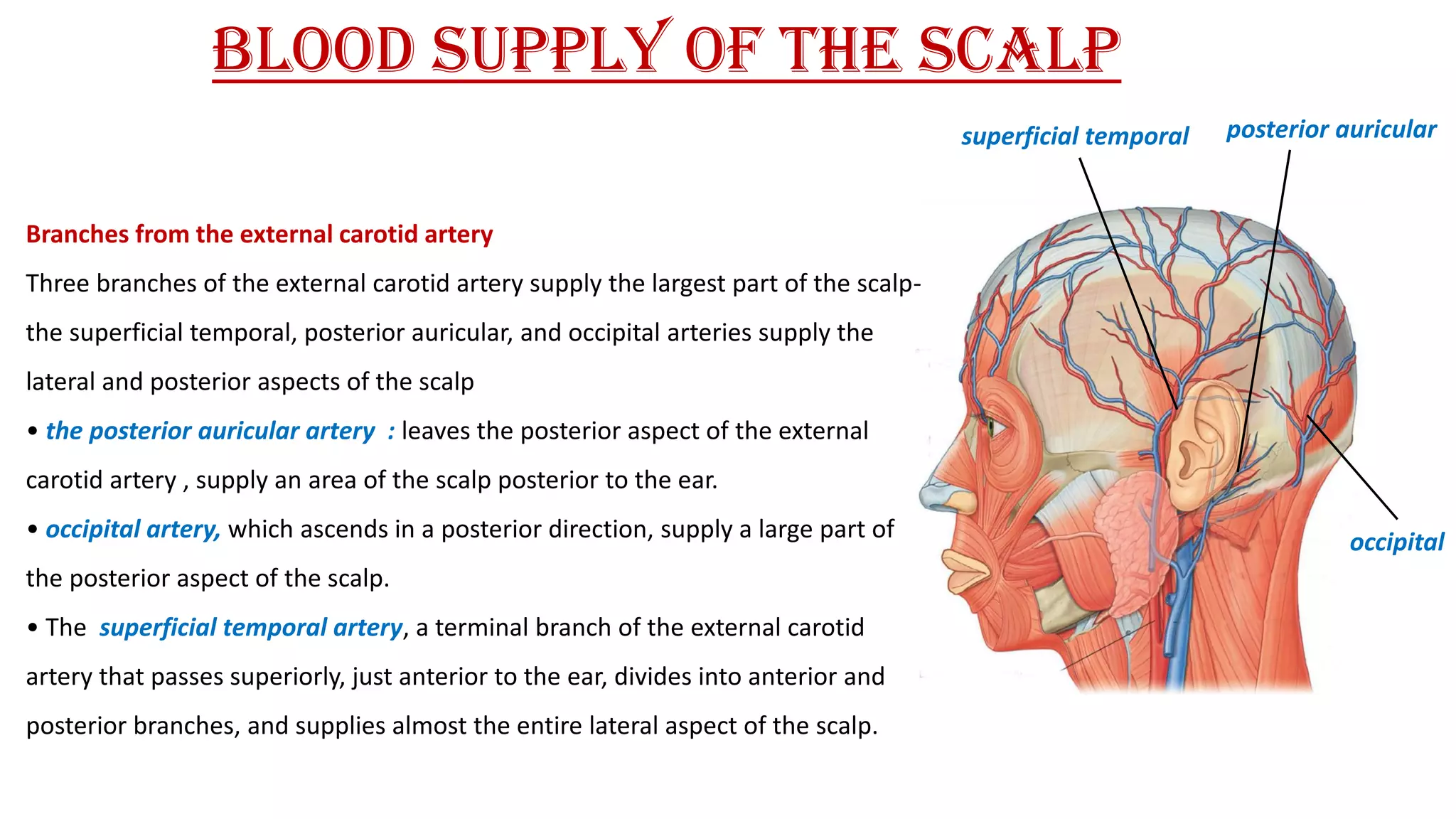
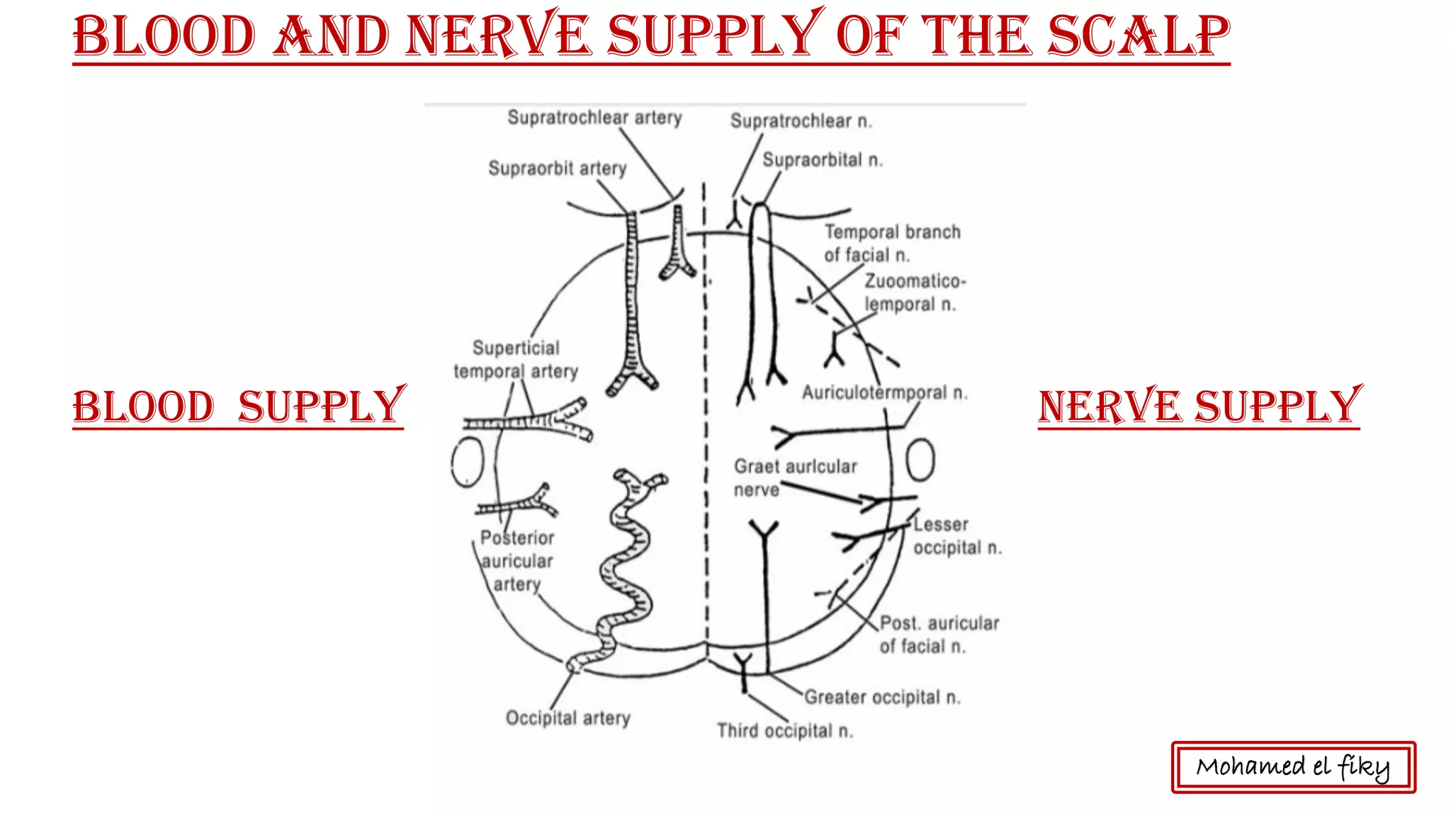
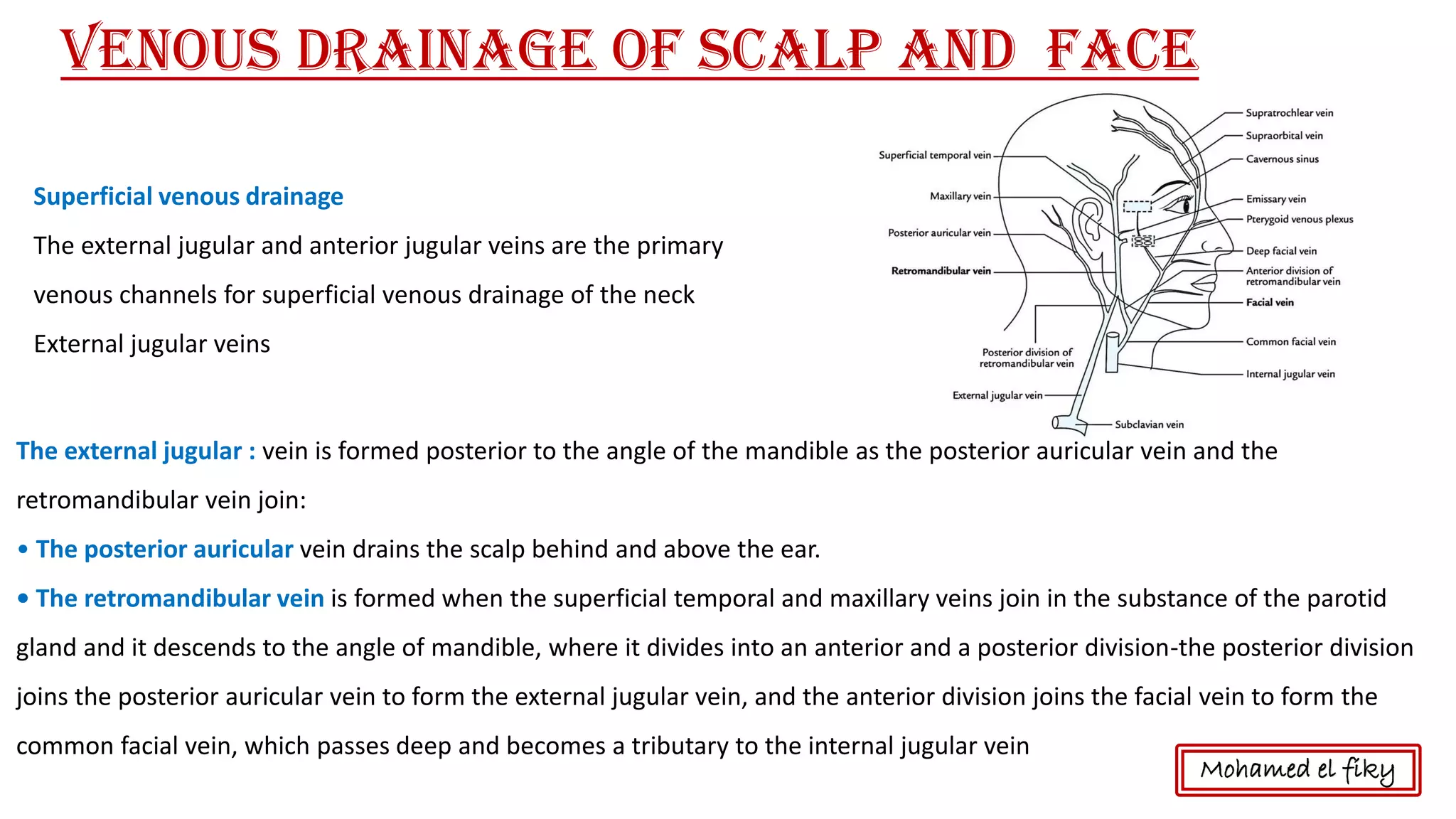
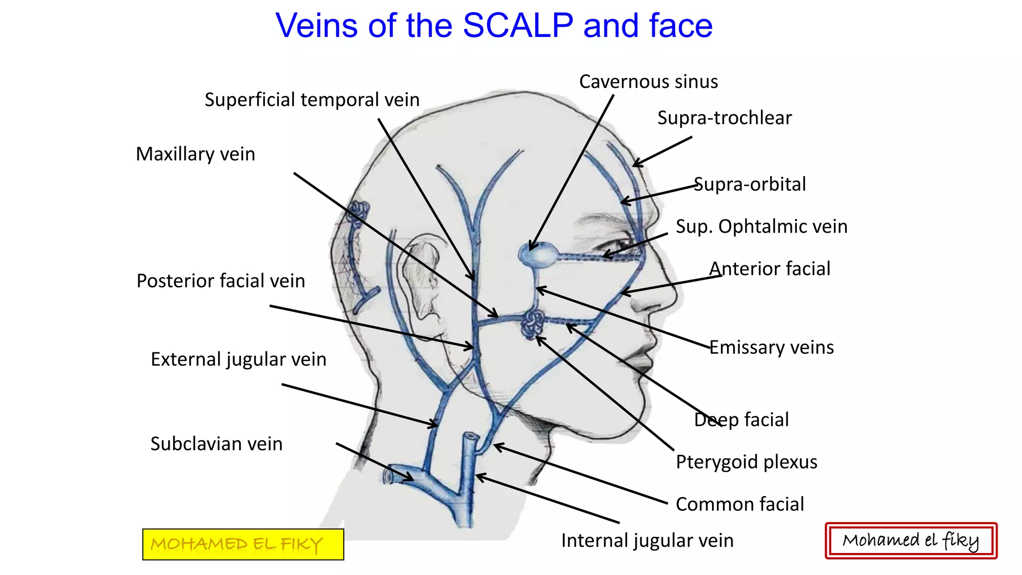
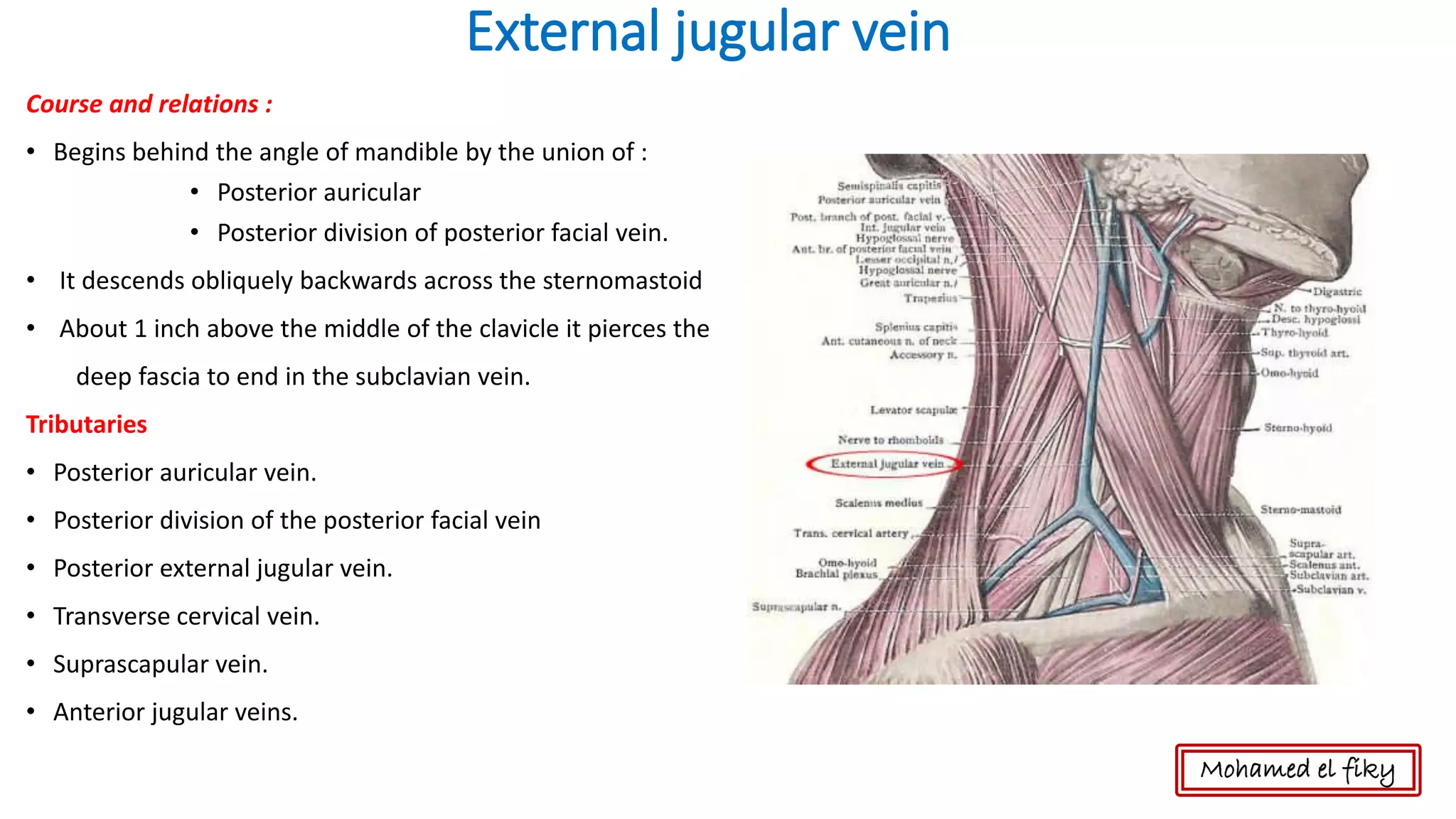
The scalp receives its blood supply from branches of the external carotid artery and the ophthalmic artery. The main arteries supplying the scalp are the superficial temporal artery, the posterior auricular artery, and the occipital artery. Venous drainage of the scalp occurs through the external jugular vein and anterior jugular veins. Sensory innervation of the scalp is provided by branches of the trigeminal nerve and cervical nerves depending on the location on the scalp.
![Scalp[1]](https://cdn.slidesharecdn.com/ss_thumbnails/scalp1-170504174806-thumbnail.jpg?width=640&height=640&fit=bounds)


![Anatomy of Male genital organs [auto saved]](https://cdn.slidesharecdn.com/ss_thumbnails/malegenitalorgansauto-saved-200818065025-thumbnail.jpg?width=640&height=640&fit=bounds)

![anatomy of Female genital organs [auto saved]](https://cdn.slidesharecdn.com/ss_thumbnails/femalegenitalorgansauto-saved-200818064612-thumbnail.jpg?width=640&height=640&fit=bounds)


