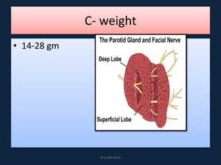
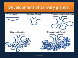
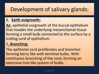
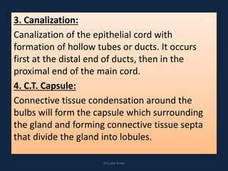
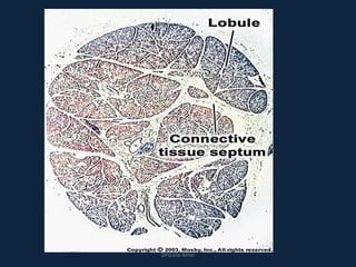
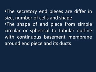
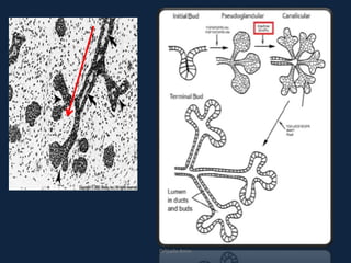
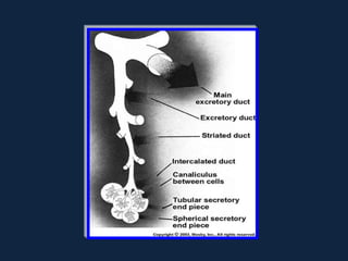
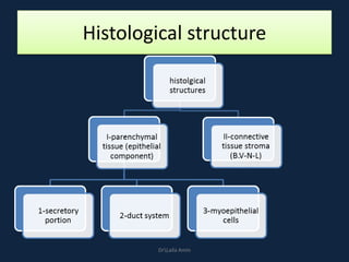
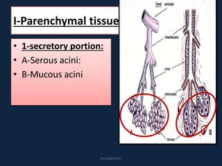
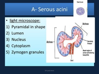
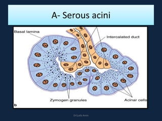
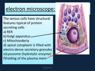
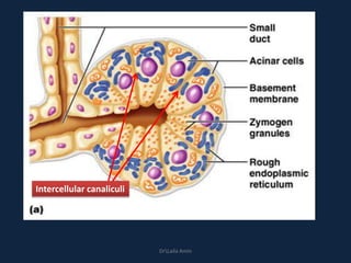
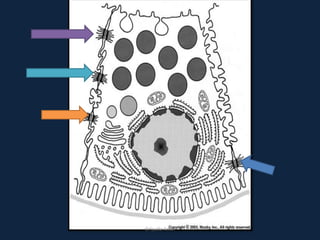
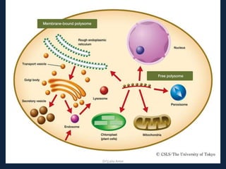
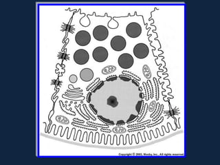
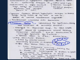
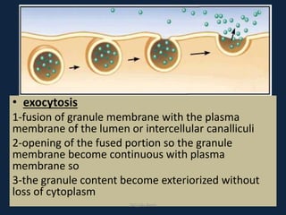
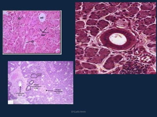
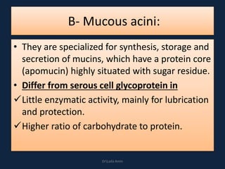
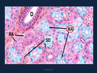
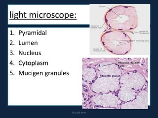
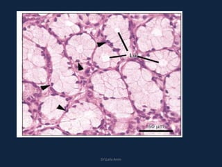
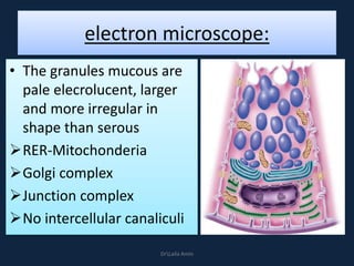
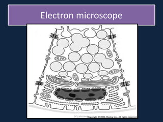
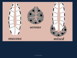
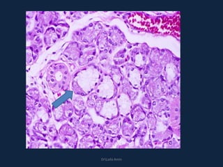
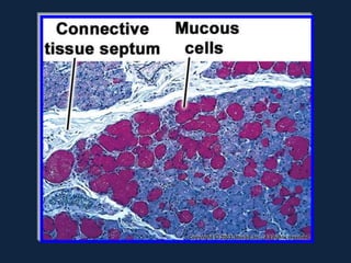
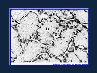
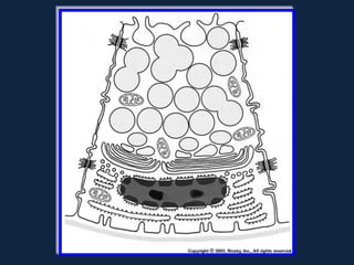
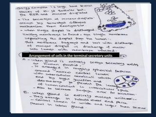
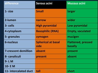
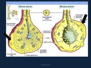
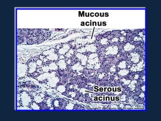
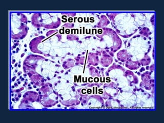
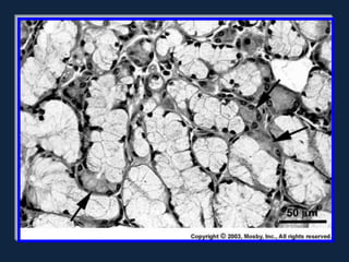
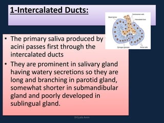
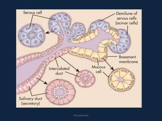
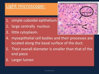
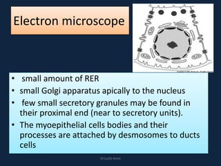
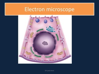
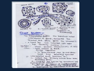
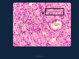
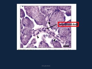
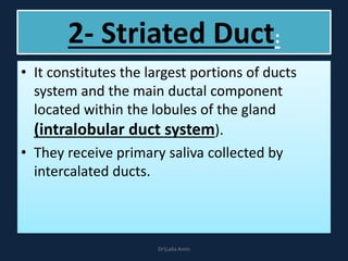
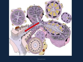
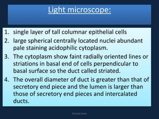
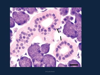
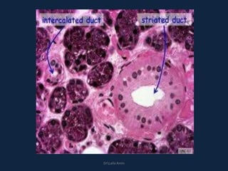
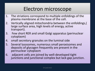
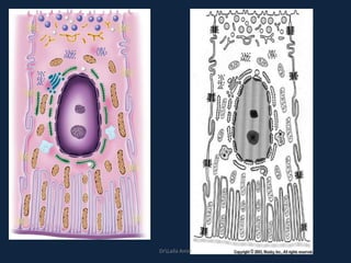
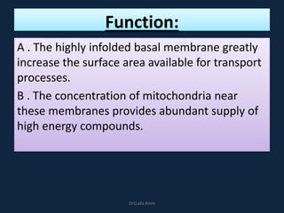
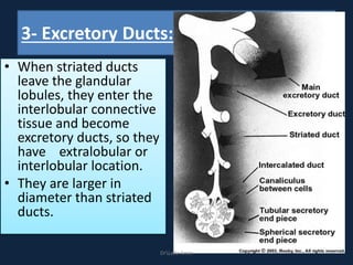
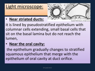
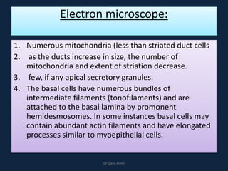
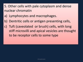
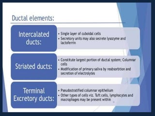
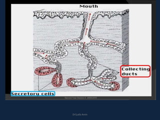
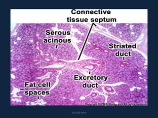
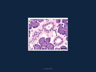
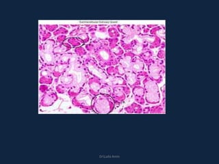
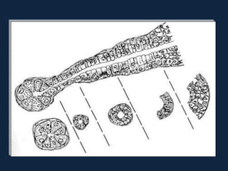
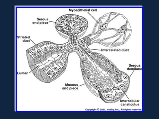
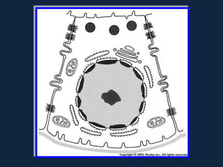
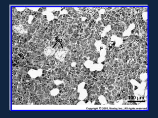
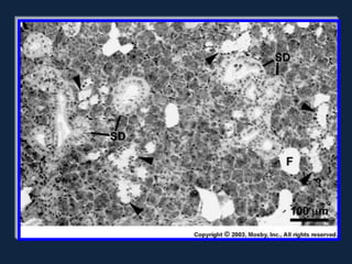
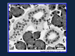
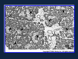
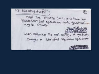
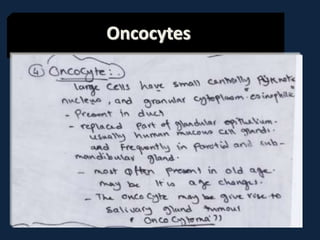
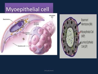
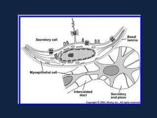
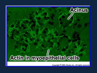
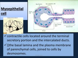
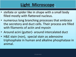
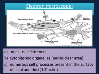
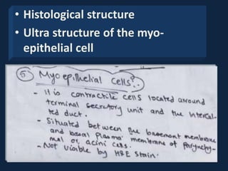
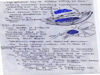
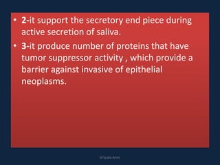
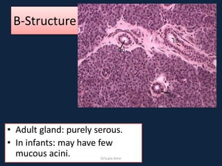
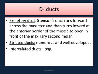
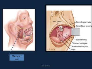
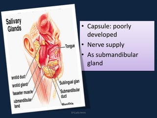
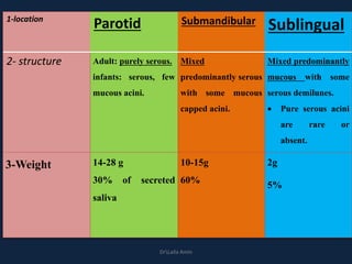
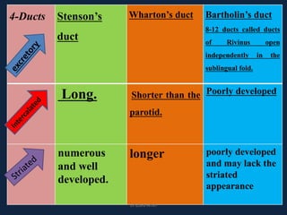
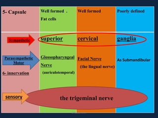
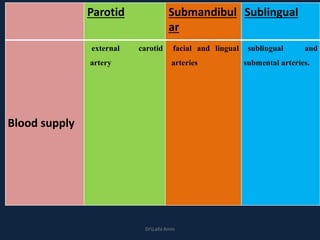
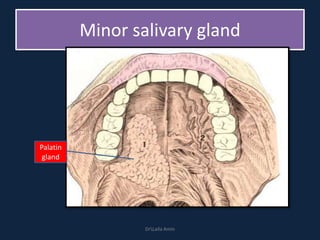
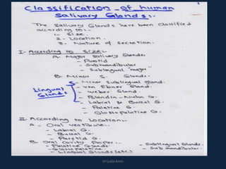
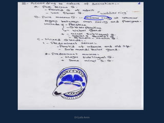
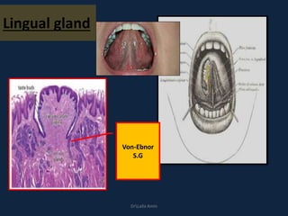
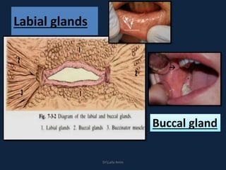
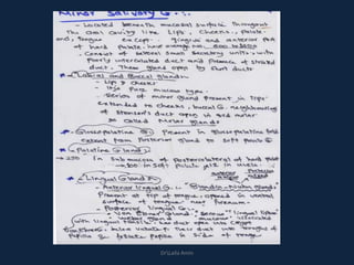
DrLaila Amin
Salivary
glands
Dr. Laila Amin
Lecturer of Oral Biology
DrLaila Amin
Development of salivary glands:
DrLaila Amin
Development of salivary glands:
1. Epith outgrowth:
An epithelial outgrowth of the buccal epithelium
that invades the underlying mesenchymal tissue
forming a small bulb connected to the surface by a
trailing cord of epithelium.
2. Branching:
The epithelial cord proliferates and branches
forming berry like with terminal bulbs. With
continuous branching of the cord, forming an
extensive tree-like system of bulbs.
DrLaila Amin
3. Canalization:
Canalization of the epithelial cord with
formation of hollow tubes or ducts. It occurs
first at the distal end of ducts, then in the
proximal end of the main cord.
4. C.T. Capsule:
Connective tissue condensation around the
bulbs will form the capsule which surrounding
the gland and forming connective tissue septa
that divide the gland into lobules.
DrLaila Amin
Development of salivary glands:
• SG arise as focal thickening of oral epithelium
that grows into the underlining mesenchymal
tissue to form small bud connected to the surface
by trailing cord of epithelium
• As it grows forming epithelium bulb it clefts to
form more buds and so on as tooth developed.
Tubular lumenization done generally form distal
end of main cord to proximal end at the surface
forming the duct system. The distal end forming
the secretory end piece.
The salivary glands are exocrine,
merocrine glands, pour their secretion
into the oral cavity.
• The oral cavity is a moist environment by saliva
coat which is a complex fluid produced by the
salivary gland. It has important role in
maintaining the well being of the mouth.
• Patient with deficiency of salivary secretion has
difficulty in eating, speaking, swallowing and
become prone to mucosal infection and
rampant caries.
• There are three pair of major SG
located outside of oral cavity
encapsulated with extended ducts to
discharge their secretion.
•Also, there are uncapsulated multiple
minor SG located in submucosa
except gingiva and anterior part of
hard palate.
DrLaila Amin
•The secretory end pieces are differ in
size, number of cells and shape
•The shape of end piece from simple
circular or spherical to tubular outline
with continuous basement membrane
around end piece and its ducts
DrLaila Amin
Histological structure
DrLaila Amin
I-Parenchymal tissue:
• 1-secretory portion:
• A-Serous acini:
• B-Mucous acini
DrLaila Amin
A- Serous acini
• light microscope:
1) Pyramidal in shape
2) Lumen
3) Nucleus
4) Cytoplasm
5) Zymogen granules
DrLaila Amin
A- Serous acini
DrLaila Amin
electron microscope:
The serous cells have structural
features typical of protein
secreting cells
a) RER
b)Golgi apparatus
c) Mitochonderia
d) apical cytoplasm is filled with
electro dense secretory granules
e)Lysosome (hydrolytic enzyme)
f)Folding of the plasma mem
DrLaila Amin
Intracellular canaliculi
The secretory cells are
separated by
intercellular canalliculi
at its lateral walls.
DrLaila Amin
DrLaila Amin
Intercellular canaliculi
DrLaila Amin
Intercellular junctions
1. tight junctions
(zonula occludense),
2. adhering junctions
(zonula adherens),
3. desmosome
(macula adherens)
junctional
complex
DrLaila Amin
DrLaila Amin
function of juctional complex
a. Prevent the leakage of the lumen content in the
intercellular spaces.
b. Regulate the passage of materials from the
lumen to the intercellular spaces and vice
versa.
c. Hold adjacent cells together.
Also, the secretory cells attached to the basement
membrane by hemidesmosomes.
DrLaila Amin
Protein synthesis:
DrLaila Amin
cell organelles of protein synthesized
cells:
DNA transcript the nuclear RNA leaving the nucleas
carrying the code messenger RNA reaching to ribosomal
RNA to coupling the appropriate amino acids to their
proper position of newly protein.
Ultrastructure of serous cell
• Nuclear RNA
• Messenger RNA
• Ribosomal RNA
DrLaila Amin
.
• exocytosis
1-fusion of granule membrane with the plasma
membrane of the lumen or intercellular canalliculi
2-opening of the fused portion so the granule
membrane become continuous with plasma
membrane so
3-the granule content become exteriorized without
loss of cytoplasm
DrLaila Amin
DrLaila Amin
Mucous cells
• Histological structure
• Electron microscopic picture
B- Mucous acini:
• They are specialized for synthesis, storage and
secretion of mucins, which have a protein core
(apomucin) highly situated with sugar residue.
• Differ from serous cell glycoprotein in
Little enzymatic activity, mainly for lubrication
and protection.
Higher ratio of carbohydrate to protein.
DrLaila Amin
DrLaila Amin
light microscope:
1. Pyramidal
2. Lumen
3. Nucleus
4. Cytoplasm
5. Mucigen granules
DrLaila Amin
DrLaila Amin
electron microscope:
• The granules mucous are
pale elecrolucent, larger
and more irregular in
shape than serous
RER-Mitochonderia
Golgi complex
Junction complex
No intercellular canaliculi
DrLaila Amin
Electron microscope
DrLaila Amin
The secretion of mucous granules differ from
exocytosis of serous granules that.
• When single mucous droplet is discharged its
limiting membrane fuses with the apical plasma
membrane resulting in single membrane
separating the granule from the lumen.
• This separating membrane may then fragment
and lost with discharge of mucous OR the
mucous granule may be discharged with the
membrane intact surrounding them.
• During rapid mucous discharge, the apical
cytoplasm may not seal itself off and the entire
mass of mucous may be spilled into the lumen.
DrLaila Amin
Separating
membrane
Granules
discharge
with
membrane
intact
Fragment,
lost
DrLaila Amin
DrLaila Amin
mucous
serous
DrLaila Amin
The mucous end piece have serous cells associated with
them in the form of demilunes or crescent called serous
demilion or crescent of Giganzi.
DrLaila Amin
DrLaila Amin
DrLaila Amin
Arrangement of cells in the terminal secretory unitsArrangement of cells in the terminal secretory units
difference Serous acini Mucous acini
1- size Small larger
2-lumen narrow wider
3- cells High pyramidal Low pyramidal
4-cytoplasm Basophilic (RNA) Empty, vaculated
5- granules zymogen mucigen
6-nucleus Spherical at basal
side
Flattened, pressed
basally
7-crescent demillion absent present
8- canaliculi present absent
9- L M
10- E M
11- intercalated duct tall short
DrLaila Amin
DrLaila Amin
Mixed salivary gland
Ducts
2- Duct system:
• 1-Intercalated Ducts:
• 2-Striated Duct:
• 3-excretory Duct:
DrLaila Amin
1-Intercalated Ducts:
• The primary saliva produced by the
acini passes first through the
intercalated ducts
• They are prominent in salivary gland
having watery secretions so they are
long and branching in parotid gland,
somewhat shorter in submandibular
gland and poorly developed in
sublingual gland.
DrLaila Amin
DrLaila Amin
Light microscope:
1. simple cuboidal epithelium
2. large centrally nucleus
3. little cytoplasm.
4. myoepithelial cell bodies and their processes are
located along the basal surface of the duct.
5. Their overall diameter is smaller than that of the
end piece
6. Larger lumen
DrLaila Amin
Electron microscope
• small amount of RER
• small Golgi apparatus apically to the nucleus
• few small secretory granules may be found in
their proximal end (near to secretory units).
• The myoepithelial cells bodies and their
processes are attached by desmosomes to ducts
cells
DrLaila Amin
Electron microscope
DrLaila Amin
Functions:
1. Collect the primary saliva formed by terminal secretory
units
2. Convey the secretion from the terminal secretory units to
the striated ducts.
3. Reserve of undifferentiated cells which proliferate and
undergo differentiation to replaced damaged or dying cells.
4. Reabsorbing proteins from the lumen by endocytosis with
the striated ducts
5. Macromolecular components stored in their secretory
granules include: lysozyme and lactoferrin (antibacterial
proteins).
6. Modify the saliva through secretory and reabsorptive
processes.
DrLaila Amin
DrLaila Amin
DrLaila Amin
Intercalated duct
2- Striated Duct:
• It constitutes the largest portions of ducts
system and the main ductal component
located within the lobules of the gland
(intralobular duct system).
• They receive primary saliva collected by
intercalated ducts.
DrLaila Amin
DrLaila Amin
Light microscope:
1. single layer of tall columnar epithelial cells
2. large spherical centrally located nuclei abundant
pale staining acidophilic cytoplasm.
3. The cytoplasm show faint radially oriented lines or
striations in basal end of cells perpendicular to
basal surface so the duct called striated.
4. The overall diameter of duct is greater than that of
secretory end piece and the lumen is larger than
those of secretory end pieces and intercalated
ducts.
DrLaila Amin
DrLaila Amin
DrLaila Amin
Electron microscope
1. The striations correspond to multiple enfoldings of the
plasma membrane at the base of the cell.
2. Vertically aligned mitochondria between the enfoldings,(
large surface area, high levels of energy, active
transport).
3. Few short RER and small Golgi apparatus (perinuclear
cytoplasm)
4. small secretory granules on the luminal side
5. Several lysosomes, numerous small peroxisomes and
deposits of glycogen frequently are present in the
perinuclear cytoplasm
6. adjacent cells are joined by well-developed tight
junctions and junctional complex but lack gap junction.
DrLaila Amin
DrLaila Amin
Function:
A . The highly infolded basal membrane greatly
increase the surface area available for transport
processes.
B . The concentration of mitochondria near
these membranes provides abundant supply of
high energy compounds.
DrLaila Amin
• Electrolyte reabsorption and secretion by
striated duct is regulated by autonomic
nervous system and by mineralocorticoids
produced by adrenal cortex.
D)Lysosome which have an antibacterial effects.
• Striated ducts contain the enzyme kallikrin
which affect the water and electrolyte
balance.
E)Amylase enzyme is added from the serum to
the saliva by the cells of the striated ducts.
DrLaila Amin
C) . Modify the primary saliva by
1) 1ry saliva (acini, intercalated
ducts)
isotonic {high Na+ ,low K+}.
1) Striated ducts (massive
folding) reflect Na+-pumping
2) Na+ diffuse into cells from
luminal fluids at the same
time active transport of K+
occur in opposite direction.
3) Hco3
- is also actively secreted.
hypotonic fluid {low Na+,Cl-}
DrLaila Amin
1ry saliva
isotonic
↑Na-↓K
Na
K
H
co
3Hypotonic
↓Na-↓Cl
Striated
duct
cl
DrLaila Amin
DrLaila Amin
3- Excretory Ducts:
• When striated ducts
leave the glandular
lobules, they enter the
interlobular connective
tissue and become
excretory ducts, so they
have extralobular or
interlobular location.
• They are larger in
diameter than striated
ducts.
DrLaila Amin
Light microscope:
• Near striated ducts:
it is lined by pseudostratified epithelium with
columnar cells extending, small basal cells that
sit on the basal lamina but do not reach the
lumen,
• Near the oral cavity:
the epithelium gradually changes to stratified
squamous epithelium that merge with the
epithelium of oral cavity at duct orifice.
DrLaila Amin
DrLaila Amin
intralobular Near the oral
cavity:
DrLaila Amin
Electron microscope:
1. Numerous mitochondria (less than striated duct cells
2. as the ducts increase in size, the number of
mitochondria and extent of striation decrease.
3. few, if any apical secretory granules.
4. The basal cells have numerous bundles of
intermediate filaments (tonofilaments) and are
attached to the basal lamina by promonent
hemidesmosomes. In some instances basal cells may
contain abundant actin filaments and have elongated
processes similar to myoepithelial cells.
DrLaila Amin
5. Other cells with pale cytoplasm and dense
nuclear chromatin
a) Lymphocytes and macrophages.
b) Dentritic cells or antigen presenting cells,
c) Tuft (caveolated or brush) cells, with long
stiff microvilli and apical vesicles are thought
to be receptor cells to some type
DrLaila Amin
Functions:
1) They convey the salivary secretion towards
the oral cavity.
2) They have the ability to reabsorb sodium and
secrete potassium and bicarbonate.
3) They are also capable of modifying saliva
through process of electrolyte reabsorption
and secretion, perhaps also by adding
mucoid compound.
DrLaila Amin
DrLaila Amin
DrLaila Amin
DrLaila Amin
The connective tissue
• includes a surrounding capsule, variably
developed, that demarcates the gland from
adjacent structures.
• Septa that extend inward from the capsule
divide the gland into lobes and lobules and
• carry the blood vessels and nerves that supply
the parenchymal components and the
excretory ducts that convey saliva to the oral
cavity
DrLaila Amin
DrLaila Amin
DrLaila Amin
• Striated ducts
–Ultra structure of the striated ducts
Excretory ducts
Oncocytes
Myo-epithelial cells
Myoepithelial cell
DrLaila Amin
Myoepithelial
cell
contractile cells located around the terminal
secretory portion and the intercalated ducts.
()the basal lamina and the plasma membrane
of parenchymal cells, joined to cells by
desmosomes. DrLaila Amin
Light Microscope:
• stellate or spider like in shape with a small body
filled mostly with flattened nucleus.
• numerous long branching processes that embrace
the secretory and duct cells. Their process are filled
with filaments of actin and myosin
• Around acini (gutter)- around intercalated duct
• H&E stain (non), special stain as adenosine
triphosphate in human and alkaline phosphatase in
animal.
DrLaila Amin
Electron microscope:
a) nucleus is flattened
b) cytoplasmic organelles (perinuclear area).
c) numerous cell processes present in the surface
of acini and duct( L.T actin).DrLaila Amin
• Histological structure
• Ultra structure of the myo-
epithelial cell
Functions:
• 1- Contractile functions to expel secretion of
saliva from the acini and duct because
• -similar to smooth muscles
a. imunoflurescent studies suggest the presence
of actin and myosin filament.
b. measurement of ductal pressure after
myoepithelial cells stimulation suggest it have a
contractile functions.
DrLaila Amin
• 2-it support the secretory end piece during
active secretion of saliva.
• 3-it produce number of proteins that have
tumor suppressor activity , which provide a
barrier against invasive of epithelial
neoplasms.
DrLaila Amin
DrLaila Amin
Classification of
salivary glands
I-Size
II-
Location
III-
Secretion
DrLaila Amin
According to size:-
1- Major salivary glands
1. Parotid gland
2. Submandibular glands
3. Sublingual glands
2- Minor salivary glands
1. Minor sublingual glands
2. Labial and Buccal glands
3. Palatine glands
4. Glossopalatine glands
5. Retromolar gland
6. Lingual glands( Blandin Nuhn, Von Ebner, Weber)
DrLaila Amin
According to location:-
1-Oral vestibule:
1) Labial glands
2) Buccal glands
3) Parotid gland
2- Oral cavity proper:
1) Submandibular gland.
2) Sublingual gland
3) Palatine glands
4) Glossopalatine glands
5) Lingual glands( Blandin Nuhn, Weber and Von Ebner).
DrLaila Amin
According to nature of secretion:-
Pure serous
Parotid gland of adult
Von Ebner
Pure mucous
Minor sublingual glands
Labial and Buccal glands
Anterior portion of
anterior lingual glands
(Blandin Nuhn)
Mucous ring
Mixed
glands:
Predominantly Serous:-
Submandibular gland
Parotid gland of infants
and old aged
Predominantly Mucous:-
Major Sublingual gland
Posterior portion of the
anterior lingual gland (Blandin
Nuhn)
DrLaila Amin
Major Salivary Glands
DrLaila Amin
I- parotid Gland
DrLaila Amin
A -Location:
Superficial part: in front of the external ear, associated
with the peripheral branches of the facial nerve.
DrLaila Amin
Deep part:
fills the retromandibular fossa behind the
ramus of the mandible.DrLaila Amin
DrLaila Amin
B-Structure
• Adult gland: purely serous.
• In infants: may have few
mucous acini. DrLaila Amin
C- weight
• 14-28 gm
DrLaila Amin
D- ducts
• Excretory duct: Stenson’s duct runs forward
across the masseter and then turns inward at
the anterior border of the muscle to open in
front of the maxillary second molar.
• Striated ducts: numerous and well developed.
• Intercalated ducts: long.
DrLaila Amin
Masseter
msc
DrLaila Amin
E-Capsule
• Well formed connective tissue capsule.
• Fat cells are found in the C.T. septa and
increase in number with age replacing parts of
the gland.
DrLaila Amin
F- Nerve supply
• Sympathetic: superior
cervical ganglia
• Parasympathetic:
glossopharyngeal
(auriculotemporal
nerve).
• Sensory: from the
trigeminal nerve.
DrLaila Amin
DrLaila Amin
G- Blood supply
exrenal carotid
artery
DrLaila Amin
DrLaila Amin
A- location
• In the
submandibular
triangle behind
and below the
free margin of
the mylohyoid
muscle.
DrLaila Amin
DrLaila Amin
B- structure
• Mixed predominantly serous with some mucous
capped acini.
DrLaila Amin
C- weight:10-15 gm
D- capsule: well formed
DrLaila Amin
E- Ducts
• Excretory duct: Wharton’s duct runs forward
above the mylohyoid muscle and opens at a
small papilla at the side of the lingual frenum
called caruncula sublingualis.
• Striated ducts: are longer
• Intercalated ducts: are relatively shorter than
the parotid.
DrLaila Amin
DrLaila Amin
Caruncula sublingualis.
DrLaila Amin
Nerve supply
• Sympathatic: as parotid
• Parasympathetic: From the facial.
• Preganglionic fibers of chorda tympani
postganglionic fibers reach the gland through
the lingual nerve.
• Sensory: the same as the parotid.
DrLaila Amin
Blood supply
facial, lingual arteries
DrLaila Amin
III-Sublingual gland
DrLaila Amin
A- Location
• Between the floor of the mouth and the
mylohyoid muscle.
DrLaila Amin
B- STRUCTURE
• Mixed predominantly mucous with some
serous demilunes.
DrLaila Amin
Ducts
• Excretory duct: Bartholin’s duct opens with or
near the submandibular duct in the sublingual
fold.
• 8-12 ducts called ducts of Rivinus open
independently in the sublingual fold.
• Striated ducts, Intercalated ducts : poorly
developed
DrLaila Amin
DrLaila Amin
DrLaila Amin
• Capsule: poorly
developed
• Nerve supply
• As submandibular
gland
DrLaila Amin
1-location
Parotid Submandibular Sublingual
2- structure Adult: purely serous.
infants: serous, few
mucous acini.
Mixed
predominantly serous
with some mucous
capped acini.
Mixed predominantly
mucous with some
serous demilunes.
 Pure serous acini
are rare or
absent.
3-Weight 14-28 g
30% of secreted
saliva
10-15g
60%
2g
5%
DrLaila Amin
4-Ducts Stenson’s
duct
Wharton’s duct Bartholin’s duct
8-12 ducts called ducts
of Rivinus open
independently in the
sublingual fold.
Long. Shorter than the
parotid.
Poorly developed
numerous
and well
developed.
longer poorly developed
and may lack the
striated
appearance
DrLaila Amin
5- Capsule Well formed .
Fat cells
Well formed Poorly defined
6- innervation
superior
Glossopharyngeal
Nerve
(auriculotemporal)
cervical
Facial Nerve
(the lingual nerve)
ganglia
As Submandibular
Sympathetic
Parasympathetic
Motor
the trigeminal nervesensory
DrLaila Amin
Parotid Submandibul
ar
Sublingual
Blood supply
external carotid
artery
facial and lingual
arteries
sublingual and
submental arteries.
DrLaila Amin
DrLaila Amin
Minor salivary gland
Palatin
gland
DrLaila Amin
DrLaila Amin
DrLaila Amin
Lingual gland
Von-Ebnor
S.G
DrLaila Amin
Labial glands
Buccal gland
DrLaila Amin
DrLaila Amin
• at the isthmus region
between the oral cavity
and the pharynx which
include:
1. Weber gland
2. palatine
3. glossopalatine glands.
The mucous
ring
Waldier Ring
DrLaila Amin
Saliva
saliva
DrLaila Amin
DrLaila Amin
• Quantity:
• Stimulation:
• Distribution:
• Consistency:
• PH:
Saliva
DrLaila Amin
Structure:
99% of water 1%
Inorganic
ions:
Organic substances:
Enzyme-Igs-mucin
DrLaila Amin
DrLaila Amin
DrLaila Amin
1. Protection
2. Buffering
3. Digestive
4. Taste
5. Tissue repair
6. Tooth integrity
7. antimicrobial
DrLaila Amin
1.Protection
– lubricant (glycoprotein)
– barrier against noxious stimuli; microbial toxins
and minor traumas
– washing non-adherent and acellular debris
– formation of salivary pellicle
• calcium-binding proteins: tooth protection; plaque
DrLaila Amin
2-Buffering
• (phosphate ions and bicarbonate)
– bacteria require specific pH conditions
– plaque microorganisms produce acids from sugars
DrLaila Amin
3-Digestion
– neutralizes esophageal contents
– dilutes gastric chyme
– forms food bolus
DrLaila Amin
4- Antimicrobial
– lysozyme hydrolyzes cell walls of some bacteria
– lactoferrin binds free iron and deprives bacteria of
this essential element
– IgA agglutinates microorganisms
DrLaila Amin
5-Maintenance of tooth integrity
– calcium and phosphate ions
• ionic exchange with tooth surface
DrLaila Amin
6-Tissue repair
– bleeding time of oral tissues shorter than other
tissues
– resulting clot less solid than normal
– remineralization
DrLaila Amin
7-Taste
– solubilizing of food substances that can be sensed
by receptors
– trophic effect on receptors
DrLaila Amin
Age changes
• By age generalized loss of salivary gland
parenchymal tissue occurs and this loss may be
replaced by adipose tissue.
• Decreased production of saliva is noted.
• Atrophy or degeneration of part or whole
terminal portion may occur with replacement
with fibrous tissue causing fibrosis.
• Accumulation of lymphocytes within the stroma
of the salivary glands.
• Secretory acini decrease in number and secretory
cells decrease in height and width.
DrLaila Amin
Oncocytes
DrLaila Amin
http://www.edewcate.com/Inner.aspx?FileName=%7B5418AFC1-96B4-4DD1-8FC5-
73FD651DC75B%7D.flv
Clinical Considerations
• Xerostomia(dry mouth):
Reduction in the amount of secreted saliva may lead to sensation of oral dryness.
Causes:
1) It occurs mostly as a side effect of other medications.
2) After radiation therapy to the head and neck region.
3) In auto-immune diseases as Sjogren syndrome.
Effects:
 Speech, eating and swallowing become difficult and painful.
 Teeth become highly susceptible to caries especially at the gingival margins.
Treatment:
 artificial saliva.
 pathomimetic drugs as pilocarpine to increase salivary flow.
 Stem cell therapy can alleviate the condition of the gland.
DrLaila Amin
Diseases:-
1- viruses such as cytomegalovirus, Epstein-Barr and human
herpes viruses infect and replicate within the salivary glands and
are shed into the saliva.
2- bacterial infections.
3- Ductal obstruction may be caused by formation of stones
(sialolith) especially in the submandibular glands
4- Severing of the duct of minor salivary glands by trauma.
5- Salivary glands may be affected by endocrine diseases like
diabetes
DrLaila Amin
Thank you
DrLaila Amin
DrLaila Amin

Salivary glands

  • 1.
  • 2.
    Salivary glands Dr. Laila Amin Lecturerof Oral Biology DrLaila Amin
  • 3.
    Development of salivaryglands: DrLaila Amin
  • 4.
    Development of salivaryglands: 1. Epith outgrowth: An epithelial outgrowth of the buccal epithelium that invades the underlying mesenchymal tissue forming a small bulb connected to the surface by a trailing cord of epithelium. 2. Branching: The epithelial cord proliferates and branches forming berry like with terminal bulbs. With continuous branching of the cord, forming an extensive tree-like system of bulbs. DrLaila Amin
  • 5.
    3. Canalization: Canalization ofthe epithelial cord with formation of hollow tubes or ducts. It occurs first at the distal end of ducts, then in the proximal end of the main cord. 4. C.T. Capsule: Connective tissue condensation around the bulbs will form the capsule which surrounding the gland and forming connective tissue septa that divide the gland into lobules. DrLaila Amin
  • 6.
    Development of salivaryglands: • SG arise as focal thickening of oral epithelium that grows into the underlining mesenchymal tissue to form small bud connected to the surface by trailing cord of epithelium • As it grows forming epithelium bulb it clefts to form more buds and so on as tooth developed. Tubular lumenization done generally form distal end of main cord to proximal end at the surface forming the duct system. The distal end forming the secretory end piece.
  • 7.
    The salivary glandsare exocrine, merocrine glands, pour their secretion into the oral cavity. • The oral cavity is a moist environment by saliva coat which is a complex fluid produced by the salivary gland. It has important role in maintaining the well being of the mouth. • Patient with deficiency of salivary secretion has difficulty in eating, speaking, swallowing and become prone to mucosal infection and rampant caries.
  • 8.
    • There arethree pair of major SG located outside of oral cavity encapsulated with extended ducts to discharge their secretion. •Also, there are uncapsulated multiple minor SG located in submucosa except gingiva and anterior part of hard palate.
  • 9.
  • 10.
    •The secretory endpieces are differ in size, number of cells and shape •The shape of end piece from simple circular or spherical to tubular outline with continuous basement membrane around end piece and its ducts
  • 11.
  • 13.
  • 14.
    I-Parenchymal tissue: • 1-secretoryportion: • A-Serous acini: • B-Mucous acini DrLaila Amin
  • 15.
    A- Serous acini •light microscope: 1) Pyramidal in shape 2) Lumen 3) Nucleus 4) Cytoplasm 5) Zymogen granules DrLaila Amin
  • 16.
  • 17.
    electron microscope: The serouscells have structural features typical of protein secreting cells a) RER b)Golgi apparatus c) Mitochonderia d) apical cytoplasm is filled with electro dense secretory granules e)Lysosome (hydrolytic enzyme) f)Folding of the plasma mem DrLaila Amin
  • 18.
    Intracellular canaliculi The secretorycells are separated by intercellular canalliculi at its lateral walls. DrLaila Amin
  • 19.
  • 20.
  • 21.
    Intercellular junctions 1. tightjunctions (zonula occludense), 2. adhering junctions (zonula adherens), 3. desmosome (macula adherens) junctional complex DrLaila Amin
  • 22.
  • 23.
    function of juctionalcomplex a. Prevent the leakage of the lumen content in the intercellular spaces. b. Regulate the passage of materials from the lumen to the intercellular spaces and vice versa. c. Hold adjacent cells together. Also, the secretory cells attached to the basement membrane by hemidesmosomes. DrLaila Amin
  • 24.
  • 25.
    cell organelles ofprotein synthesized cells: DNA transcript the nuclear RNA leaving the nucleas carrying the code messenger RNA reaching to ribosomal RNA to coupling the appropriate amino acids to their proper position of newly protein. Ultrastructure of serous cell • Nuclear RNA • Messenger RNA • Ribosomal RNA
  • 26.
  • 30.
    . • exocytosis 1-fusion ofgranule membrane with the plasma membrane of the lumen or intercellular canalliculi 2-opening of the fused portion so the granule membrane become continuous with plasma membrane so 3-the granule content become exteriorized without loss of cytoplasm DrLaila Amin
  • 31.
  • 32.
  • 33.
    • Histological structure •Electron microscopic picture
  • 34.
    B- Mucous acini: •They are specialized for synthesis, storage and secretion of mucins, which have a protein core (apomucin) highly situated with sugar residue. • Differ from serous cell glycoprotein in Little enzymatic activity, mainly for lubrication and protection. Higher ratio of carbohydrate to protein. DrLaila Amin
  • 35.
  • 36.
    light microscope: 1. Pyramidal 2.Lumen 3. Nucleus 4. Cytoplasm 5. Mucigen granules DrLaila Amin
  • 37.
  • 38.
    electron microscope: • Thegranules mucous are pale elecrolucent, larger and more irregular in shape than serous RER-Mitochonderia Golgi complex Junction complex No intercellular canaliculi DrLaila Amin
  • 39.
  • 40.
    The secretion ofmucous granules differ from exocytosis of serous granules that. • When single mucous droplet is discharged its limiting membrane fuses with the apical plasma membrane resulting in single membrane separating the granule from the lumen. • This separating membrane may then fragment and lost with discharge of mucous OR the mucous granule may be discharged with the membrane intact surrounding them. • During rapid mucous discharge, the apical cytoplasm may not seal itself off and the entire mass of mucous may be spilled into the lumen. DrLaila Amin
  • 41.
  • 42.
  • 43.
  • 44.
    The mucous endpiece have serous cells associated with them in the form of demilunes or crescent called serous demilion or crescent of Giganzi. DrLaila Amin
  • 45.
  • 46.
  • 50.
    Arrangement of cellsin the terminal secretory unitsArrangement of cells in the terminal secretory units
  • 51.
    difference Serous aciniMucous acini 1- size Small larger 2-lumen narrow wider 3- cells High pyramidal Low pyramidal 4-cytoplasm Basophilic (RNA) Empty, vaculated 5- granules zymogen mucigen 6-nucleus Spherical at basal side Flattened, pressed basally 7-crescent demillion absent present 8- canaliculi present absent 9- L M 10- E M 11- intercalated duct tall short DrLaila Amin
  • 52.
  • 54.
  • 58.
  • 59.
    2- Duct system: •1-Intercalated Ducts: • 2-Striated Duct: • 3-excretory Duct: DrLaila Amin
  • 60.
    1-Intercalated Ducts: • Theprimary saliva produced by the acini passes first through the intercalated ducts • They are prominent in salivary gland having watery secretions so they are long and branching in parotid gland, somewhat shorter in submandibular gland and poorly developed in sublingual gland. DrLaila Amin
  • 61.
  • 62.
    Light microscope: 1. simplecuboidal epithelium 2. large centrally nucleus 3. little cytoplasm. 4. myoepithelial cell bodies and their processes are located along the basal surface of the duct. 5. Their overall diameter is smaller than that of the end piece 6. Larger lumen DrLaila Amin
  • 63.
    Electron microscope • smallamount of RER • small Golgi apparatus apically to the nucleus • few small secretory granules may be found in their proximal end (near to secretory units). • The myoepithelial cells bodies and their processes are attached by desmosomes to ducts cells DrLaila Amin
  • 64.
  • 65.
    Functions: 1. Collect theprimary saliva formed by terminal secretory units 2. Convey the secretion from the terminal secretory units to the striated ducts. 3. Reserve of undifferentiated cells which proliferate and undergo differentiation to replaced damaged or dying cells. 4. Reabsorbing proteins from the lumen by endocytosis with the striated ducts 5. Macromolecular components stored in their secretory granules include: lysozyme and lactoferrin (antibacterial proteins). 6. Modify the saliva through secretory and reabsorptive processes. DrLaila Amin
  • 67.
  • 68.
  • 69.
    2- Striated Duct: •It constitutes the largest portions of ducts system and the main ductal component located within the lobules of the gland (intralobular duct system). • They receive primary saliva collected by intercalated ducts. DrLaila Amin
  • 70.
  • 71.
    Light microscope: 1. singlelayer of tall columnar epithelial cells 2. large spherical centrally located nuclei abundant pale staining acidophilic cytoplasm. 3. The cytoplasm show faint radially oriented lines or striations in basal end of cells perpendicular to basal surface so the duct called striated. 4. The overall diameter of duct is greater than that of secretory end piece and the lumen is larger than those of secretory end pieces and intercalated ducts. DrLaila Amin
  • 72.
  • 73.
  • 74.
    Electron microscope 1. Thestriations correspond to multiple enfoldings of the plasma membrane at the base of the cell. 2. Vertically aligned mitochondria between the enfoldings,( large surface area, high levels of energy, active transport). 3. Few short RER and small Golgi apparatus (perinuclear cytoplasm) 4. small secretory granules on the luminal side 5. Several lysosomes, numerous small peroxisomes and deposits of glycogen frequently are present in the perinuclear cytoplasm 6. adjacent cells are joined by well-developed tight junctions and junctional complex but lack gap junction. DrLaila Amin
  • 75.
  • 76.
    Function: A . Thehighly infolded basal membrane greatly increase the surface area available for transport processes. B . The concentration of mitochondria near these membranes provides abundant supply of high energy compounds. DrLaila Amin
  • 77.
    • Electrolyte reabsorptionand secretion by striated duct is regulated by autonomic nervous system and by mineralocorticoids produced by adrenal cortex. D)Lysosome which have an antibacterial effects. • Striated ducts contain the enzyme kallikrin which affect the water and electrolyte balance. E)Amylase enzyme is added from the serum to the saliva by the cells of the striated ducts. DrLaila Amin
  • 78.
    C) . Modifythe primary saliva by 1) 1ry saliva (acini, intercalated ducts) isotonic {high Na+ ,low K+}. 1) Striated ducts (massive folding) reflect Na+-pumping 2) Na+ diffuse into cells from luminal fluids at the same time active transport of K+ occur in opposite direction. 3) Hco3 - is also actively secreted. hypotonic fluid {low Na+,Cl-} DrLaila Amin
  • 79.
  • 80.
  • 84.
    3- Excretory Ducts: •When striated ducts leave the glandular lobules, they enter the interlobular connective tissue and become excretory ducts, so they have extralobular or interlobular location. • They are larger in diameter than striated ducts. DrLaila Amin
  • 85.
    Light microscope: • Nearstriated ducts: it is lined by pseudostratified epithelium with columnar cells extending, small basal cells that sit on the basal lamina but do not reach the lumen, • Near the oral cavity: the epithelium gradually changes to stratified squamous epithelium that merge with the epithelium of oral cavity at duct orifice. DrLaila Amin
  • 86.
  • 87.
  • 88.
    Electron microscope: 1. Numerousmitochondria (less than striated duct cells 2. as the ducts increase in size, the number of mitochondria and extent of striation decrease. 3. few, if any apical secretory granules. 4. The basal cells have numerous bundles of intermediate filaments (tonofilaments) and are attached to the basal lamina by promonent hemidesmosomes. In some instances basal cells may contain abundant actin filaments and have elongated processes similar to myoepithelial cells. DrLaila Amin
  • 89.
    5. Other cellswith pale cytoplasm and dense nuclear chromatin a) Lymphocytes and macrophages. b) Dentritic cells or antigen presenting cells, c) Tuft (caveolated or brush) cells, with long stiff microvilli and apical vesicles are thought to be receptor cells to some type DrLaila Amin
  • 90.
    Functions: 1) They conveythe salivary secretion towards the oral cavity. 2) They have the ability to reabsorb sodium and secrete potassium and bicarbonate. 3) They are also capable of modifying saliva through process of electrolyte reabsorption and secretion, perhaps also by adding mucoid compound. DrLaila Amin
  • 91.
  • 92.
  • 93.
  • 94.
    The connective tissue •includes a surrounding capsule, variably developed, that demarcates the gland from adjacent structures. • Septa that extend inward from the capsule divide the gland into lobes and lobules and • carry the blood vessels and nerves that supply the parenchymal components and the excretory ducts that convey saliva to the oral cavity DrLaila Amin
  • 95.
  • 96.
  • 100.
    • Striated ducts –Ultrastructure of the striated ducts
  • 104.
  • 107.
  • 108.
  • 109.
  • 113.
    Myoepithelial cell contractile cells locatedaround the terminal secretory portion and the intercalated ducts. ()the basal lamina and the plasma membrane of parenchymal cells, joined to cells by desmosomes. DrLaila Amin
  • 114.
    Light Microscope: • stellateor spider like in shape with a small body filled mostly with flattened nucleus. • numerous long branching processes that embrace the secretory and duct cells. Their process are filled with filaments of actin and myosin • Around acini (gutter)- around intercalated duct • H&E stain (non), special stain as adenosine triphosphate in human and alkaline phosphatase in animal. DrLaila Amin
  • 115.
    Electron microscope: a) nucleusis flattened b) cytoplasmic organelles (perinuclear area). c) numerous cell processes present in the surface of acini and duct( L.T actin).DrLaila Amin
  • 116.
    • Histological structure •Ultra structure of the myo- epithelial cell
  • 119.
    Functions: • 1- Contractilefunctions to expel secretion of saliva from the acini and duct because • -similar to smooth muscles a. imunoflurescent studies suggest the presence of actin and myosin filament. b. measurement of ductal pressure after myoepithelial cells stimulation suggest it have a contractile functions. DrLaila Amin
  • 120.
    • 2-it supportthe secretory end piece during active secretion of saliva. • 3-it produce number of proteins that have tumor suppressor activity , which provide a barrier against invasive of epithelial neoplasms. DrLaila Amin
  • 121.
  • 122.
  • 123.
    According to size:- 1-Major salivary glands 1. Parotid gland 2. Submandibular glands 3. Sublingual glands 2- Minor salivary glands 1. Minor sublingual glands 2. Labial and Buccal glands 3. Palatine glands 4. Glossopalatine glands 5. Retromolar gland 6. Lingual glands( Blandin Nuhn, Von Ebner, Weber) DrLaila Amin
  • 124.
    According to location:- 1-Oralvestibule: 1) Labial glands 2) Buccal glands 3) Parotid gland 2- Oral cavity proper: 1) Submandibular gland. 2) Sublingual gland 3) Palatine glands 4) Glossopalatine glands 5) Lingual glands( Blandin Nuhn, Weber and Von Ebner). DrLaila Amin
  • 125.
    According to natureof secretion:- Pure serous Parotid gland of adult Von Ebner Pure mucous Minor sublingual glands Labial and Buccal glands Anterior portion of anterior lingual glands (Blandin Nuhn) Mucous ring Mixed glands: Predominantly Serous:- Submandibular gland Parotid gland of infants and old aged Predominantly Mucous:- Major Sublingual gland Posterior portion of the anterior lingual gland (Blandin Nuhn) DrLaila Amin
  • 126.
  • 127.
  • 128.
    A -Location: Superficial part:in front of the external ear, associated with the peripheral branches of the facial nerve. DrLaila Amin
  • 129.
    Deep part: fills theretromandibular fossa behind the ramus of the mandible.DrLaila Amin
  • 130.
  • 131.
    B-Structure • Adult gland:purely serous. • In infants: may have few mucous acini. DrLaila Amin
  • 132.
    C- weight • 14-28gm DrLaila Amin
  • 133.
    D- ducts • Excretoryduct: Stenson’s duct runs forward across the masseter and then turns inward at the anterior border of the muscle to open in front of the maxillary second molar. • Striated ducts: numerous and well developed. • Intercalated ducts: long. DrLaila Amin
  • 134.
  • 135.
    E-Capsule • Well formedconnective tissue capsule. • Fat cells are found in the C.T. septa and increase in number with age replacing parts of the gland. DrLaila Amin
  • 136.
    F- Nerve supply •Sympathetic: superior cervical ganglia • Parasympathetic: glossopharyngeal (auriculotemporal nerve). • Sensory: from the trigeminal nerve. DrLaila Amin
  • 137.
  • 138.
    G- Blood supply exrenalcarotid artery DrLaila Amin
  • 139.
  • 140.
    A- location • Inthe submandibular triangle behind and below the free margin of the mylohyoid muscle. DrLaila Amin
  • 141.
  • 142.
    B- structure • Mixedpredominantly serous with some mucous capped acini. DrLaila Amin
  • 143.
    C- weight:10-15 gm D-capsule: well formed DrLaila Amin
  • 144.
    E- Ducts • Excretoryduct: Wharton’s duct runs forward above the mylohyoid muscle and opens at a small papilla at the side of the lingual frenum called caruncula sublingualis. • Striated ducts: are longer • Intercalated ducts: are relatively shorter than the parotid. DrLaila Amin
  • 145.
  • 146.
  • 147.
    Nerve supply • Sympathatic:as parotid • Parasympathetic: From the facial. • Preganglionic fibers of chorda tympani postganglionic fibers reach the gland through the lingual nerve. • Sensory: the same as the parotid. DrLaila Amin
  • 148.
    Blood supply facial, lingualarteries DrLaila Amin
  • 149.
  • 150.
    A- Location • Betweenthe floor of the mouth and the mylohyoid muscle. DrLaila Amin
  • 151.
    B- STRUCTURE • Mixedpredominantly mucous with some serous demilunes. DrLaila Amin
  • 152.
    Ducts • Excretory duct:Bartholin’s duct opens with or near the submandibular duct in the sublingual fold. • 8-12 ducts called ducts of Rivinus open independently in the sublingual fold. • Striated ducts, Intercalated ducts : poorly developed DrLaila Amin
  • 153.
  • 154.
  • 155.
    • Capsule: poorly developed •Nerve supply • As submandibular gland DrLaila Amin
  • 156.
    1-location Parotid Submandibular Sublingual 2-structure Adult: purely serous. infants: serous, few mucous acini. Mixed predominantly serous with some mucous capped acini. Mixed predominantly mucous with some serous demilunes.  Pure serous acini are rare or absent. 3-Weight 14-28 g 30% of secreted saliva 10-15g 60% 2g 5% DrLaila Amin
  • 157.
    4-Ducts Stenson’s duct Wharton’s ductBartholin’s duct 8-12 ducts called ducts of Rivinus open independently in the sublingual fold. Long. Shorter than the parotid. Poorly developed numerous and well developed. longer poorly developed and may lack the striated appearance DrLaila Amin
  • 158.
    5- Capsule Wellformed . Fat cells Well formed Poorly defined 6- innervation superior Glossopharyngeal Nerve (auriculotemporal) cervical Facial Nerve (the lingual nerve) ganglia As Submandibular Sympathetic Parasympathetic Motor the trigeminal nervesensory DrLaila Amin
  • 159.
    Parotid Submandibul ar Sublingual Blood supply externalcarotid artery facial and lingual arteries sublingual and submental arteries. DrLaila Amin
  • 160.
  • 161.
  • 162.
  • 163.
  • 164.
  • 165.
  • 166.
  • 167.
    • at theisthmus region between the oral cavity and the pharynx which include: 1. Weber gland 2. palatine 3. glossopalatine glands. The mucous ring Waldier Ring DrLaila Amin
  • 168.
  • 169.
  • 170.
    • Quantity: • Stimulation: •Distribution: • Consistency: • PH: Saliva DrLaila Amin
  • 171.
    Structure: 99% of water1% Inorganic ions: Organic substances: Enzyme-Igs-mucin DrLaila Amin
  • 172.
  • 173.
  • 174.
    1. Protection 2. Buffering 3.Digestive 4. Taste 5. Tissue repair 6. Tooth integrity 7. antimicrobial DrLaila Amin
  • 175.
    1.Protection – lubricant (glycoprotein) –barrier against noxious stimuli; microbial toxins and minor traumas – washing non-adherent and acellular debris – formation of salivary pellicle • calcium-binding proteins: tooth protection; plaque DrLaila Amin
  • 176.
    2-Buffering • (phosphate ionsand bicarbonate) – bacteria require specific pH conditions – plaque microorganisms produce acids from sugars DrLaila Amin
  • 177.
    3-Digestion – neutralizes esophagealcontents – dilutes gastric chyme – forms food bolus DrLaila Amin
  • 178.
    4- Antimicrobial – lysozymehydrolyzes cell walls of some bacteria – lactoferrin binds free iron and deprives bacteria of this essential element – IgA agglutinates microorganisms DrLaila Amin
  • 179.
    5-Maintenance of toothintegrity – calcium and phosphate ions • ionic exchange with tooth surface DrLaila Amin
  • 180.
    6-Tissue repair – bleedingtime of oral tissues shorter than other tissues – resulting clot less solid than normal – remineralization DrLaila Amin
  • 181.
    7-Taste – solubilizing offood substances that can be sensed by receptors – trophic effect on receptors DrLaila Amin
  • 182.
    Age changes • Byage generalized loss of salivary gland parenchymal tissue occurs and this loss may be replaced by adipose tissue. • Decreased production of saliva is noted. • Atrophy or degeneration of part or whole terminal portion may occur with replacement with fibrous tissue causing fibrosis. • Accumulation of lymphocytes within the stroma of the salivary glands. • Secretory acini decrease in number and secretory cells decrease in height and width. DrLaila Amin
  • 183.
  • 184.
  • 185.
    Clinical Considerations • Xerostomia(drymouth): Reduction in the amount of secreted saliva may lead to sensation of oral dryness. Causes: 1) It occurs mostly as a side effect of other medications. 2) After radiation therapy to the head and neck region. 3) In auto-immune diseases as Sjogren syndrome. Effects:  Speech, eating and swallowing become difficult and painful.  Teeth become highly susceptible to caries especially at the gingival margins. Treatment:  artificial saliva.  pathomimetic drugs as pilocarpine to increase salivary flow.  Stem cell therapy can alleviate the condition of the gland. DrLaila Amin
  • 186.
    Diseases:- 1- viruses suchas cytomegalovirus, Epstein-Barr and human herpes viruses infect and replicate within the salivary glands and are shed into the saliva. 2- bacterial infections. 3- Ductal obstruction may be caused by formation of stones (sialolith) especially in the submandibular glands 4- Severing of the duct of minor salivary glands by trauma. 5- Salivary glands may be affected by endocrine diseases like diabetes DrLaila Amin
  • 187.
  • 188.