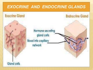
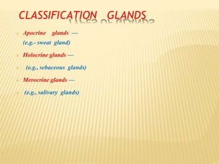
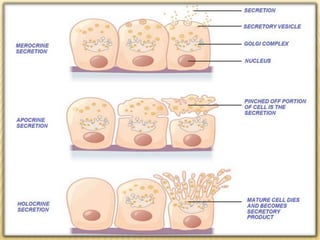
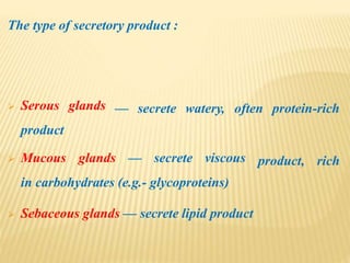
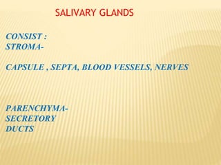
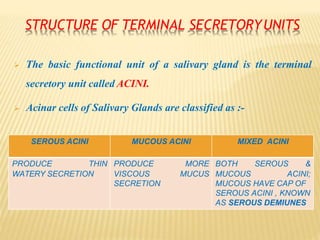
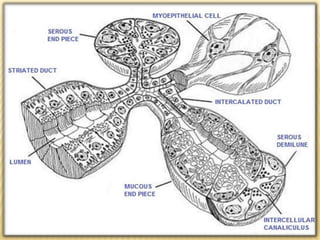
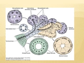
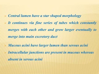
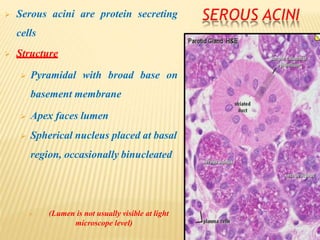
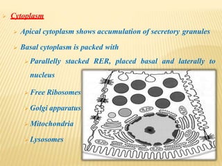
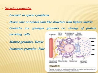
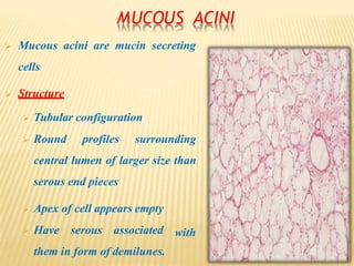
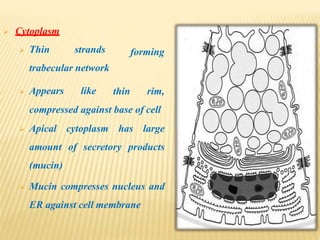
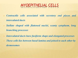
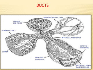
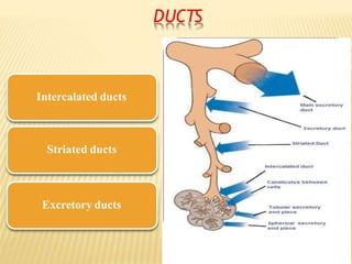
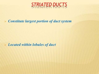
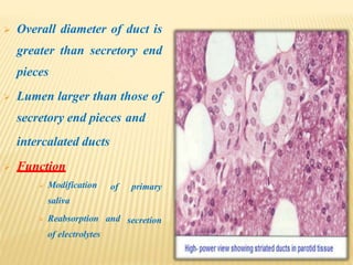
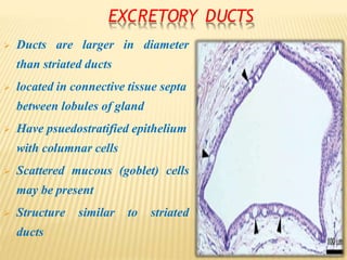
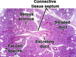
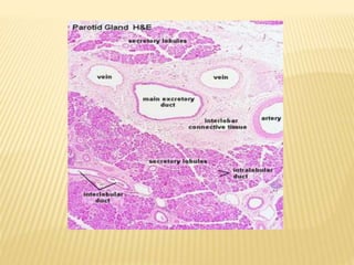
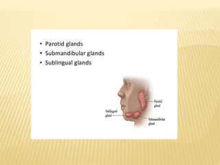
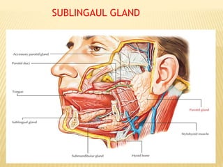
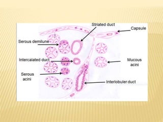
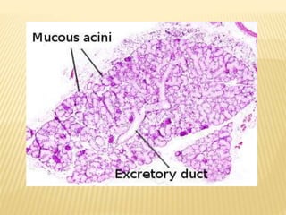
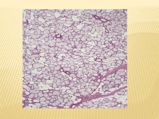
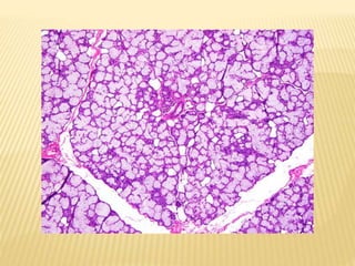
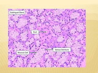
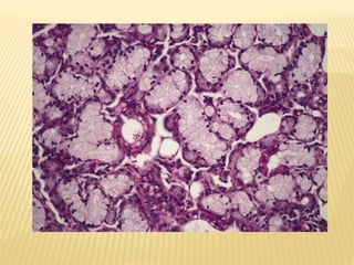
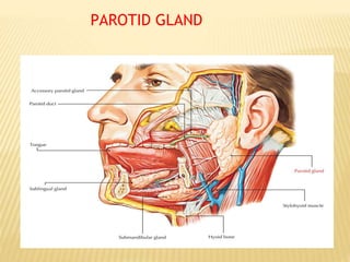
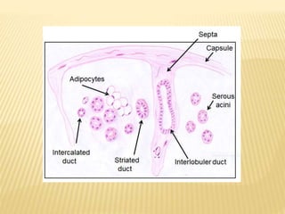
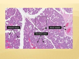
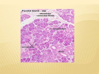
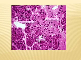
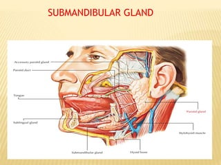
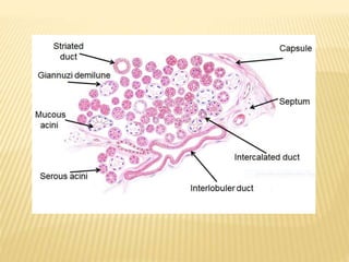
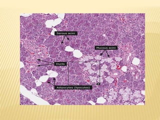
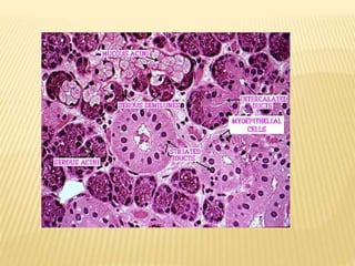
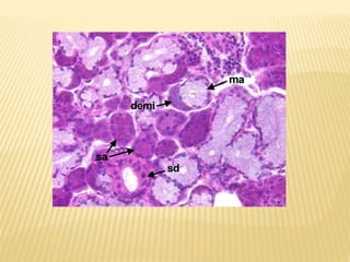
SALIVARY GLANDS : HISTOLOGY
DR D B PATIL
GLANDS
 Gland is an organ in body that synthesizes substance
for release of substances such as hormones often into
bloodstream(endocrine gland) or into cavities inside
body or its outer surface (exocrine gland)
CLASSIFICATION OF GLANDS :
 Endocrine glands — are glands that secrete their
products and lack duct system
 Exocrine glands — secrete their products through
duct or directly onto apical surface, glands in this
group can be divided into three groups :
EXOCRINE AND ENDOCRINE GLANDS
CLASSIFICATION GLANDS
 Apocrine glands —
(e.g.- sweat gland)
 Holocrine glands —
 (e.g., sebaceous glands)
 Merocrine glands —
 (e.g., salivary glands)
The type of secretory product :
— secrete watery, often protein-rich Serous glands
product
product, rich Mucous glands — secrete viscous
in carbohydrates (e.g.- glycoproteins)
 Sebaceous glands — secrete lipid product
SALIVARY GLANDS
CONSIST :
STROMA-
CAPSULE , SEPTA, BLOOD VESSELS, NERVES
PARENCHYMA-
SECRETORY
DUCTS
STRUCTURE OF TERMINAL SECRETORYUNITS
 The basic functional unit of a salivary gland is the terminal
secretory unit called ACINI.
 Acinar cells of Salivary Glands are classified as :-
SEROUS ACINI MUCOUS ACINI MIXED ACINI
PRODUCE THIN PRODUCE MORE BOTH SEROUS &
WATERY SECRETION VISCOUS MUCUS MUCOUS ACINI;
SECRETION MUCOUS HAVE CAP OF
SEROUS ACINI , KNOWN
AS SEROUS DEMIUNES
ACINI
 Rest on basement
 Arranged in single layer
 Intercellular spaces of apical ends of cells separated
from lumen by Junctional complexes
 Junctional complexes hold cells together in acini
 Regulate permeability
 Myoepithelial cells are located on surface of acini
 Central lumen have a star shaped morphology
 It continues via fine series of tubes which constantly
merges with each other and grow larger eventually to
merge into main excretory duct
 Mucous acini have larger lumen than serous acini
 Intracellular junctions are present in mucous whereas
absent in serous acini
SEROUS ACINI Serous acini are protein secreting
cells
 Structure
 Pyramidal with broad base on
basement membrane
 Apex faces lumen
 Spherical nucleus placed at basal
region, occasionally binucleated
 (Lumen is not usually visible at light
microscope level)
 Cytoplasm
 Apical cytoplasm shows accumulation of secretory granules
 Basal cytoplasm is packed with
 Parallelly stacked RER, placed basal and laterally to
nucleus
 Free Ribosomes
 Golgi apparatus
 Mitochondria
 Lysosomes
 Secretory granules
 Located in apical cytoplasm
 Dense core or twisted skin like structure with lighter matrix
 Granules are zymogen granules i.e. storage of protein
secreting cells
 Mature granules- Dense
 Immature granules- Pale
MUCOUS ACINI
 Mucous acini are mucin secreting
cells
 Structure
 Tubular configuration
 Round profiles surrounding
central lumen of larger size than
serous end pieces
with
 Apex of cell appears empty
 Have serous associated
them in form of demilunes.
forming
 Cytoplasm
 Thin strands
trabecular network
 Appears like thin rim,
compressed against base of cell
 Apical cytoplasm has large
amount of secretory products
(mucin)
 Mucin compresses nucleus and
ER against cell membrane
MYOEPITHELIAL CELLS
 Contractile cells associated with secretory end pieces and
intercalated ducts
 Stellate shaped with flattened nuclei, scanty cytoplasm, long
branching processes
 Intercalated ducts have fusiform shape and elongated processes
 These cells lies between basal lamina and joined to each other by
desmosomes
DUCTS
DUCTS
 Ductal system is varied network of tubules that progressively
increase in diameter
 Begins from secretory end pieces to oral cavity
 Ducts are for passage of saliva; it actively participates in
production and modification of saliva
DUCTS
Intercalated ducts
Striated ducts
Excretory ducts
INTERCALATED DUCTS
 Primary saliva produced by secretory end pieces passes first
through it
 First cell of duct is adjacent to end pieces, lumen is continuous
with lumen of intercalated duct
 Lined by simple cuboidal epithelium, myoepithelial cell bodies
 Diameter of duct is smaller than end pieces but their lumen is
larger than end pieces
 Constitute largest portion of duct system
 Located within lobules of duct
STRIATED DUCTS
 Overall diameter of duct is
greater than secretory end
pieces
 Lumen larger than those of
secretory end pieces and
of primary
intercalated ducts
 Function
 Modification
saliva
secretion Reabsorption and
of electrolytes
EXCRETORY DUCTS
 Ducts are larger in diameter
than striated ducts
 located in connective tissue septa
between lobules of gland
 Have psuedostratified epithelium
with columnar cells
 Scattered mucous (goblet) cells
may be present
 Structure similar to striated
ducts
SUBLINGAUL GLAND
PAROTID GLAND
SUBMANDIBULAR GLAND
APPLIED ASPECT
PLEOMORPHIC ADEMONA
MUMPS
SIALORHIA
XEROSTOMIA
SIALOGRAPHY
FROIN SYNDROME
Histologyofsalivaryglands ppt converted

Histologyofsalivaryglands ppt converted

  • 1.
    SALIVARY GLANDS :HISTOLOGY DR D B PATIL
  • 2.
    GLANDS  Gland isan organ in body that synthesizes substance for release of substances such as hormones often into bloodstream(endocrine gland) or into cavities inside body or its outer surface (exocrine gland)
  • 3.
    CLASSIFICATION OF GLANDS:  Endocrine glands — are glands that secrete their products and lack duct system  Exocrine glands — secrete their products through duct or directly onto apical surface, glands in this group can be divided into three groups :
  • 4.
  • 5.
    CLASSIFICATION GLANDS  Apocrineglands — (e.g.- sweat gland)  Holocrine glands —  (e.g., sebaceous glands)  Merocrine glands —  (e.g., salivary glands)
  • 7.
    The type ofsecretory product : — secrete watery, often protein-rich Serous glands product product, rich Mucous glands — secrete viscous in carbohydrates (e.g.- glycoproteins)  Sebaceous glands — secrete lipid product
  • 8.
    SALIVARY GLANDS CONSIST : STROMA- CAPSULE, SEPTA, BLOOD VESSELS, NERVES PARENCHYMA- SECRETORY DUCTS
  • 9.
    STRUCTURE OF TERMINALSECRETORYUNITS  The basic functional unit of a salivary gland is the terminal secretory unit called ACINI.  Acinar cells of Salivary Glands are classified as :- SEROUS ACINI MUCOUS ACINI MIXED ACINI PRODUCE THIN PRODUCE MORE BOTH SEROUS & WATERY SECRETION VISCOUS MUCUS MUCOUS ACINI; SECRETION MUCOUS HAVE CAP OF SEROUS ACINI , KNOWN AS SEROUS DEMIUNES
  • 12.
    ACINI  Rest onbasement  Arranged in single layer  Intercellular spaces of apical ends of cells separated from lumen by Junctional complexes  Junctional complexes hold cells together in acini  Regulate permeability  Myoepithelial cells are located on surface of acini
  • 13.
     Central lumenhave a star shaped morphology  It continues via fine series of tubes which constantly merges with each other and grow larger eventually to merge into main excretory duct  Mucous acini have larger lumen than serous acini  Intracellular junctions are present in mucous whereas absent in serous acini
  • 14.
    SEROUS ACINI Serousacini are protein secreting cells  Structure  Pyramidal with broad base on basement membrane  Apex faces lumen  Spherical nucleus placed at basal region, occasionally binucleated  (Lumen is not usually visible at light microscope level)
  • 15.
     Cytoplasm  Apicalcytoplasm shows accumulation of secretory granules  Basal cytoplasm is packed with  Parallelly stacked RER, placed basal and laterally to nucleus  Free Ribosomes  Golgi apparatus  Mitochondria  Lysosomes
  • 16.
     Secretory granules Located in apical cytoplasm  Dense core or twisted skin like structure with lighter matrix  Granules are zymogen granules i.e. storage of protein secreting cells  Mature granules- Dense  Immature granules- Pale
  • 17.
    MUCOUS ACINI  Mucousacini are mucin secreting cells  Structure  Tubular configuration  Round profiles surrounding central lumen of larger size than serous end pieces with  Apex of cell appears empty  Have serous associated them in form of demilunes.
  • 18.
    forming  Cytoplasm  Thinstrands trabecular network  Appears like thin rim, compressed against base of cell  Apical cytoplasm has large amount of secretory products (mucin)  Mucin compresses nucleus and ER against cell membrane
  • 19.
    MYOEPITHELIAL CELLS  Contractilecells associated with secretory end pieces and intercalated ducts  Stellate shaped with flattened nuclei, scanty cytoplasm, long branching processes  Intercalated ducts have fusiform shape and elongated processes  These cells lies between basal lamina and joined to each other by desmosomes
  • 20.
  • 21.
    DUCTS  Ductal systemis varied network of tubules that progressively increase in diameter  Begins from secretory end pieces to oral cavity  Ducts are for passage of saliva; it actively participates in production and modification of saliva
  • 22.
  • 23.
    INTERCALATED DUCTS  Primarysaliva produced by secretory end pieces passes first through it  First cell of duct is adjacent to end pieces, lumen is continuous with lumen of intercalated duct  Lined by simple cuboidal epithelium, myoepithelial cell bodies  Diameter of duct is smaller than end pieces but their lumen is larger than end pieces
  • 25.
     Constitute largestportion of duct system  Located within lobules of duct STRIATED DUCTS
  • 26.
     Overall diameterof duct is greater than secretory end pieces  Lumen larger than those of secretory end pieces and of primary intercalated ducts  Function  Modification saliva secretion Reabsorption and of electrolytes
  • 27.
    EXCRETORY DUCTS  Ductsare larger in diameter than striated ducts  located in connective tissue septa between lobules of gland  Have psuedostratified epithelium with columnar cells  Scattered mucous (goblet) cells may be present  Structure similar to striated ducts
  • 33.
  • 40.
  • 45.
  • 50.