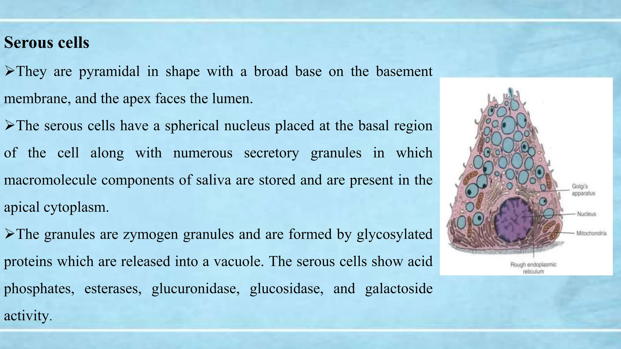
HISTOLOGY OF SALIVARY GLANDS
Presented By,
Dr. Nitha Willy
1st Year PG
Department Of Oral Medicine And Radiology
EPITOME
I. Terminal secretory units
II. Ducts
III. Connective tissue elements
IV. Histology of major salivary glands
V. Histology of minor salivary glands
Terminal secretory units
The functional unit of a salivary gland is the terminal secretory unit called acini .
Regardless of size and location, the terminal secretory unit is made up of epithelial secretory
cells, namely, serous and mucous acini.
The serous and mucous cells along with myoepithelial cells are arranged in an acinus or acini
with a roughly spherical or tubular shape and a central lumen.
Serous cells
They are pyramidal in shape with a broad base on the basement
membrane, and the apex faces the lumen.
The serous cells have a spherical nucleus placed at the basal region
of the cell along with numerous secretory granules in which
macromolecule components of saliva are stored and are present in the
apical cytoplasm.
The granules are zymogen granules and are formed by glycosylated
proteins which are released into a vacuole. The serous cells show acid
phosphates, esterases, glucuronidase, glucosidase, and galactoside
activity.
Cytoplasm: Deeply basophilic (due to
numerous RER), with apical acidophilic secretory
granules (rich in salivary amylase).
The central lumen usually has fingerlike
extensions located between adjacent cells called
intercellular canaliculi that increase the size of the
luminal surface of the cells.
Mucous cells
The secretory endpieces that are composed of mucous cells
typically have a tubular configuration; when cut in cross section,
these tubules appear as round profiles with mucous cells
surrounding central lumen of larger size than that of serous
endpieces.
The nucleus is oval or flattened in shape and located above the
basal plasma membrane.
Sometimes, mucous cells have bonnet- or crescent-shaped
appearance, which is made up of serous cells and are also known as
demilunes first described by Giuseppe Oronzo Giannuzzi in 1865.
Cytoplasm: Pale basophilic and vacuolated (foamy)
Due to dissolved mucinogen secretory granules.
The presence of demilunes is not clearly known, but
these demilunes occur as a result of artifact during
tissue preparation.
Nowadays, recent studies like rapid freezing, freeze substitution, and three-dimensional
reconstruction techniques have shown that serous cells align with mucous cells to surround a
common lumen.
The mucous cells show accumulation of large amounts of secretory product that pushes the
nucleus and endoplasmic reticulum against the basal cell membrane.
The mucous secretion differs from secretion of serous in two important aspects:
• The secretion of mucous cells has little or no enzymatic activity and is responsible mainly
for lubrication and protection of the oral tissues.
• The ratio of carbohydrates to protein is greater, and large amount of sialic acid and
occasionally sulfated sugars are present
In routine histological sections, the secretion of mucous cell appears unstained, and they are
strongly stained when special stains like PAS, alcian blue, mucicarmine, etc. are used
Myoepithelial cells
These are the contractile cells associated with secretory endpiece and intercalated duct of
the salivary glands.
These cells are present between the basal lamina and the secretory or duct cells and are
joined to the cells by desmosomes.
They appear similar to smooth muscle but are derived from the epithelium. They are also
known as basket cells or octopus sitting on a rock.
These cells accelerate the initial flow of saliva from the acini, reduce luminal volume,
support the underlying parenchyma, reduce the back permeation of fluid, and also help to
maintain the patency.
They maintain the cell polarity and structural organization of cells.
They secrete various tumor suppressor proteins such as protease inhibitors and
antiangiogenesis factors which provide a barrier against invasive epithelial neoplasm.
The myoepithelial cells located around the secretory endpieces have stellate-shaped,
numerous branching processes with a flattened nucleus and scanty perinuclear cytoplasm, but
the cells associated with intercalated ducts have more fusiform shape and are elongated with
fewer processes.
Ducts
It consists of hollow tubes that connect initially with
the acinus, i.e., secretory endpieces, and extends to
the oral cavity.
It is not a pipeline or conduit for the passageway for
the saliva, but it actively participates in the production
and modification of saliva.
On the basis of location, ducts are of two types:
Intralobular ducts: Those ducts which are within the lobule. The intercalated and striated ducts
are intralobular ducts.
Interlobular ducts: Those ducts which lie within the connective tissue within the lobules of the
gland. The excretory ducts are interlobular ducts.
Intercalated duct
These are lined by single layer of cuboidal epithelium and are
surrounded by myoepithelial cell bodies, and their processes
typically are found along the basal surface of the duct.
Under the light microscope, the intercalated ducts are difficult
to identify as they are compressed between the secretory units.
Under the electron microscope, the intercalated ducts have
centrally placed nuclei and a small amount of cytoplasm
containing some rough endoplasmic reticulum and a small Golgi
complex.
A few secretory granules may be found in the apical cytoplasm, especially in the cells
located near the endpieces.
The apical cell surface has a few short microvilli projecting into the lumen, and lateral
surfaces are joined by junctional complexes.
The macromolecule components, i.e., lysozyme and lactoferrin, are stored in the
secretory granules of the intercalated duct and contribute to the saliva.
Striated ducts
The striated ducts receive the primary saliva from the intercalated ducts which constitute the
largest portion of the duct system and are lined by columnar cells with a centrally placed large,
spherical nucleus and pale, acidophilic cytoplasm.
Under the electron microscope, the basal cytoplasm of the striated duct cells is
partitioned by deep in foldings of the plasma membrane producing numerous sheet like
folds that extend beyond the lateral boundaries of the cell and interdigitate with similar
folds of adjacent cells.
Between the membrane in foldings, a large amount of radially oriented mitochondria are
located in the portion of the cytoplasm.
The combination of in foldings and
mitochondria accounts for the striations seen in
the light microscope.
These ducts are involved in active transport and
are considered as site of electrolyte reabsorption
especially of sodium and chloride and secretion of
potassium and bicarbonate.
They also synthesize and secrete glycoproteins
such as kallikrein and epidermal growth factor.
Excretory ducts
These ducts are located in the connective tissue septa between the lobules
of the gland and are larger in diameter than striated duct.
These ducts are lined by pseudostratified epithelium with columnar cells
extending from the basal lamina to the ductal lumen and small basal cells
that sit on the basal lamina.
 As the smaller ducts join to form large excretory ducts, the number of
basal cell increases, and scattered mucous (goblet) cells may be present.
The main excretory duct may become stratified near the oral opening.
Tuft or brush cells with long stiff microvilli and apical vesicles are seen and are considered
as receptor cells as they show nerve endings adjacent to the basal portion of the cell.
Dendritic cells are also seen and play an important role in immune surveillance.
Connective tissue elements
The cells that are found in the connective tissue of the salivary glands are similar to those in
other connective tissues of the body and include fibroblasts, macrophages, mast cells,
occasional leukocytes, fat cells, and plasma cells.
Collagen and reticular fibers are also embedded in a ground substance which is composed
of proteoglycans and glycoproteins.
It consists of a surrounding capsule that delineates the gland from the adjacent structures.
Blood vessels and nerves are also present that supply the parenchymal components, i.e.,
glandular components and excretory ducts.
The plasma cells present in the connective tissue produce immunoglobulins that are
secreted into saliva by transcytosis.
Parotid glands
The parotid gland is a purely serous gland, and all the acinar cells
are similar in structure to the serous cells.
Under the electron microscope, serous granules may have a dense
central core, and the intercalated ducts are long branching along with
pale-staining striated ducts, are numerous, and stand out evidently
against the more densely stained acini.
The connective tissue septa contain numerous fat cells which
increase in number with age and leave an empty space in histologic
sections.
Submandibular glands
The submandibular gland is a mixed gland with both serous
and mucous secretory units, but the serous units predominate.
The mucous terminal portions are capped by demilunes of
serous cells.
Under the electron microscope, the intercalated ducts appear
shorter in submandibular gland than those of the parotid,
whereas the striated ducts are usually longer.
Sublingual glands
The sublingual gland is also a mixed gland, but the mucous
secretory units predominate.
The mucous cells are present in tubular pattern along with
serous, demilunes and may be present at the blind ends of the
tubules. The intercalated and striated ducts are poorly
developed, and mucous tubules open directly into ducts lined
with cuboidal or columnar cells without typical basal
striations.
Minor salivary glands
Labial and buccal glands
The glands of the lips and cheeks are a mixed gland consisting of
mucous tubules with serous demilunes.
The intercalated ducts appear variable in length, and the
intralobular ducts possess only a few cells with basal striations.
Glossopalatine glands
The glands present in the region of isthmus in the glossopalatine fold are purely mucous
gland.
Palatine glands
Palatine glands are a purely mucous gland, and
the excretory ducts may have an irregular contour
with large distensions as they course through the
lamina propria.
Lingual glands
In anterior lingual glands, the anterior portion of the glands is chiefly mucous in nature,
whereas the posterior portions are mixed.
The posterior lingual glands are purely mucous glands, but von Ebne’sr glands are purely
serous gland.
PART 2- SALIVARY GLANDS second psrt.pptx

PART 2- SALIVARY GLANDS second psrt.pptx

  • 1.
    HISTOLOGY OF SALIVARYGLANDS Presented By, Dr. Nitha Willy 1st Year PG Department Of Oral Medicine And Radiology
  • 2.
    EPITOME I. Terminal secretoryunits II. Ducts III. Connective tissue elements IV. Histology of major salivary glands V. Histology of minor salivary glands
  • 3.
    Terminal secretory units Thefunctional unit of a salivary gland is the terminal secretory unit called acini . Regardless of size and location, the terminal secretory unit is made up of epithelial secretory cells, namely, serous and mucous acini. The serous and mucous cells along with myoepithelial cells are arranged in an acinus or acini with a roughly spherical or tubular shape and a central lumen.
  • 4.
    Serous cells They arepyramidal in shape with a broad base on the basement membrane, and the apex faces the lumen. The serous cells have a spherical nucleus placed at the basal region of the cell along with numerous secretory granules in which macromolecule components of saliva are stored and are present in the apical cytoplasm. The granules are zymogen granules and are formed by glycosylated proteins which are released into a vacuole. The serous cells show acid phosphates, esterases, glucuronidase, glucosidase, and galactoside activity.
  • 5.
    Cytoplasm: Deeply basophilic(due to numerous RER), with apical acidophilic secretory granules (rich in salivary amylase). The central lumen usually has fingerlike extensions located between adjacent cells called intercellular canaliculi that increase the size of the luminal surface of the cells.
  • 6.
    Mucous cells The secretoryendpieces that are composed of mucous cells typically have a tubular configuration; when cut in cross section, these tubules appear as round profiles with mucous cells surrounding central lumen of larger size than that of serous endpieces. The nucleus is oval or flattened in shape and located above the basal plasma membrane. Sometimes, mucous cells have bonnet- or crescent-shaped appearance, which is made up of serous cells and are also known as demilunes first described by Giuseppe Oronzo Giannuzzi in 1865.
  • 7.
    Cytoplasm: Pale basophilicand vacuolated (foamy) Due to dissolved mucinogen secretory granules. The presence of demilunes is not clearly known, but these demilunes occur as a result of artifact during tissue preparation.
  • 8.
    Nowadays, recent studieslike rapid freezing, freeze substitution, and three-dimensional reconstruction techniques have shown that serous cells align with mucous cells to surround a common lumen. The mucous cells show accumulation of large amounts of secretory product that pushes the nucleus and endoplasmic reticulum against the basal cell membrane.
  • 9.
    The mucous secretiondiffers from secretion of serous in two important aspects: • The secretion of mucous cells has little or no enzymatic activity and is responsible mainly for lubrication and protection of the oral tissues. • The ratio of carbohydrates to protein is greater, and large amount of sialic acid and occasionally sulfated sugars are present In routine histological sections, the secretion of mucous cell appears unstained, and they are strongly stained when special stains like PAS, alcian blue, mucicarmine, etc. are used
  • 10.
    Myoepithelial cells These arethe contractile cells associated with secretory endpiece and intercalated duct of the salivary glands. These cells are present between the basal lamina and the secretory or duct cells and are joined to the cells by desmosomes. They appear similar to smooth muscle but are derived from the epithelium. They are also known as basket cells or octopus sitting on a rock.
  • 11.
    These cells acceleratethe initial flow of saliva from the acini, reduce luminal volume, support the underlying parenchyma, reduce the back permeation of fluid, and also help to maintain the patency. They maintain the cell polarity and structural organization of cells. They secrete various tumor suppressor proteins such as protease inhibitors and antiangiogenesis factors which provide a barrier against invasive epithelial neoplasm. The myoepithelial cells located around the secretory endpieces have stellate-shaped, numerous branching processes with a flattened nucleus and scanty perinuclear cytoplasm, but the cells associated with intercalated ducts have more fusiform shape and are elongated with fewer processes.
  • 12.
    Ducts It consists ofhollow tubes that connect initially with the acinus, i.e., secretory endpieces, and extends to the oral cavity. It is not a pipeline or conduit for the passageway for the saliva, but it actively participates in the production and modification of saliva.
  • 13.
    On the basisof location, ducts are of two types: Intralobular ducts: Those ducts which are within the lobule. The intercalated and striated ducts are intralobular ducts. Interlobular ducts: Those ducts which lie within the connective tissue within the lobules of the gland. The excretory ducts are interlobular ducts.
  • 14.
    Intercalated duct These arelined by single layer of cuboidal epithelium and are surrounded by myoepithelial cell bodies, and their processes typically are found along the basal surface of the duct. Under the light microscope, the intercalated ducts are difficult to identify as they are compressed between the secretory units. Under the electron microscope, the intercalated ducts have centrally placed nuclei and a small amount of cytoplasm containing some rough endoplasmic reticulum and a small Golgi complex.
  • 15.
    A few secretorygranules may be found in the apical cytoplasm, especially in the cells located near the endpieces. The apical cell surface has a few short microvilli projecting into the lumen, and lateral surfaces are joined by junctional complexes. The macromolecule components, i.e., lysozyme and lactoferrin, are stored in the secretory granules of the intercalated duct and contribute to the saliva.
  • 16.
    Striated ducts The striatedducts receive the primary saliva from the intercalated ducts which constitute the largest portion of the duct system and are lined by columnar cells with a centrally placed large, spherical nucleus and pale, acidophilic cytoplasm.
  • 17.
    Under the electronmicroscope, the basal cytoplasm of the striated duct cells is partitioned by deep in foldings of the plasma membrane producing numerous sheet like folds that extend beyond the lateral boundaries of the cell and interdigitate with similar folds of adjacent cells. Between the membrane in foldings, a large amount of radially oriented mitochondria are located in the portion of the cytoplasm.
  • 18.
    The combination ofin foldings and mitochondria accounts for the striations seen in the light microscope. These ducts are involved in active transport and are considered as site of electrolyte reabsorption especially of sodium and chloride and secretion of potassium and bicarbonate. They also synthesize and secrete glycoproteins such as kallikrein and epidermal growth factor.
  • 19.
    Excretory ducts These ductsare located in the connective tissue septa between the lobules of the gland and are larger in diameter than striated duct. These ducts are lined by pseudostratified epithelium with columnar cells extending from the basal lamina to the ductal lumen and small basal cells that sit on the basal lamina.  As the smaller ducts join to form large excretory ducts, the number of basal cell increases, and scattered mucous (goblet) cells may be present.
  • 20.
    The main excretoryduct may become stratified near the oral opening. Tuft or brush cells with long stiff microvilli and apical vesicles are seen and are considered as receptor cells as they show nerve endings adjacent to the basal portion of the cell. Dendritic cells are also seen and play an important role in immune surveillance.
  • 21.
    Connective tissue elements Thecells that are found in the connective tissue of the salivary glands are similar to those in other connective tissues of the body and include fibroblasts, macrophages, mast cells, occasional leukocytes, fat cells, and plasma cells. Collagen and reticular fibers are also embedded in a ground substance which is composed of proteoglycans and glycoproteins. It consists of a surrounding capsule that delineates the gland from the adjacent structures.
  • 22.
    Blood vessels andnerves are also present that supply the parenchymal components, i.e., glandular components and excretory ducts. The plasma cells present in the connective tissue produce immunoglobulins that are secreted into saliva by transcytosis.
  • 23.
    Parotid glands The parotidgland is a purely serous gland, and all the acinar cells are similar in structure to the serous cells. Under the electron microscope, serous granules may have a dense central core, and the intercalated ducts are long branching along with pale-staining striated ducts, are numerous, and stand out evidently against the more densely stained acini. The connective tissue septa contain numerous fat cells which increase in number with age and leave an empty space in histologic sections.
  • 24.
    Submandibular glands The submandibulargland is a mixed gland with both serous and mucous secretory units, but the serous units predominate. The mucous terminal portions are capped by demilunes of serous cells. Under the electron microscope, the intercalated ducts appear shorter in submandibular gland than those of the parotid, whereas the striated ducts are usually longer.
  • 25.
    Sublingual glands The sublingualgland is also a mixed gland, but the mucous secretory units predominate. The mucous cells are present in tubular pattern along with serous, demilunes and may be present at the blind ends of the tubules. The intercalated and striated ducts are poorly developed, and mucous tubules open directly into ducts lined with cuboidal or columnar cells without typical basal striations.
  • 26.
    Minor salivary glands Labialand buccal glands The glands of the lips and cheeks are a mixed gland consisting of mucous tubules with serous demilunes. The intercalated ducts appear variable in length, and the intralobular ducts possess only a few cells with basal striations.
  • 27.
    Glossopalatine glands The glandspresent in the region of isthmus in the glossopalatine fold are purely mucous gland.
  • 28.
    Palatine glands Palatine glandsare a purely mucous gland, and the excretory ducts may have an irregular contour with large distensions as they course through the lamina propria.
  • 29.
    Lingual glands In anteriorlingual glands, the anterior portion of the glands is chiefly mucous in nature, whereas the posterior portions are mixed. The posterior lingual glands are purely mucous glands, but von Ebne’sr glands are purely serous gland.