https://userupload.net/3ppacneii1wj
Toxicologic Pathology (Second Edition), 2010
INTRODUCTION
The oral mucosa is, in many ways, similar to the skin in its architecture, function, and reaction patterns. This section only emphasizes those characteristics of the oral mucosa that influence or result in a distinct group of pathologic entities.
Because of its location at the entrance of the digestive and respiratory tracts and its proximity to the teeth, the oral mucosa is subjected to numerous natural and man-made xenobiotics. The peculiar architecture and absorption characteristics of the oral mucosa, especially in areas of extreme thinness, coupled with the rich microorganism flora of the mouth, makes the oral mucosa a peculiar site deserving separate discussion.
Amelogenesis is the formation of enamel. During amelogenesis, the ameloblast (enamel-forming cells) undergo various stages i.e the life cycle of ameloblast.
For more content check out my blog: www.rkharitha.wordpress.com "a little about everything dental"
I prepared this presentation during the first year of my MDS. This will give you a basic idea and necessary information about the pulp of the teeth and its histology. Hope you guys find it useful.
It is a presentation in detail about the strongest structure of the oral cavity "ENAMEL". It is a simple topic but people find it difficult to learn about it. I hope my presentation is a simple method to learn about it. I would like to thank my professors for assign me this project and i learn't a lot from it and still learning my basics daily.
Definition
Classification Of Salivary Glands
Anatomy of salivary glands
Development of salivary glands
Structure Of Salivary Glands
Histology of major and minor salivary glands
salivary gland lecture
oral biology department fayoum university
Prof.Dr. Sahar shawkat ppt - Profesessor and head of the department -cairo university
presented by Dr. Dina Hassouna
lecturer at oral biology department fayoum university
Classification of glands.
Detailed microscopic structure of exocrine glands.
differences between serous and mucus acini.
Microscopic structure of Parotid, submandibular and sublingual glands.
Indian Dental Academy: will be one of the most relevant and exciting training center with best faculty and flexible training programs for dental professionals who wish to advance in their dental practice,Offers certified courses in Dental implants,Orthodontics,Endodontics,Cosmetic Dentistry, Prosthetic Dentistry, Periodontics and General Dentistry.
The mucose membrane lining of gastrointestinal tract is stratified squamous epithelium at the esophagus which slowly convert into simple columnar epithelium at the stomach until the anus it converts back into the stratified squamous epithelium at the lower half of the anal canal. The stratified epithelium is a wear and tear epithelium.
As it passes down from the small to large intestine, goblet cells increase because as it passes down water was absorb, goblet cells function to produce mucous.
This is just a rough idea, for better slides with more reference please PM the author at davidgqf@gmail.com.
Synthetic Fiber Construction in lab .pptxPavel ( NSTU)
Synthetic fiber production is a fascinating and complex field that blends chemistry, engineering, and environmental science. By understanding these aspects, students can gain a comprehensive view of synthetic fiber production, its impact on society and the environment, and the potential for future innovations. Synthetic fibers play a crucial role in modern society, impacting various aspects of daily life, industry, and the environment. ynthetic fibers are integral to modern life, offering a range of benefits from cost-effectiveness and versatility to innovative applications and performance characteristics. While they pose environmental challenges, ongoing research and development aim to create more sustainable and eco-friendly alternatives. Understanding the importance of synthetic fibers helps in appreciating their role in the economy, industry, and daily life, while also emphasizing the need for sustainable practices and innovation.
Instructions for Submissions thorugh G- Classroom.pptxJheel Barad
This presentation provides a briefing on how to upload submissions and documents in Google Classroom. It was prepared as part of an orientation for new Sainik School in-service teacher trainees. As a training officer, my goal is to ensure that you are comfortable and proficient with this essential tool for managing assignments and fostering student engagement.
How to Create Map Views in the Odoo 17 ERPCeline George
The map views are useful for providing a geographical representation of data. They allow users to visualize and analyze the data in a more intuitive manner.
Palestine last event orientationfvgnh .pptxRaedMohamed3
An EFL lesson about the current events in Palestine. It is intended to be for intermediate students who wish to increase their listening skills through a short lesson in power point.
The Art Pastor's Guide to Sabbath | Steve ThomasonSteve Thomason
What is the purpose of the Sabbath Law in the Torah. It is interesting to compare how the context of the law shifts from Exodus to Deuteronomy. Who gets to rest, and why?
How to Split Bills in the Odoo 17 POS ModuleCeline George
Bills have a main role in point of sale procedure. It will help to track sales, handling payments and giving receipts to customers. Bill splitting also has an important role in POS. For example, If some friends come together for dinner and if they want to divide the bill then it is possible by POS bill splitting. This slide will show how to split bills in odoo 17 POS.
We all have good and bad thoughts from time to time and situation to situation. We are bombarded daily with spiraling thoughts(both negative and positive) creating all-consuming feel , making us difficult to manage with associated suffering. Good thoughts are like our Mob Signal (Positive thought) amidst noise(negative thought) in the atmosphere. Negative thoughts like noise outweigh positive thoughts. These thoughts often create unwanted confusion, trouble, stress and frustration in our mind as well as chaos in our physical world. Negative thoughts are also known as “distorted thinking”.
The French Revolution, which began in 1789, was a period of radical social and political upheaval in France. It marked the decline of absolute monarchies, the rise of secular and democratic republics, and the eventual rise of Napoleon Bonaparte. This revolutionary period is crucial in understanding the transition from feudalism to modernity in Europe.
For more information, visit-www.vavaclasses.com
Students, digital devices and success - Andreas Schleicher - 27 May 2024..pptxEduSkills OECD
Andreas Schleicher presents at the OECD webinar ‘Digital devices in schools: detrimental distraction or secret to success?’ on 27 May 2024. The presentation was based on findings from PISA 2022 results and the webinar helped launch the PISA in Focus ‘Managing screen time: How to protect and equip students against distraction’ https://www.oecd-ilibrary.org/education/managing-screen-time_7c225af4-en and the OECD Education Policy Perspective ‘Students, digital devices and success’ can be found here - https://oe.cd/il/5yV
Students, digital devices and success - Andreas Schleicher - 27 May 2024..pptx
Handout of Salivary Glands Histology
1. 1| P a g e
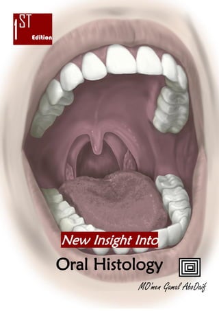
1ST
Edition
New Insight Into
Oral Histology
MO'men Gamal AboDaif
2. 2| P a g e
Salivary Glands
Definition: Multicellular, Merocrine & Exocrine glands whose secretion (Saliva) flow into the oral cavity
••Multicellular: Multi cells Organ ••Merocrine: Secreting Organ ••Exocrine: Discharge their secretion into Ducts
Classification (according to Size)
•Major: ••Parotid ••Submandibular •• Sublingual
•Minor: The rest of Salivary Glands
Classification (according to Natural of Secretion)
•Pure Serous: ••Parotid “Adult” ••Von Ebner
•Pure Mucous: ••Mucous Ring (Palatine - Glossopalatine -
Weber)
••Labial
•Mixed: ••Predominantly Serous (Parotid “Newborn-Older people” – Submandibular)
••Predominantly Mucous (Sublingual – Blandin Nuhn – Buccal)
Classification (according to Location)
•Oral Vestibule: ••Parotid ••Labial ••Buccal
•Oral Cavity Proper: ••Mouth Floor (Submandibular – Sublingual “Major & Minor” – Glossopalatine)
••Tongue (Von Ebner – Weber – Blandin Nuhn)
••Palatine (Hard palate – Soft palate – Uvula)
•Mouth Ring
Site: at Isthmus Region between Oral cavity & Pharynx
Contain: ••Palatine ••Glossopalatine ••Weber
Development
•During fetal life, Salivary Glands growth as an Epithelial Buds into underlying
mesenchyme at:
••4th
weak i.u in Parotid ••6th
weak i.u in Submandibular
••8th
weak i.u in Sublingual ••3rd
month in Minor Glands
•The Buds grow into Branched System of solid cords which gradually develop a
lumen
3. 3| P a g e
Macroanatomy of Salivary Glands Composed of:
I-Parenchymal Elements
Origin: epithelial outgrowth which invades
the underlying mesenchyme
Structure: Lobes &Lobules
*Secretory Portion
Unit Structure: Tubular end pieces resemble a
grape cluster called acini
Acini: Tubular end piece, either mucous or serous
Function: Saliva Secretion
*Ductal System
Structure: Confluence of small ducts into larger ones
They may be -Intralobular ducts are (intercalated & striated)
-Interlobular ducts are excretory ducts
Function: 1.Transport (Convey) Primary Saliva to the oral cavity (main function)
2. Participate in Saliva production & modulation, as they modify the primary saliva by
••Secretion & reabsorption of electrolytes ••Secretion of proteins
*Myoepithelial cells:
Structure: Star shape cells partially surround the acini
Function: Contract to expel secretions from acini to the lumens & then ducts toward the oral cavity
II-Connective Tissue Stroma
Origin: arise from the mesenchyme
Structure:
Capsule: C.T around the gland
Septa: C.T that extends into the gland dividing it into
lobes & lobules
Function: Carry the nerves, blood vessels & lymph
vessels
4. 4| P a g e
Serous acinus “end piece” Mucous acinus
Fun. Synthesis, storage & secretion of Protein Synthesis, storage & secretion of Carbohydrate
Secretion Watery For enzymic activity Viscous For lubrication
Shape •Spherical •Tubular
Size Smaller Larger
Lumen Narrow Wide
Canaliculi Present Absent (Except: Human Labial gland)
Serous Cell Mucous Cell
LightMicroscope
Shape
•High Pyramidal
Connect together by Junctional Complexes
•Base: resting on B.M
•Apex: toward lumen
•Triangular (low Pyramidal)
•Base: resting on B.M
•Apex: toward lumen
(Wider than those of serous cell)
Nuc Spherical & Basally located (deeply stained) Flat & Compressed Basally
Cytoplasm
•Apically: Zymogen Granules: Eosinophilic
secretory granules about 1 um in diameter
•Basally: Chromophils or Ergastoplasm: large
concentration of basophilic substances arranged
in parallel rods that give Vertical Striations to
the basal portions
Folded B.M with Mitochondria in-between
•Apically: Trabecular Network: Basophilic thin
strands of cytoplasm
•Basally: Thin Rim between Nucleus & B.M
ElectronicMicroscope
Protein Synthetic Cells Carbohydrate Synthetic Cells
Nu. Large & Open faced Flat & Basally located
RER More
located along base & lateral borders of the cell in
parallel stacks packed arrangement
Less
located along base & lateral borders of the cell
More Ribosomes Content Less Ribosomes Content
Mit. More
located along base & lateral borders of the cell
Less
located along base & lateral borders of the cell
Golgi
Less
Prominent & With 4-6 Stacks
located apically or laterally to nucleus
More
Large & With 10 to 12 Stacks of saccules
sandwiched between basal RER & mucous droplets
NB: Golgi apparatus plays an important role in
these cells because of large amount of
carbohydrate that it adds to the secretory products
Granules
Zymogen: Secretory granules fill Apical
cytoplasm & surrounded by a thin membrane
Mucigen: Irregular & compressed Droplets
containing scattered flocculent material & Apically
(larger than serous one)
NB: Adjacent mucous droplets are separated by thin
cytoplasm strands or may be fused
Other Few peroxisomes, Microfilaments & Microtubules
5. 5| P a g e
NB: Secretory products of mucous cells differ from those of serous cells in two important aspects;
1-They have little or no enzymatic activity, so they serve mainly for lubrication & protection of the oral tissue
2-The ratio of carbohydrate to protein is greater
Junctional Complexes
Structure: Tight junction, Intermediate junction & 1 or more desmosomes
Site: Between adjoining cells
Tight junction is at the apical end of adjoining cells
Function: 1. Hold the cells together
2. Sealed lumen off from lateral intercellular spaces. So, they Prevent
leakage of luminal contents into intercellular spaces
Canaliculi: Intercellular Branches of lumen extend between adjacent cells
almost to their base
Mixed Glands
Definition: Secretory units formed of both secretory acini, serous & mucous
Structure: Demilune: Crescent of several serous cells cap the tubular portion
of the mucous acini
NB: -Their secretion reaches the lumen through Intercellular Canaliculi
-Separate serous & mucous units may exist
Serous – Mixed >> With Canaliculi
Mucous >> Without Canaliculi
Myoepithelial cells
Shape: Branched stellate cells
Origin: epithelial
Site: between B.L & B.M of the Parenchymal cells, close to
the secretory acini & intercalated ducts cells
•Not usually present along the striated ducts
Function: Contract to expel secretions from acini to the lumen &
ducts
Structure: •By L.M, only the nuclei of the cells are Visible
By E.M: contains Actin & Myosin: longitudinally oriented fine filaments similar to those in smooth muscle
6. 6| P a g e
Oncocytes
Site: Parotid & Submandibular glands of older individuals
Shape: large cells with •Nucleus: small Pyknotic & Centrally placed
•Cytoplasm: Abundant & Strongly eosinophilic
7. 7| P a g e
Lining cell Intercalated ducts Striated ducts Excretory ducts
Type Intra-tubular Inter-tubular
Size Small Large (lager or same as acini) The largest
Function
1. Convey saliva to straight
ducts
2. Sharing in secretion of saliva
as they contain secretory
granules
3. Modify secretion by form 2
antibacterial portions
(lysozyme & lacto-ferrin)
4. Acts as Stem cells for
regeneration of secretory
portion cells
1. Convey saliva to excretory duct
2. Modify secretions passing through
them from isotonic to hypotonic
solution By
•Absorb of Na & K ions
•Secret Bi-carbonate ions
3. Pumping capacity to saliva Due to
the Basal deep infolding
4. Secret Kallikrein enzyme which
found in saliva & synthesize Glyco-
proteins (stored in the apical granules)
1. Convey saliva to main
duct
1. Modify the final saliva
by
•adding mucoid
components
•altering electrolyte
concentration of saliva
Cells
LightMicroscope
Shape Simple layer of low Cuboid cell Simple layer of tall Columnar cells •Near the striated
ducts, Pseudo-stratified
epith. With Goblet
cells
(Nucleus on more than
one layer)
•Near the ductal orifice,
stratified squamous
epith. merging with
that of the oral cavity
(Main Duct)
Nu. Large, Round & Centrally placed Large, Round & Centrally placed
Cytoplasm
Empty-appearing Abundant & Eosinophilic
With Basal Striations, perpendicular to
the basal surface (L.M)
Because of Folding of plasma
membrane with elongated
Mitochondria in-between (E.M)
ElectronicMicroscope
Nu. Round & Centrally(more basally Round & Centrally placed
RER Few & Basally Few & Short (with SER apically)
Golgi Moderate size & Apically Small & in Peri-nuclear Cytoplasm
Mito. Around the nucleus Around the nucleus
Between basal infolding
Sec. Few & apically More, Small & Apically
Other *Protein synthesis cell (in low rate)
NB: Adjacent cells are joined
apically by desmosomes
*Apically;
-Free Ribosomes, Lysosomes (several)
-Peroxisomes (numerous)
-Filaments bundles
-Glycogen (moderate amount)
-Microvilli (Numerous, short project into the
lumen)
NB: Adjacent cells are joined by
junctional complex
8. 8| P a g e
II-Connective Tissue Stroma
Cells •Fibroblasts •Macrophages •Mast cells •Plasma cells •Adipose cells
Which are embedded in ••Extracellular matrix of Collagen & Oxytalin fibers
••Ground substances of Glyco-proteins & Proteo-glycans
Blood supply
Course ••enters the gland along the excretory ducts
••branches to follow them into the individual lobules
NB: Extensive blood supply is required for rapid secretion of saliva
Nerve supply
Course ••enter the salivary glands following the blood vessels
••break up into smaller bundles unit they form a Final Plexus adjacent to the terminal parenchyma
There are 2 types of innervations are established
Sub-epithelial Supply Intra-epithelial Supply
(Epi-lemmal) (Hypo-lemmal)
Axon
Site
(Remain in the C.T)
-Separated from the secretory cells by B.M by a gap
of 100 to 200 nm
(Penetrate the B.M)
-Run between the secretory cells separated from
them by a gap of 10 to 20 nm
Lose Schwann cell covering Lose Schwann cell covering during penetration
Contain
Neuro-transmitters (No-repin-ephrine & Acetyl-
chloline) which are •Released when a nerve impulse
passes
•Diffuse across the space & basal lamina
NB:
•Saliva flow is controlled by Nervous Stimulation
•Both divisions of Autonomic Nervous System participate in the secretory cells innervation
•In some glands, both Sympathetic & Parasympathetic terminals are seen near the secretory cells
••Parasympathetic stimulation produces watery saliva
••Sympathetic stimulation produces thicker, higher organic content & less quantity saliva
9. 9| P a g e
Major Salivary Glands
Site: Extra-orally & their secretions reach the mouth by variable ducts
Parotid “The largest” Submandibular Sublingual
Site •Superficial Portion;
in Front of External Ear
•Deeper Portion;
fills Retro-mandibular Fossa
•Submandibular triangle
•Behind & below Mylohyoid
muscle free border
(with a Tongue-like extension
above the mylohyoid muscle)
•Between Mouth Floor &
Mylohyoid muscle
Capsule Encapsulated Encapsulated Poor /Non Capsulated
Type •Pure serous in adult
•Mixed serous in infant & Old
(with few mucous secreting units)
NB: In older gland, Fat cells are
seen
•Mixed “Mostly Serous”
(80% serous & 20% Mixed)
(with few mucous terminal
portions which are capped by
serous demilunes)
•Mixed “Mostly mucous”
(Mucous tubules are capped by
serous demilunes)
Saliva Se. 30% 60% 5%
Main
Excretory
Duct
“Stensen’s Duct”
Open in a Papilla on the buccal
mucosa opposite to Upper 7
“Wharton’s Ducts”
Open in a Papilla on the
mouth floor
(on the side of lingual frenum)
“Bartholin's Duct” (Major)
Open in a Papilla on the mouth
floor
(with or near submandibular duct)
•Rivinian Ducts (Minor): 8-20
small ducts of smaller glands
open along the sublingual fold
independently
lntercalated
Ducts
Numerous & Elongated Shorter than in Parotid Extremely Short or absent
Striated
Ducts
Easy recognized Well developed & Longer
than in Parotid
Extremely Short or absent
10. 11| P a g e
Minor Salivary Glands
Site: Beneath the epith. in almost all parts of the oral cavity
Structure: Several small groups of secretary units opening via short ducts directly
into the mouth
Characters: 1. Lack the distinct capsule
2. Focal accumulation of lymphocytes around ductal walls
Function: 1. Saliva Secretion
2. Have a role in the mouth immune surveillance
Group
Labial & Buccal Palatine Glosso-
palatine
Lingual
Anterior Posterior
Blandin Nuhn Von Ebner Weber
Site
•Lips & Cheeks •Hard palate, Soft
palate & Uvula
Submucosa of
Postero-lateral
region
•Isthmus
region in
Glosso-
palatine
fold
•Near Tongue
apex
•Between
tongue
muscle fibers
•Below
vallate
papillae
•Lateral &
Posterior to
vallate papillae
•Associated with
the lingual
tonsil)
Type
•Mixed
(Mostly mucous)
•Mucous •Mucous •Mostly Mucous
(Anterior region)
•Mixed
(Posterior region)
•Serous •Mucous
Ducts
Intercalated duets:
variable in length
Striated ducts:
show few cells with
basal striations
Large & recognized
Openings open on
Palatal mucosa
Open on tongue
ventral surface
(near lingual
frenum)
Open in
vallate
papillae
trough &
foliate
papillae
Open in tongue
dorsal surface
NB: •Recent ultra-structural studies of labial glands have revealed:
“Presence of only mucous cells & intercellular canaliculi in-between“
•Function of the Von Ebner Salivary Gland
1. Wash tongue papillae, so prepare the taste receptors for a new stimulus
2. Secret anti-bacterial enzymes (Per-oxidase & Lyso-zyme) “Histo-chemical
Studies”
3. Secret lingual lipase enzyme (by lipolytic activity) “Biochemical Studies”
Which ••Hydrolyze stomach tri-glyceride
••Plays a significant role in lipid digestion in newborn when
•Fat intake is high •Levels of pancreatic lipase are low
4. Protective & Digestive function