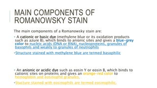
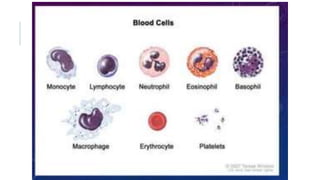
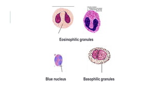
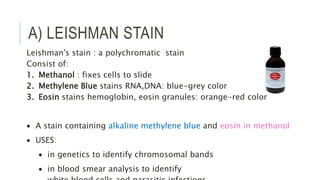
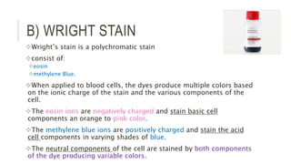
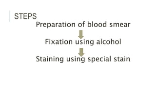
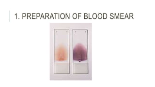
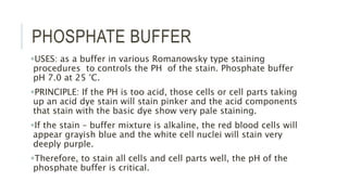
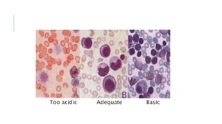
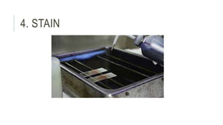
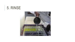
BT 1103 HEMATOLOGY 1
TOPIC 10: ROMANOWSKY
STAINING
LEARNING OUTCOME
Explain the principle of Romanowsky stain
Understand the usage of Romanowsky stain
Explain the principle of Leishman staining
Perform the Leishman staining
Differentiate the cells after staining
ROMANOWSKY STAINING
Romanowsky stains are universally employed for staining blood films
and are generally very satisfactory.
There are a number of different combinations of these dyes, which vary,
in their staining characteristics.
1. May-Grunwald-Giemsa is a good method for routine work.
2. Giemsa stain is thought to produce more delicate staining
characteristics.
3. Wright's stain is a simpler method.
4. Leishman's is also a simple method, which is especially suitable when
a stained blood film is required urgently or the routine stain is not
available (e.g. at night).
5. Field's stain is a rapid stain used primarily on thin films for malarial
MAIN COMPONENTS OF
ROMANOWSKY STAIN
The main components of a Romanowsky stain are:
 A cationic or basic dye (methylene blue or its oxidation products
such as azure B), which binds to anionic sites and gives a blue-grey
color to nucleic acids (DNA or RNA), nucleoproteins, granules of
basophils and weakly to granules of neutrophils
Structure stained with methylene blue are termed basophilic
 An anionic or acidic dye such as eosin Y or eosin B, which binds to
cationic sites on proteins and gives an orange-red color to
hemoglobin and eosinophil granules.
Stucture stained with eosinophils are termed eosinophilic.
A) LEISHMAN STAIN
Leishman's stain : a polychromatic stain
Consist of:
1. Methanol : fixes cells to slide
2. Methylene Blue stains RNA,DNA: blue-grey color
3. Eosin stains hemoglobin, eosin granules: orange-red color
 A stain containing alkaline methylene blue and eosin in methanol
 USES:
 in genetics to identify chromosomal bands
 in blood smear analysis to identify
B) WRIGHT STAIN
Wright’s stain is a polychromatic stain
consist of:
eosin
methylene Blue.
When applied to blood cells, the dyes produce multiple colors based
on the ionic charge of the stain and the various components of the
cell.
The eosin ions are negatively charged and stain basic cell
components an orange to pink color.
The methylene blue ions are positively charged and stain the acid
cell components in varying shades of blue.
The neutral components of the cell are stained by both components
of the dye producing variable colors.
STEPS
Preparation of blood smear
Fixation using alcohol
Staining using special stain
1. PREPARATION OF BLOOD SMEAR
2. FIXATION USING ABSOLUTE
ALCOHOL
3. PHOSPHATE BUFFER
PHOSPHATE BUFFER
USES: as a buffer in various Romanowsky type staining
procedures to controls the PH of the stain. Phosphate buffer
pH 7.0 at 25 °C.
PRINCIPLE: If the PH is too acid, those cells or cell parts taking
up an acid dye stain will stain pinker and the acid components
that stain with the basic dye show very pale staining.
If the stain – buffer mixture is alkaline, the red blood cells will
appear grayish blue and the white cell nuclei will stain very
deeply purple.
Therefore, to stain all cells and cell parts well, the pH of the
phosphate buffer is critical.
Too acidic Adequate Basic
4. STAIN
PROCEDURE
1. Thin smear are air dried.
2. Flood the smear with stain.
3. Stain for 1-5 min.
4. Add an equal amount of buffer solution and mix the stain by
blowing an eddy in the fluid.
5. Leave the mixture on the slide for 10-15 min.
6. Wash off by running water directly to the centre of the slide to
prevent a residue of precipitated stain.
7. Stand slide on end, and let dry in air.
5. RINSE
PRECAUTIONS
1. The staining rack must be exactly level to guard against uneven
staining of the smear.
2. Insufficient washing of the smears when removing the stain and
buffer mixture may cause stain precipitate on the dried smear.
3. Excessive rinsing of the stained smear will cause the stain to fade.
4. Whichever method is used, it is important to select dyes that are not
contaminated with other dyes or metallic salts.
5. Staining time must be specific for each lot of stains and so we must
follow the kit procedure.
6. Bone marrow time staining must be increased.
STAINING FAULTS
Too faint:
Staining time too short.
Excessive washing after staining.
Stain deposit:
Stain solution left in uncovered jar.
Stain solution not filtered.
Dirty slides.
TOPIC 10 ROMANOWSKY STAINING.pptx

TOPIC 10 ROMANOWSKY STAINING.pptx

  • 1.
    BT 1103 HEMATOLOGY1 TOPIC 10: ROMANOWSKY STAINING
  • 2.
    LEARNING OUTCOME Explain theprinciple of Romanowsky stain Understand the usage of Romanowsky stain Explain the principle of Leishman staining Perform the Leishman staining Differentiate the cells after staining
  • 3.
    ROMANOWSKY STAINING Romanowsky stainsare universally employed for staining blood films and are generally very satisfactory. There are a number of different combinations of these dyes, which vary, in their staining characteristics. 1. May-Grunwald-Giemsa is a good method for routine work. 2. Giemsa stain is thought to produce more delicate staining characteristics. 3. Wright's stain is a simpler method. 4. Leishman's is also a simple method, which is especially suitable when a stained blood film is required urgently or the routine stain is not available (e.g. at night). 5. Field's stain is a rapid stain used primarily on thin films for malarial
  • 4.
    MAIN COMPONENTS OF ROMANOWSKYSTAIN The main components of a Romanowsky stain are:  A cationic or basic dye (methylene blue or its oxidation products such as azure B), which binds to anionic sites and gives a blue-grey color to nucleic acids (DNA or RNA), nucleoproteins, granules of basophils and weakly to granules of neutrophils Structure stained with methylene blue are termed basophilic  An anionic or acidic dye such as eosin Y or eosin B, which binds to cationic sites on proteins and gives an orange-red color to hemoglobin and eosinophil granules. Stucture stained with eosinophils are termed eosinophilic.
  • 7.
    A) LEISHMAN STAIN Leishman'sstain : a polychromatic stain Consist of: 1. Methanol : fixes cells to slide 2. Methylene Blue stains RNA,DNA: blue-grey color 3. Eosin stains hemoglobin, eosin granules: orange-red color  A stain containing alkaline methylene blue and eosin in methanol  USES:  in genetics to identify chromosomal bands  in blood smear analysis to identify
  • 8.
    B) WRIGHT STAIN Wright’sstain is a polychromatic stain consist of: eosin methylene Blue. When applied to blood cells, the dyes produce multiple colors based on the ionic charge of the stain and the various components of the cell. The eosin ions are negatively charged and stain basic cell components an orange to pink color. The methylene blue ions are positively charged and stain the acid cell components in varying shades of blue. The neutral components of the cell are stained by both components of the dye producing variable colors.
  • 9.
    STEPS Preparation of bloodsmear Fixation using alcohol Staining using special stain
  • 10.
    1. PREPARATION OFBLOOD SMEAR
  • 11.
    2. FIXATION USINGABSOLUTE ALCOHOL
  • 12.
  • 13.
    PHOSPHATE BUFFER USES: asa buffer in various Romanowsky type staining procedures to controls the PH of the stain. Phosphate buffer pH 7.0 at 25 °C. PRINCIPLE: If the PH is too acid, those cells or cell parts taking up an acid dye stain will stain pinker and the acid components that stain with the basic dye show very pale staining. If the stain – buffer mixture is alkaline, the red blood cells will appear grayish blue and the white cell nuclei will stain very deeply purple. Therefore, to stain all cells and cell parts well, the pH of the phosphate buffer is critical.
  • 14.
  • 15.
  • 16.
    PROCEDURE 1. Thin smearare air dried. 2. Flood the smear with stain. 3. Stain for 1-5 min. 4. Add an equal amount of buffer solution and mix the stain by blowing an eddy in the fluid. 5. Leave the mixture on the slide for 10-15 min. 6. Wash off by running water directly to the centre of the slide to prevent a residue of precipitated stain. 7. Stand slide on end, and let dry in air.
  • 17.
  • 18.
    PRECAUTIONS 1. The stainingrack must be exactly level to guard against uneven staining of the smear. 2. Insufficient washing of the smears when removing the stain and buffer mixture may cause stain precipitate on the dried smear. 3. Excessive rinsing of the stained smear will cause the stain to fade. 4. Whichever method is used, it is important to select dyes that are not contaminated with other dyes or metallic salts. 5. Staining time must be specific for each lot of stains and so we must follow the kit procedure. 6. Bone marrow time staining must be increased.
  • 19.
    STAINING FAULTS Too faint: Stainingtime too short. Excessive washing after staining. Stain deposit: Stain solution left in uncovered jar. Stain solution not filtered. Dirty slides.