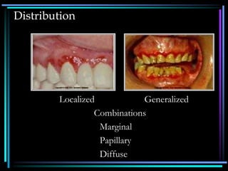
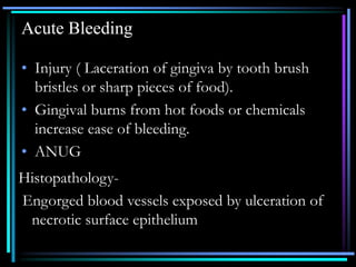
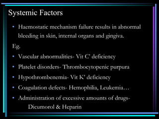
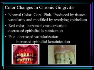
CLINICAL FEATURES OF
GINGIVITIS
Dr.D.Navya, M.D.S.
Course And Duration
• Acute Gingivitis- Sudden onset, Short
duration, Painful
• Chronic Gingivitis- Slow onset, Long
duration, Painless
• Recurrent Gingivitis- Reappear after
having been eliminated
Distribution
Localized Generalized
Combinations
Marginal
Papillary
Diffuse
Clinical Findings
Gingival Bleeding On Probing
• Gingival bleeding varies in
severity, duration and the ease
with which it is provoked.
• Bleeding on probing is easily
detectable clinically & therefore
it is of value for the early
diagnosis and prevention of
advanced gingivitis.
Causes
• Local Factors
• Systemic Factors
Local Factors
• Chronic recurrent bleeding
• Acute bleeding
Chronic Recurrent Bleeding
• Chronic inflammation.
• Provoked by mechanical trauma( Tooth
brushing, tooth picks, food impaction).
Histopathology-
• Dilation of capillaries & thinning or ulceration
of epithelium.
Acute Bleeding
• Injury ( Laceration of gingiva by tooth brush
bristles or sharp pieces of food).
• Gingival burns from hot foods or chemicals
increase ease of bleeding.
• ANUG
Histopathology-
Engorged blood vessels exposed by ulceration of
necrotic surface epithelium
Systemic Factors
• Haemostatic mechanism failure results in abnormal
bleeding in skin, internal organs and gingiva.
Eg.
• Vascular abnormalities- Vit C’ deficiency
• Platelet disorders- Thrombocytopenic purpura
• Hypothrombenemia- Vit K’ deficiency
• Coagulation defects- Hemophilia, Leukemia…
• Administration of excessive amounts of drugs-
Dicumorol & Heparin
Color Changes Of Gingival
Chronic Gingivitis
Acute Gingivitis
Color Changes In Chronic Gingivitis
• Normal Color- Coral Pink- Produced by tissues
vascularity and modified by overlying epithelium
• Red color- increased vascularization
decreased epithelial keratinization
• Pale- decreased vascularization
increased epithelial keratinization
Chronic Inflammation
• Red or bluish red color coz of proliferation and
reduction of keratinization due to epithelial
compression by inflamed tissue.
• Venous Stasis - Bluish hue
Color Changes In Acute Gingivitis
• ANUG – Marginal Involvement
• Herpetic gingivostomatitis – Diffuse
Chemical Irritation – Patch like or
Diffuse
Metallic Pigmentation
• Heavy metals absorbed systemically from therapeutic
use or occupational or household environments may
discolor gingiva (lead, bismuth, mercury, arsenic etc)
• Produce a black or bluish line along gingival margin
that follows the contour.
• Amalgam tattoo ??
Color Changes Associated With
Systemic Factors
• Endogenous oral pigmentation – Melanin, Iron,
Bilirubin
• Exogenous oral pigmentation- atmospheric
agents such as coal, metal dust, and colouring
agents in food, tobacco causes hyperkeratosis
and increased melanin pigmentation.
Melanin Pigmentation
• Addison's disease-isolated bluish black to brown patches
• Peutz Jeghers Syndrome- pigmentation in mucosa and
lips
• Albrights Syndrome
• Von recklinghausen’s disease
Iron- Hemochromatosis-Bluegray pigmentation
Changes In Consistency
• Normal Gingiva – Firm & Resilient
• Chronic Gingivitis – both Edematous
(Destructive)
and Fibrous (Reparative) changes coexist, and
the consistency of the gingiva is determined
by their relative predominance.
Calcified masses in gingiva- root remnants,
cementum fragments, cementicles.
Acute Gingivitis
Diffuse puffiness and
softening
Sloughing with grayish, flake-
like particles of debris
adhering to eroded surface
Vesicle formation
Diffuse edema of acute
inflammatory origin, fatty
infiltration in xanthomatosis.
Necrosis with formation of
pseudomembrane composed
of bacteria, PMNs, and
degenerated epithelial cells in a
fibrinous meshwork.
Inter and intra-cellular edema
with degeneration of nucleus
and cytoplasm and rupture of
cell wall
Changes In Surface Texture
• Loss of stippling is an early sign of gingivitis.
• Chronic Inflammation-
Smooth & Shiny
Firm & Nodular
• Desquamative gingivitis- Peeling of the surface
• Hyperkeratosis – Leathery Texture
• Drug induced – Nodular Surface
Changes In Position Of Gingiva
• Actual Position
• Apparent Position
Gingival Recession
• Recession is exposure of the root surface by an apical
shift in the position of the gingiva.
• Actual position
• Apparent position
• Visible
• Hidden
Causes Of Recession
• Faulty tooth brushing
• Tooth malposition
• Abnormal frenum attachment
• Periodontitis
Susceptibility to recession is influenced by
• Position of teeth in the arch
• Root – Bone Angle
• Mesiodistal curvature of tooth surface
Clinical Significance
• Sensitivity
• Food Lodgment
• Root caries
Changes In Contour
• Gingival Enlargement
• STILLMANS CLEFTS-
Specific type of gingival recession
consisting of a narrow triangular
shaped gingival recession
• MC CALL FESTOONS- Rolled
thickened band of gingiva usually
seen adjacent to the cuspids when
recession approaches the
mucogingival junction.

2.clinical features of gingivitis.ppt

  • 1.
  • 2.
    Course And Duration •Acute Gingivitis- Sudden onset, Short duration, Painful • Chronic Gingivitis- Slow onset, Long duration, Painless • Recurrent Gingivitis- Reappear after having been eliminated
  • 3.
  • 4.
  • 5.
    Gingival Bleeding OnProbing • Gingival bleeding varies in severity, duration and the ease with which it is provoked. • Bleeding on probing is easily detectable clinically & therefore it is of value for the early diagnosis and prevention of advanced gingivitis.
  • 6.
  • 7.
    Local Factors • Chronicrecurrent bleeding • Acute bleeding
  • 8.
    Chronic Recurrent Bleeding •Chronic inflammation. • Provoked by mechanical trauma( Tooth brushing, tooth picks, food impaction). Histopathology- • Dilation of capillaries & thinning or ulceration of epithelium.
  • 9.
    Acute Bleeding • Injury( Laceration of gingiva by tooth brush bristles or sharp pieces of food). • Gingival burns from hot foods or chemicals increase ease of bleeding. • ANUG Histopathology- Engorged blood vessels exposed by ulceration of necrotic surface epithelium
  • 10.
    Systemic Factors • Haemostaticmechanism failure results in abnormal bleeding in skin, internal organs and gingiva. Eg. • Vascular abnormalities- Vit C’ deficiency • Platelet disorders- Thrombocytopenic purpura • Hypothrombenemia- Vit K’ deficiency • Coagulation defects- Hemophilia, Leukemia… • Administration of excessive amounts of drugs- Dicumorol & Heparin
  • 11.
    Color Changes OfGingival Chronic Gingivitis Acute Gingivitis
  • 12.
    Color Changes InChronic Gingivitis • Normal Color- Coral Pink- Produced by tissues vascularity and modified by overlying epithelium • Red color- increased vascularization decreased epithelial keratinization • Pale- decreased vascularization increased epithelial keratinization
  • 13.
    Chronic Inflammation • Redor bluish red color coz of proliferation and reduction of keratinization due to epithelial compression by inflamed tissue. • Venous Stasis - Bluish hue
  • 14.
    Color Changes InAcute Gingivitis • ANUG – Marginal Involvement • Herpetic gingivostomatitis – Diffuse Chemical Irritation – Patch like or Diffuse
  • 15.
    Metallic Pigmentation • Heavymetals absorbed systemically from therapeutic use or occupational or household environments may discolor gingiva (lead, bismuth, mercury, arsenic etc) • Produce a black or bluish line along gingival margin that follows the contour. • Amalgam tattoo ??
  • 16.
    Color Changes AssociatedWith Systemic Factors • Endogenous oral pigmentation – Melanin, Iron, Bilirubin • Exogenous oral pigmentation- atmospheric agents such as coal, metal dust, and colouring agents in food, tobacco causes hyperkeratosis and increased melanin pigmentation.
  • 17.
    Melanin Pigmentation • Addison'sdisease-isolated bluish black to brown patches • Peutz Jeghers Syndrome- pigmentation in mucosa and lips • Albrights Syndrome • Von recklinghausen’s disease Iron- Hemochromatosis-Bluegray pigmentation
  • 18.
    Changes In Consistency •Normal Gingiva – Firm & Resilient • Chronic Gingivitis – both Edematous (Destructive) and Fibrous (Reparative) changes coexist, and the consistency of the gingiva is determined by their relative predominance. Calcified masses in gingiva- root remnants, cementum fragments, cementicles.
  • 20.
    Acute Gingivitis Diffuse puffinessand softening Sloughing with grayish, flake- like particles of debris adhering to eroded surface Vesicle formation Diffuse edema of acute inflammatory origin, fatty infiltration in xanthomatosis. Necrosis with formation of pseudomembrane composed of bacteria, PMNs, and degenerated epithelial cells in a fibrinous meshwork. Inter and intra-cellular edema with degeneration of nucleus and cytoplasm and rupture of cell wall
  • 21.
    Changes In SurfaceTexture • Loss of stippling is an early sign of gingivitis. • Chronic Inflammation- Smooth & Shiny Firm & Nodular • Desquamative gingivitis- Peeling of the surface • Hyperkeratosis – Leathery Texture • Drug induced – Nodular Surface
  • 22.
    Changes In PositionOf Gingiva • Actual Position • Apparent Position
  • 23.
    Gingival Recession • Recessionis exposure of the root surface by an apical shift in the position of the gingiva. • Actual position • Apparent position • Visible • Hidden
  • 24.
    Causes Of Recession •Faulty tooth brushing • Tooth malposition • Abnormal frenum attachment • Periodontitis
  • 25.
    Susceptibility to recessionis influenced by • Position of teeth in the arch • Root – Bone Angle • Mesiodistal curvature of tooth surface
  • 26.
    Clinical Significance • Sensitivity •Food Lodgment • Root caries
  • 27.
    Changes In Contour •Gingival Enlargement • STILLMANS CLEFTS- Specific type of gingival recession consisting of a narrow triangular shaped gingival recession • MC CALL FESTOONS- Rolled thickened band of gingiva usually seen adjacent to the cuspids when recession approaches the mucogingival junction.