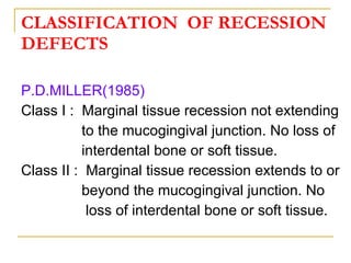
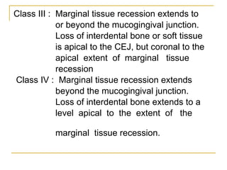
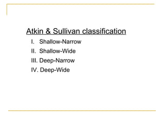
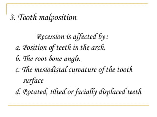
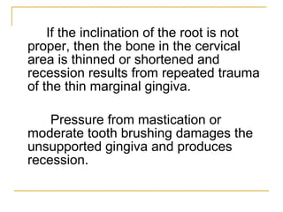
GINGIVAL RECESSION
CONTENTS  : 1. Definition 2. Classification 3. Etiology 4. Clinical examination 5. Clinical significance 6. Treatment
DEFINITION It  is  the  exposure  of  root surface  by  an apical  shift  in the  position  of gingiva.
3 The recession is determined by the actual position of the gingiva not by its apparent position
5. Recession can be studied   as, VISIBLE HIDDEN
Also it may be   Localised  Generalised
CLASSIFICATION  OF RECESSION DEFECTS P.D.MILLER(1985) Class I :  Marginal tissue recession not extending  to the mucogingival junction. No loss of interdental bone or soft tissue. Class II :  Marginal tissue recession extends to or beyond the mucogingival junction. No loss of interdental bone or soft tissue.
Class III :  Marginal tissue recession extends to or beyond the mucogingival junction.  Loss of interdental bone or soft tissue is apical to the CEJ, but coronal to the apical  extent  of  marginal  tissue recession Class IV :  Marginal tissue recession extends  beyond the mucogingival junction.  Loss of interdental bone extends to a  level  apical  to  the  extent  of  the  marginal  tissue recession.
 
Atkin & Sullivan classification I.  Shallow-Narrow II.  Shallow-Wide III. Deep-Narrow  IV. Deep-Wide
Etiology: 1. Age 2. Faulty tooth brushing technique 3. Tooth malposition 4. Gingival inflammation 5. Abnormal frenal attachment 6. Trauma from occlusion 7. Masochistic habits
1.  Age  It inraeses with age. 8% in children  50%, above age of 50 yrs  Reason being , (a) Cummulative effect of minor  pathological involvement  (b) Repeated, minor  direct trauma
2.  Faulty tooth brushing technique Brush with hard bristles Excessive or Aggressive brushing in horizontal direction. When used with highly abrasive dentrifice A Wedge shaped defect  Surface – clear, smooth , polished.
 
3. Tooth malposition   Recession is affected by : a. Position of teeth in the arch. b. The root bone angle. c. The mesiodistal curvature of the tooth surface  d. Rotated, tilted or facially displaced teeth
 
If the inclination of the root is not proper, then the bone in the cervical area is thinned or shortened and recession results from repeated trauma of the thin marginal gingiva. Pressure from mastication or moderate tooth brushing damages the unsupported gingiva and produces recession.
Malpositioned teeth
4.   Gingival inflammation
5. Abnormal frenal attachment
6. Masochistic Habits
Clinical examination Measurement of amount of gingival recession is made by Periodontal probe from CEJ to the gingival crest
Clinical significance: 1. Exposed root surfaces are susceptible to caries. 2. Abrasion or erosion of the cementum Underlying dentinal suface  Sensitivity
3. Hyperemia of pulp may also result from excessive exposure of root surfaces. 4. Interproximal recession creates oral  hygiene problems & resulting plaque  accumulation
Treatment It can be treated Non-surgically Surgically
NON – SURGICAL  METHOD 1. Correction of tooth brushing technique 2. Removal of masochistic habits 3. Correction of malocclusion 4. Treating the dentinal sensitivity
Surgically treated by two procedures : 1.  Pedicle soft tissue graft procedures : Flaps used : Rotational flap Advanced flap 2.  Free soft tissue graft procedures :  Epithelialised graft  Sub epithelial connective tissue graft
ROTATIONAL FLAP PROCEDURES Lateral sliding flap Double papilla flap ADVANCED FLAP Coronally Advanced flap  Semilunar Coronally Advanced flap
Lateral sliding flap
Clinical view :
Double papilla flap procedure
ADVANCED  FLAP  PROCEDURES
Coronally advanced flap
 
Semilunar coronally repositioned flap
Guided tissue regeneration
 
 
FREE SOFT TISSUE GRAFT PROCEDURES  Epithelialised graft  Sub epithelial connective tissue graft
Epithelialized free soft tissue graft procedure
Free connective tissue graft combined with a coronally advanced flap procedure
REFRENCES : 1.  Carranza : Textbook of Periodontics 2.  Jan Lindhe : Clinical periodontology and Implant Dentistry

Gingival recession

  • 1.
  • 2.
    CONTENTS :1. Definition 2. Classification 3. Etiology 4. Clinical examination 5. Clinical significance 6. Treatment
  • 3.
    DEFINITION It is the exposure of root surface by an apical shift in the position of gingiva.
  • 4.
    3 The recessionis determined by the actual position of the gingiva not by its apparent position
  • 5.
    5. Recession canbe studied as, VISIBLE HIDDEN
  • 6.
    Also it maybe Localised Generalised
  • 7.
    CLASSIFICATION OFRECESSION DEFECTS P.D.MILLER(1985) Class I : Marginal tissue recession not extending to the mucogingival junction. No loss of interdental bone or soft tissue. Class II : Marginal tissue recession extends to or beyond the mucogingival junction. No loss of interdental bone or soft tissue.
  • 8.
    Class III : Marginal tissue recession extends to or beyond the mucogingival junction. Loss of interdental bone or soft tissue is apical to the CEJ, but coronal to the apical extent of marginal tissue recession Class IV : Marginal tissue recession extends beyond the mucogingival junction. Loss of interdental bone extends to a level apical to the extent of the marginal tissue recession.
  • 9.
  • 10.
    Atkin & Sullivanclassification I. Shallow-Narrow II. Shallow-Wide III. Deep-Narrow IV. Deep-Wide
  • 11.
    Etiology: 1. Age2. Faulty tooth brushing technique 3. Tooth malposition 4. Gingival inflammation 5. Abnormal frenal attachment 6. Trauma from occlusion 7. Masochistic habits
  • 12.
    1. Age It inraeses with age. 8% in children 50%, above age of 50 yrs Reason being , (a) Cummulative effect of minor pathological involvement (b) Repeated, minor direct trauma
  • 13.
    2. Faultytooth brushing technique Brush with hard bristles Excessive or Aggressive brushing in horizontal direction. When used with highly abrasive dentrifice A Wedge shaped defect Surface – clear, smooth , polished.
  • 14.
  • 15.
    3. Tooth malposition Recession is affected by : a. Position of teeth in the arch. b. The root bone angle. c. The mesiodistal curvature of the tooth surface d. Rotated, tilted or facially displaced teeth
  • 16.
  • 17.
    If the inclinationof the root is not proper, then the bone in the cervical area is thinned or shortened and recession results from repeated trauma of the thin marginal gingiva. Pressure from mastication or moderate tooth brushing damages the unsupported gingiva and produces recession.
  • 18.
  • 19.
    4. Gingival inflammation
  • 20.
  • 21.
  • 22.
    Clinical examination Measurementof amount of gingival recession is made by Periodontal probe from CEJ to the gingival crest
  • 23.
    Clinical significance: 1.Exposed root surfaces are susceptible to caries. 2. Abrasion or erosion of the cementum Underlying dentinal suface Sensitivity
  • 24.
    3. Hyperemia ofpulp may also result from excessive exposure of root surfaces. 4. Interproximal recession creates oral hygiene problems & resulting plaque accumulation
  • 25.
    Treatment It canbe treated Non-surgically Surgically
  • 26.
    NON – SURGICAL METHOD 1. Correction of tooth brushing technique 2. Removal of masochistic habits 3. Correction of malocclusion 4. Treating the dentinal sensitivity
  • 27.
    Surgically treated bytwo procedures : 1. Pedicle soft tissue graft procedures : Flaps used : Rotational flap Advanced flap 2. Free soft tissue graft procedures : Epithelialised graft Sub epithelial connective tissue graft
  • 28.
    ROTATIONAL FLAP PROCEDURESLateral sliding flap Double papilla flap ADVANCED FLAP Coronally Advanced flap Semilunar Coronally Advanced flap
  • 29.
  • 30.
  • 31.
  • 32.
    ADVANCED FLAP PROCEDURES
  • 33.
  • 34.
  • 35.
  • 36.
  • 37.
  • 38.
  • 39.
    FREE SOFT TISSUEGRAFT PROCEDURES Epithelialised graft Sub epithelial connective tissue graft
  • 40.
    Epithelialized free softtissue graft procedure
  • 41.
    Free connective tissuegraft combined with a coronally advanced flap procedure
  • 42.
    REFRENCES : 1. Carranza : Textbook of Periodontics 2. Jan Lindhe : Clinical periodontology and Implant Dentistry