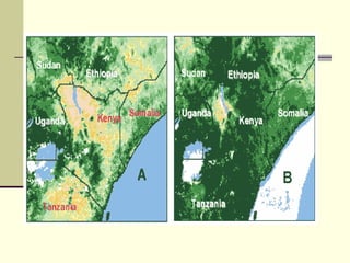
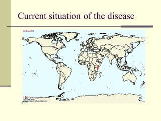
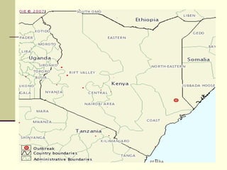
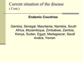
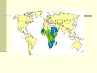
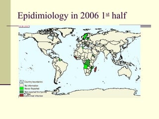
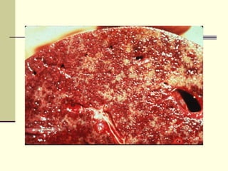
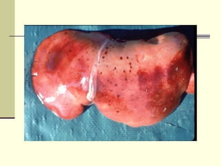
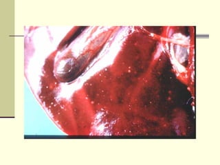
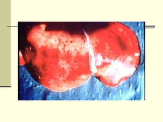
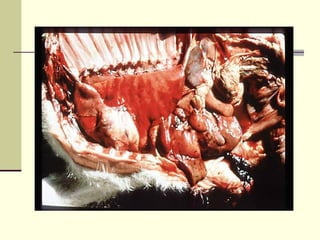
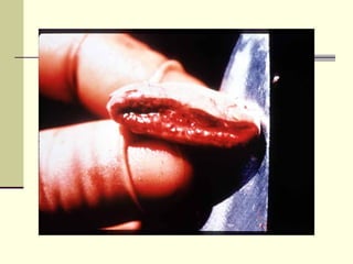
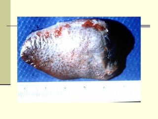
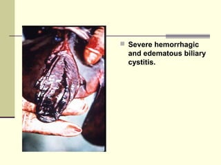
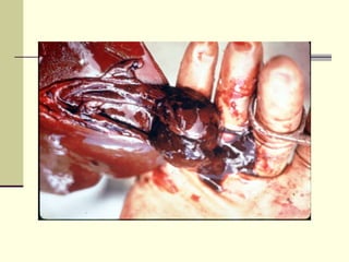
Rift Valley fever is an arthropod-borne viral disease that affects various mammals. It is characterized by abortions in pregnant animals and liver damage. The disease was first described in Kenya in 1931. It is endemic in many African and Middle Eastern countries. Transmission occurs via mosquito bites or contact with infected animal tissues. Symptoms in animals include fever, vomiting, and abortions. The virus can be diagnosed by isolating it from blood or tissues of infected hosts. Controlling mosquito populations and vaccinating susceptible animal species are important for prevention.
Tuberculosis hardly excuse anyone irrespective of its shape, size, colour, cast, creed, breed, species or genus having a little warmth in blood. Therefore, elephants e not exception, rather very prone for this disease which have taken many times more lives than any of the war.
Developing countries like Nepal, the disease like fascioliasis has made its existence and is prevailing in every shed of the ruminants. Untimely deworming practices and herding the ruminants together have made an economic loss. So routine examination of the ruminants, timely deworming, and good pasture management are necessary to be practiced to decrease the prevalence of the fascioliasis.
West Nile fever is an infection by the West Nile virus, which is typically spread by mosquitoes. It causes disease in humans, horses, and several species of birds
Brucellosis is an important re-emerging zoonosis with a worldwide distribution, in India was recognised first in 1942.
It is still an uncontrolled serious public health problem in many developing countries including India. Brucellosis in India is yet a very common but often neglected disease.
Zoonotic disease caused by Bacillus anthracis
Infects primarily herbivores- goats, sheep, cattle, horses and swine
Human infections - contact with infected animals or contaminated animal products
Human infections rarely via the respiratory or gastrointestinal tracts
Brucellosis: Epidemiology and Control in indiaBhoj Raj Singh
Brucellosis is an important endemic infectious disease in animals in India. In India brucellosis was first recognized in 1942 by Polding. It causes economic loss to the tune of nearly Rs. 350 million/year. Bovine brucellosis is caused by the bacterium Brucella abortus. In countries where cattles are kept in close association with sheep and goat it can also be caused by B. melitensis. Occasionally B. suis may also cause disease in mammary gland of cattle but it has not been reported to cause abortion and usually does not spread to other animals. Principal manifestations of animal brucellosis are reproductive failure, i.e., abortion, still births and birth of unthrifty offspring in females, and orchitis and epididymitis in males. Genus Brucella has six recognized species on the basis of host specificity viz. B. abortus, B. melitensis, B. ovis, B. suis, B. canis and B. neotome, infecting cattle, goats and sheep, sheep, pig, dog and rats, respectively. All Brucella species may also infect wildlife species. Classical Brucella species have been isolated from a great variety of wildlife species such as bison, elk, feral swine, wild boar, fox, hare, African buffalo, reindeer, and caribou. Infection in wildlife can hinder eradication efforts in cattle.
The classical species viz., B. abortus, B. melitensis, and B. suis have been identified as category B bioterrorism agents (Rotz et al. 2002, CDC 2005) because they are zoonotic and capable of causing considerable morbidity with low mortality if used in a mass event.
Epidemiology of African Swine Fever: A prerequisite to controlILRI
Presentation by Richard Bishop, Edward Okoth, Jocelyn Davies at the 'BecA-CSIRO Partnership Review meeting' held on 10-14 September 2012 at ILRI Nairobi, Kenya Campus.
Impact of Emerging Transboundary Diseases, using African Swine Fever in Ugand...SIANI
This study was presented during the conference “Production and Carbon Dynamics in Sustainable Agricultural and Forest Systems in Africa” held in September, 2010.
Using ecological niche modelling for mapping the risk of Rift Valley fever in...ILRI
Presented by PN Kiunga, PM Kitala, KA Kipronoh, G Mosomtai, J Kiplimo and B Bett at the Regional Conference on Zoonoses in Eastern Africa, Naivasha, Kenya, 9-12 March 2015.
Rift Valley fever virus: Diagnosis and vaccinesmarketsblog
Presentation by Dr Kariuki Njenga of the Centers of Disease Control & Prevention, at the Enhancing Safe Inter-regional Livestock Trade held at Dubai, UAE, 13-16 June 2011.
Fiebre de Oropuche es una enfermedad causada por el virus de Oropuche en zonas tropicales y rurales cuya manifestación son cefalea, fiebre, dolores osteo-musculares y síntomas gastrointestinales con gran porcentaje de recurrencia pero de evolución benigna.
Presentation by Michael Healy, Student Intern from Sydney University at the Milton Pink Eye Workshop held on the 18th June 2009. This Presentation covers:
- What is Bovine Viral Diarrhoea (BVDV) or Pertivirus
- Case Study on a South Coast Diary Herd
- Control Methods
etiology, local names, definition, transmission, source of infection, epidemiology, pathogenesis, clinical signs, diagnosis, differential diagnosis, treatment prevention and control
Malignant catarrhal fever (MCF) is an infectious systemic disease that presents as a variable complex of lesions affecting mainly ruminants and rarely swine. It is principally a disease of domestic cattle, water buffalo, Bali cattle (banteng), American bison, and deer. In addition to these farmed animals, MCF has been described in a variety of captive ruminants in mixed zoologic collections.MCF results from infection by one of several members of a group of closely related ruminant gammaherpesviruses of the Rhadinovirus genus.
An approache for different kinds of sampling for diagnosis of animal diseases
( Scientific activity done at Al Ain , UAE . Under the supervesion of Department of Agriculture and livestock
1. Rift Valley Fever
Dr.Tariq Mustafa Mohamed Ali,
Department of Municipalities and Agriculture,
Agriculture Sector,
Veterinary Laboratory , Al Ain
2. Definition
Rift Valley fever (RVF) is an arthropod-borne viral
disease affecting wide variety of mammals .
characterized by abortions among pregnant animals,
high mortality in neonates, and hepatic necrosis.
The Human beings are highly susceptible to the
disease .
The Disease Classified as an OIE List A disease
(A080)
3. Geographic Distribution
RVF has been recognised as an exclusive
disease in African countries, with an
underlying association with high rainfall and
dense populations of vector mosquitoes.
4. History of the disease
First described by Daubny in 1931 in Rift
valley area In kenya .
Antibodies recorded in human cases from
Central africa and the virus isolated from
animals raised in Uganda ,Mali, Congo and
Gabon by 1936
5. History of the disease (Cont.)
In 1951 reported in South Africa
In 1954 reported in French Equatorial Africa
In 1956 reported in Cattle in Kenya
In 1957 reported in Cattle in Togo
In 1958 reported in Rhodesia (Nambia)
In 1973 in sudan in Kosti District
6. History of the disease (Cont.)
The only epizootic outbreaks of RVF outside
sub-Saharan Africa were recorded in animals
and humans in Egypt in 1977-78,
Mauritania in 1987 and again in Egypt in
1993.
7. History of the disease (Cont.)
The first confirmed Rift Valley fever outbreak
outside Africa was reported in September
2000, in the Arabian Peninsula
8. History of the disease (Cont.)
Laboratory infections have been recorded in
other parts of the world
15. Current situation of the disease
( Cont.)
Endemic Countries
Gambia, Senegal, Mauritania, Namibia, South
Africa, Mozambique, Zimbabwe, Zambia,
Kenya, Sudan, Egypt, Madagascar, Saudi
Arabia, Yemen
16.
17. Epidemiology ( Cont.)
Countries known to have some cases with
periodic virus isolation
Botswana, Angola , Democratic Republic of
the Congo, Congo, Gabon, Cameroon,
Nigeria, Central African Republic, Chad,
Niger, Burkina Faso, Mali, Guinea, Tanzania,
Malawi, Uganda, Ethiopia, Somalia
24. Family Bunyaviridae
The largest family of viruses.
It includes the most arthropod-born
viruses
It include more than 350 membres with
large diversity
Genetic reassortment in infected
mosquitoes migt be the cause of this
diversity.
25. Bunyaviruses
Group V: (-)sense RNA Viruses
Genus Type Species Hosts
Family
Orthobunyavirus Bunyamwera virus Vertebrates
Hantavirus Hantaan virus Vertebrates
Nairobi sheep disease
Bunyaviridae Nairovirus Vertebrates
virus
Sandfly fever Sicilian
Phlebovirus Vertebrates
virus , RFV
Tomato spotted wilt
Tospovirus Plants
virus
26. Cryptogram of RVF virus
R/1: Σ 3 6 / L- : Se/ E : I,V/C,I,Ve(C)/Ac,Di
27. Cryptogram
R/1: Σ 3 6 / L- :Se/ E :I,V/C,I,Ve(C)/Ac,Di
RNAV , SS,
3 molecules/ - ve strand
Spherical,elongated NC / Enveloped
90 - 100 um , heat labile , ether sensitive .
All isolates are serologically similar
Host range /mode of transmission / kind of vector
if present
28. Micrograph of RVFV
The virion is budding into
membrane vesicles of Golgi
vesicles in the cytoplasm of
a liver cell of an infected rat.
The virion is about
100 nm (nanometer)
29. NA of RVF virus
Single-stranded RNA
Negative sense / ambi-sense
Each virion contains, 3 linear segments :
L 2.7 X 10 6
M 1.6 X 10 6
S 0.6 X 10 6
Not present in equimolar amounts
5' ends not capped; 3' ends not
polyadenylated;
Genomic RNA not infectious.
30.
31. Protein structure of RFV
L ~8.5kb / RNA dep.RNA polymerase
( Transcriptase)
M ~5.7kb / G1, G2, NSM
S ~0.9kb / N, NSS
32. Effect of Temperature
The virus can remain viable for up to 4
months at 4o C.
Specimens stored below 0o C will retain
infectivity for 8 years .
The virus in serum, inactivated by 56°C for
120 minutes
Rift Valley fever virus in aerosols has a half-
life in excess of 77 minutes at 25o C and 30
percent relative humidity .
33. Effect of Chemical factors on RFV
virus
Rift Valley fever virus is inactivated by lipid
solvents (ether and chloroform ),
detergents, and low pH.
.
34. Effect of Chemical factors on RFV
virus ( Cont.)
At neutral or alkaline pH in the presence of
protein such as serum, the virus can remain
viable for up to 4 months at 4o C.
Solutions having a pH of 6.2 (acetic acid) or
lower are also effective.
35. Effect of Disinfectants
Inactivated by strong solutions of sodium or
calcium hypochlorite (residual chlorine should
exceed 5000 ppm)
37. Host range
Rift Valley fever virus infects many species of
animals and humans .
Sheep and cattle are the primary species
affected and the primary amplifiers of the
virus.
38. Host Range
Neonatal lambs, kids, calves, and puppies
are highly susceptible and have a high
mortality.
39. Human being
Humans are highly susceptible to RVF virus
infection and are readily infected by
mosquitoes and aerosols.
Humans develop a sufficient viremia to be a
source of infection for mosquitoes and thus
could introduce the disease into uninfected
areas.
40. Transmission
Haematophagous mosquitoes of many genera
(Aedes, Anopheles, Culex, Eretmapodites, Mansonia,
etc.) can transmit fever as biological, competent
vectors.
Can replicate extensively in insects - transovarian
passage allows overwintering.
Mosquitoes (Aedes) migt be the reservoir host
Direct contamination: occurs in humans when
handling infected animals and meat
42. Infection of human being
Direct and indirect contact with infected
animals through nasal discharge, blood,
vaginal secretions after abortion in animals,
mosquitoes, and infected meat.
Aerosol and consumption of raw milk is also
possible
43.
44. Incubation Period
Experimentally, the incubation period in
newborn lambs, kids, calves, and puppies, is
about 12 hours.
In adult sheep, cattle, goats, and dogs the
incubation period may be as long as 3 days.
In humans, the incubation period is 4 to 6
days.
46. Morbidity and Mortality in an Outbreak
Susceptibl Morbdity Mortality Case
Species Cases Deaths
e cases rate rate fatality rate
Sheep 9000 1500 105 16.7% 1.2% 7%
Goats 10000 1500 95 15% 0.95% 6.3%
Cattle 4000 500 30 13% 0.75% 6%
Camelidae 4000 500 5 13% 0.13% 1%
Animals belong to different herds.70 people have died
47. Clinical Signs in cattle
Adults: fever (40-41°C), excessive salivation,
anorexia, weakness, fetid diarrhoea, fall in
milk yield.
Calves: fever (40-41°C), depression. Mortality
rate: 10-70%
Abortion may reach 85% in the herd.
Mortality rate is usually less than 10%
52. Clinical Signs in lambs and Kids
fever (40-42°C), anorexia, weakness, death
within 36 hours after inoculation.
Mortality rate: for animals under 1 week of
age - up to 90%; for animals over 1 week of
age - up to 20%
53. Clinical Signs in other animals
Inapparent infections are quite frequent in other
species than sheep
54. PM lesions.
Focal or generalised hepatic necrosis (white
necrotic foci of about 1 mm in diameter)
55.
56.
57.
58. PM Lesions ( cont.)
Congestion, enlargement, and discoloration of
liver with subcapsular haemorrhages
59.
60.
61. PM Lesions ( cont.)
Brown-yellowish colour of liver in aborted
fetuses
62.
63. PM Lesions ( cont.)
Widespread cutaneous haemorrhages,
petechial to ecchymotic haemorrhages on
parietal and visceral serosal membranes
64.
65.
66.
67.
68. PM Lesions ( cont.)
Enlargement, oedema, haemorrhages and
necrosis of lymph nodes
69.
70.
71. PM Lesions ( cont.)
Congestion and cortical haemorrhages of
kidneys and gallbladder
80. Specimens for Laboratory
Heparinised or clotted blood
Plasma or serum
Tissue samples of liver, spleen, kidney, lymph
node, heart blood and brain from aborted
fetus.
Specimens should be submitted preserved in
10% buffered formalin and in glycerol/saline
and transported at 4°C
82. Laboratory diagnosis
Virus isolation:
Inoculation of mice or hamsters - preferred
method
Inoculation of 1-2-day-old lambs
Inoculation of embryonated chicken eggs
Tissue culture inoculation (Vero, CER, BHK-
21, mosquito line cells or primary calf, lamb
and goat kidney and testis cells)
83. Laboratory diagnosis (cont.)
Viral antigen detection by :
1. Immunofluorescence in cryostat sections or
in impression smears of liver, spleen and
brain.
2. Complement fixation and immunodiffusion
on tissue suspensions
85. Laboratory diagnosis by Serological tests
Enyzme-linked immunosorbent assay - IgG and IgM
and for Ag.detection
Virus neutralisation
Fluorescent antibody test
Haemagglutination inhibition
Plaque reduction neutralisation
Complement fixation
Immunodiffusion
86. Control and Eradication
Vaccination is the only practical method of
preventing low-level losses.
Animal movement of from endemic areas to
RVF-free areas should be discouraged.
Mosquito control during an epizootic is logical
but not practical for large areas;
Slaughter of sick animals is not
recommended
87. Vaccination
Vaccination of all susceptible animals to
prevent infection of amplifying hosts and thus
infection of vectors is the only way to prevent
infection of animals and man
Inactivated vaccine containing saponin or
peanut oil