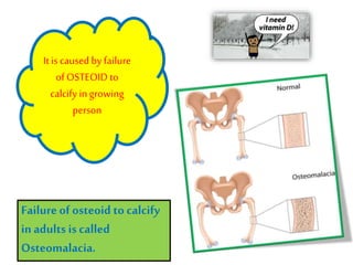
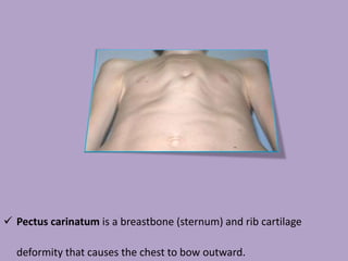
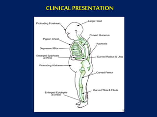
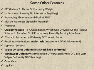
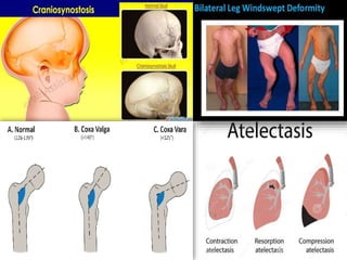
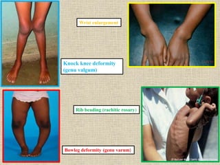
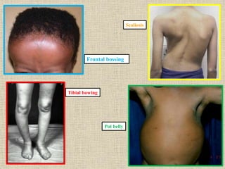
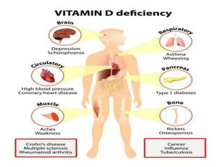
Detailed information about rickets.....everything you wanna know including causes, symptoms, diagnosis, prevention and treatment... i hope you will like it
Download it to view properly with all the effects
This document discusses rickets, a disease characterized by soft, weak, and deformed bones in children caused by vitamin D deficiency or disorders of vitamin D and phosphate metabolism. It defines rickets as occurring in growing children before bone growth plate closure, while osteomalacia occurs after closure in adults. The key roles and mechanisms of vitamin D in calcium and phosphate homeostasis are described. Causes, signs and symptoms, diagnosis using biochemical markers and radiology, treatment using large vitamin D doses, and prevention through diet and sunlight exposure are summarized. Other types of rickets including genetic and renal forms are also outlined.
This document summarizes rickets, a childhood bone disease caused by vitamin D deficiency or impaired vitamin D metabolism. It discusses the pathology of rickets, which involves abnormal cartilage cell maturation and failed bone mineralization. This leads to bone deformities and fractures. The document outlines various causes of rickets, including nutritional deficiencies, malabsorption, and genetic disorders affecting vitamin D and phosphate metabolism. Diagnosis involves x-rays showing characteristic bone changes. Treatment involves vitamin D, calcium, phosphate supplementation as needed, and sometimes surgery to correct deformities once the disease is resolved.
Rickets and osteomalacia are diseases characterized by impaired mineralization of bone and cartilage. Rickets occurs in children and affects growing bones and cartilage, while osteomalacia occurs in adults after growth is complete. Both are primarily caused by vitamin D deficiency or disturbances in its metabolism. The diseases result in softened, deformed bones and increased fracture risk. Diagnosis involves clinical features, elevated alkaline phosphatase levels, and radiographic signs of impaired mineralization. Treatment focuses on vitamin D supplementation, calcium, and addressing any underlying causes.
INTRODUCTION — Normal bone growth and mineralization require adequate calcium and phosphate, the two major constituents of the crystalline component of bone. Deficient mineralization can result in rickets and/or osteomalacia. Rickets refers to deficient mineralization at the growth plate, as well as architectural disruption of this structure. Osteomalacia refers to impaired mineralization of the bone matrix. Rickets and osteomalacia usually occur together as long as the growth plates are open; only osteomalacia occurs after the growth plates have fused.
rickets is a nutritional deficiency disease that involves mainly calcium, vitamin d, or phosphate resulting in decreased bone stability and strength, Delayed closure of the fontanelles,Parietal and frontal bossing. Craniotabes (soft skull bones).
Enlargement of the costochondral junction visible as beading along the anterolateral aspects of the chest (the "rachitic rosary") . Formation of Harrison sulcus (or groove),Widening of the wrist and bowing of the distal radius and ulna, Progressive lateral bowing of the femur and tibia and causes defects in teeth.
there is two types of rickets: phosphopenic and calcipenic.
pathogenesis: Growth plate thickness is determined by two opposing processes: o chondrocyte proliferation and hypertrophy on the one hand. o vascular invasion of the growth plate followed by conversion into primary bone spongiosa on the other. • Vascular invasion requires mineralization of the growth plate cartilage and is delayed or prevented by deficiency of calcium or phosphorus growth plate cartilage accumulates and the growth plate thickens. • In addition, the chondrocytes of the growth plate become disorganized, losing their columnar orientation with characteristic expansion of the hypertrophic zone. • In the bone tissue below the growth plate (metaphysis), the mineralization defect leads to the accumulation of osteoid.
Rickets is a disease characterized by softening and weakening of bones in children, caused by a vitamin D deficiency or impaired metabolism of calcium or phosphorus. It most commonly affects children between 6-24 months old and can lead to fractures and bone deformities. While historically more prevalent, rickets decreased in developed countries in the 20th century but remains common in developing nations and populations with dark skin where access to vitamin D-fortified foods is limited. Treatment involves increasing dietary intake of calcium, phosphorus and vitamin D through supplements, exposure to sunlight, or vitamin D-fortified foods and formula. Physical therapy can help reduce bone and muscle pain through stretching and strengthening exercises.
This document discusses rickets, a disease characterized by weak and malformed bones in children due to a lack of vitamin D or calcium. It defines rickets and describes the various types, including nutritional rickets caused by vitamin D deficiency, vitamin D dependent rickets involving the kidneys or end organs, and genetic or malabsorption related forms. Symptoms, biochemical markers, x-ray findings and treatments are outlined. Management involves vitamin D or calcium supplementation depending on the type, with the goal of restoring normal bone growth and structure over 6 months to 2 years.
This document summarizes rickets, a disease characterized by softening and weakening of bones in children due to a lack of vitamin D or calcium and phosphate. It discusses calcium, phosphate, and vitamin D metabolism and how deficiencies can lead to inadequate mineralization of bone. Symptoms include bone pain, deformities like bowed legs, and fractures. Diagnosis involves biochemical testing showing low calcium, phosphate, and vitamin D levels as well as characteristic findings on x-rays of bones. Treatment is supplementation to correct nutritional deficiencies.
The document discusses the causes and presentation of rickets. The main causes are vitamin D disorders, calcium deficiency, phosphorus deficiency, renal losses, and distal renal tubular acidosis. Clinical features include bone deformities, softening of the skull, and leg pain. Diagnosis involves physical exam, x-rays showing bone changes, and lab tests showing abnormalities in calcium, phosphorus, vitamin D, and parathyroid hormone levels. Nutritional vitamin D deficiency is the most common cause globally. Treatment involves vitamin D, calcium, and phosphorus supplementation.
This document provides an overview of rickets presented by Lyudmyla Olexandrivna Rakovska from the Department of Pediatrics at V.N. Karazin Kharkiv National University. The 10-point plan discusses the definition, etiology, pathogenesis, classification, clinical manifestations, laboratory/radiological findings, differential diagnosis, treatment and prevention of rickets. Key points include that rickets is caused by vitamin D deficiency impairing bone mineralization, especially in children under 2 years old. Symptoms involve soft, deformed bones and skeletal abnormalities. Treatment focuses on vitamin D supplementation through either low daily doses or high single doses.
This document provides information on rickets, a metabolic bone disease caused by vitamin D deficiency or impaired mineralization. It discusses the following key points:
- Rickets mainly affects children under 2 years old and causes soft, weak bones and skeletal deformities from imperfect bone mineralization.
- It is most commonly caused by nutritional deficiencies, especially of vitamin D, but can also be caused by genetic or other medical conditions.
- Symptoms include bone pain, softening of the skull and ribs, bowed legs, fractures, and delayed growth. Radiographs show widened growth plates and fraying of the metaphysis.
- Treatment involves high-dose vitamin D and calcium supplementation to promote bone mineralization and
This document discusses rickets, a disease caused by defective bone mineralization in children due to vitamin D or calcium/phosphate deficiencies. It defines rickets and describes the different types (nutritional, vitamin D dependent types 1 and 2, and vitamin D resistant). Nutritional rickets is most common, caused by inadequate sunlight exposure or vitamin D/calcium intake. The document outlines vitamin D metabolism and the pathogenesis of rickets. It then describes clinical features like bone deformities and laboratory abnormalities seen in a case of rickets. The recommended approach is to obtain a detailed history, perform a physical exam including skeletal assessment, order biochemical and radiological tests, and initiate vitamin D, calcium, and plastic surgery treatments as needed.
Rickets is a childhood bone disease caused by vitamin D deficiency and a lack of calcium and phosphate. It results in soft, weak bones that can lead to skeletal deformities. Symptoms include bowed legs, knock-knees, frontal bossing of the skull, and chest deformities like pigeon chest. Laboratory findings often include low calcium, phosphorus, and vitamin D levels as well as elevated alkaline phosphatase and parathyroid hormone levels. Radiographs show widened growth plates and fraying and cupping of the bone metaphysis. The condition is treated by supplementing with vitamin D and calcium.
This document discusses rickets, a disease characterized by defective bone mineralization due to vitamin D deficiency or abnormal metabolism. It covers the sources and types of vitamin D, how vitamin D is metabolized in the body, and its actions in increasing calcium and phosphate absorption and bone mineralization. Symptoms of rickets are described including bone deformities, craniotabes, and rosary beads. Diagnosis involves physical exam findings, elevated alkaline phosphatase levels, and x-rays showing metaphyseal changes. Treatment is aimed at correcting the vitamin D deficiency through sunlight exposure or vitamin D supplementation. Prognosis is generally good with treatment but deformities may require orthopedic correction.
This document discusses rickets, a disease caused by vitamin D deficiency or impaired mineralization that affects growing bones in children. It defines rickets and outlines various types including nutritional, vitamin D dependent, and vitamin D resistant rickets. Clinical features like softening of bones and skeletal deformities are described. Laboratory findings of low calcium and phosphorus and high alkaline phosphatase are also summarized. The document outlines prevention through diet, supplementation, and sunlight exposure as well as treatment with high dose vitamin D or calcium therapy.
Vitamin D deficiency is a common cause of rickets in children. It results from inadequate sunlight exposure, diet low in vitamin D, or malabsorption issues. Clinical features include bone pain, skeletal deformities, and hypocalcemia. Laboratory tests show low calcium and phosphorus levels and elevated alkaline phosphatase. Treatment involves high dose vitamin D supplementation and calcium. Rickets can also be caused by inherited disorders that impact calcium, phosphorus, or vitamin D metabolism.
Rickets is a childhood bone disease caused by vitamin D deficiency and lack of calcium and phosphate. It results in soft and weak bones in children. Symptoms include bone pain, bowed legs, and skull deformities. Risk factors include lack of sunlight exposure, dark skin pigmentation, malnutrition, and liver or kidney diseases. Diagnosis involves blood tests showing low calcium, phosphate, and vitamin D levels and x-rays showing bone changes. Treatment consists of vitamin D and calcium supplementation to strengthen and repair bones. Prevention involves adequate sunlight exposure, vitamin D supplementation, and calcium-rich nutrition during childhood bone growth.
Nutritional rickets is caused by defective bone growth and mineralization due to vitamin D deficiency or low calcium intake. It is diagnosed based on history, physical exam, biochemical testing and confirmed by radiographs. Serum 25-hydroxyvitamin D levels below 30 nmol/L indicate deficiency and levels below 50 nmol/L increase risk of rickets. Treatment involves high dose vitamin D supplementation of 2000 IU/day for at least 3 months along with calcium supplementation. Prevention involves adequate dietary vitamin D and calcium intake.
This document provides information on rickets in children. It begins with an overview of vitamin D physiology and the sources of vitamin D. It then discusses the introduction, etiology, clinical features, radiology, diagnosis, and treatment of rickets. The main causes of rickets are outlined as vitamin D deficiencies, calcium deficiencies, phosphorus deficiencies, and renal losses. Symptoms and physical exam findings of rickets are explained. Laboratory tests useful in diagnosis are provided. Treatment involves high dose vitamin D supplementation either orally or via injection. Prognosis is typically excellent with treatment, though severe cases can cause permanent deformities.
Rickets is a disease of growing bones caused by inadequate mineralization due to vitamin D deficiency or disorders of calcium and phosphorus. It occurs in children before bone growth plate fusion. Common causes include nutritional deficiencies of vitamin D, calcium, or phosphorus. Clinical features include bone softening and deformities, muscle weakness, and hypocalcemic symptoms. Diagnosis is confirmed by blood tests and radiographic findings of widened growth plates. Treatment involves high dose vitamin D supplementation along with adequate calcium and phosphorus intake.
Definition of Rickets &Osteomalacia.
Etiology of Rickets & Osteomalacia
Rickets :
is defective mineralization of bones before epiphyseal closure (at growth plate) due to deficiency or impaired metabolism of vitamin D, phosphorus or calcium ,potentially leading to fractures and deformity.
Osteomalacia :
is a similar condition occurring in adults, generally due to impaired mineralization of the bone matrix. Due to a deficiency of vitamin D.
Etiology:
Vit D deficiency :
low intake plus inadequate sunlight exposure
malabsorption
Abnormal Vit D metabolism:
Impaired 25 OH Vitamin D production (Liver diseases)
Impaired 1,25 OH2 Vitamin D production (chronic renal failure or vitamin D-resistant rickets type I ).
Vitamin D receptor defects (vitaminD-resistant rickets type II)
Low calcium intake
Hypophosphatemia :
Low intake.
Inherited or acquired defects in renal tubular phosphate reabsorption.
Tumors that secrete phosphaturic substance.
Rickets and Osteomalacia are impaired memorization of the bone.
Causes of Rickets and Osteomalacia :
Vit D deficiency
Low calcium intake
Abnormal Vit D metabolism
Hypophosphatemia
Rickets is a disease caused by vitamin D deficiency or a lack of calcium or phosphorus in children, which results in soft, weakened bones. It occurs when osteoid, or bone matrix, fails to calcify during bone growth. Symptoms include bowed legs, bone pain, and fractures. Treatment involves high dose vitamin D supplementation orally or via injection to restore calcium and phosphorus levels.
Vitamin D is essential for calcium absorption and bone health. It is produced in the skin upon exposure to sunlight and is also obtained through dietary sources. Vitamin D must undergo hydroxylation in the liver and kidneys to become its active form. Deficiency can result in rickets in children or osteomalacia in adults due to impaired bone mineralization. Risk groups include those with inadequate sun exposure or dietary intake, as well as patients with liver or kidney disease.
The document outlines the signs and symptoms of rickets, including abdominal protrusion, bone bowing, widened growth plates, delayed development, and joint deformities. It discusses the different types of rickets caused by calcium or phosphate deficiency. Blood tests typically show low calcium and phosphate levels and high alkaline phosphatase. Treatment involves high dose vitamin D3 to stimulate bone mineralization and healing, followed by maintenance doses of vitamin D3. Surgery is only performed once healing via vitamin D3 treatment begins.
18 Rickets Of Vitamin D Deficiency,Tetany Of Vitamin D Deficiencyghalan
This document summarizes rickets of vitamin D deficiency, including its pathogenesis, clinical manifestations, diagnosis, prevention and treatment. Rickets results from failure of mineralization of growing bone due to vitamin D deficiency. Key symptoms include bone pain, softening of the skull, and bowed legs. Diagnosis is based on low vitamin D levels and x-ray findings. Treatment involves vitamin D supplementation to promote bone mineralization and prevent deformities.
Rickets is a childhood bone disease caused by vitamin D deficiency and a lack of calcium and phosphate. It results in soft, weak bones that can lead to skeletal deformities. The disease develops when vitamin D levels are too low to absorb calcium and phosphate from the intestines, causing these minerals to be lost from the bones. Symptoms include bowed legs, soft skull, and bone pain. Diagnosis involves blood tests showing low calcium and vitamin D levels and high alkaline phosphatase. X-rays show widened growth plates and fraying and cupping of the bone. Treatment involves high dose vitamin D and calcium supplementation to strengthen the bones.
Vitamin D is a fat-soluble vitamin obtained through exposure to sunlight, food sources like fatty fish and supplements. It plays an important role in calcium absorption and bone health. Deficiency can lead to rickets in children and osteomalacia in adults characterized by soft, weak bones and skeletal deformities. Recommended daily intake amounts vary from 200-600IU depending on age. Both deficiency and excess can impact multiple body systems.
Rickets is caused by vitamin D deficiency which leads to low calcium and phosphate levels. This weakens the bones and causes skeletal deformities. Vitamin D is obtained from sunlight exposure and diet. Lack of sunlight, inadequate diet, dark skin pigmentation, liver or kidney problems can cause vitamin D deficiency and subsequent rickets. Symptoms include bowed legs, soft skull bones, spinal curvature and bone pain. Diagnosis involves blood tests showing low calcium, vitamin D and high alkaline phosphatase levels along with characteristic bone changes on x-ray. Treatment is vitamin D supplementation and ensuring adequate calcium and vitamin D in the diet.
Rickets is a disease of growing bones in children caused by vitamin D deficiency or lack of calcium and phosphate. It results in soft, weak bones that can deform. Symptoms include bowed legs, fractures, and delayed growth. Diagnosis involves lab tests showing low calcium, vitamin D, and phosphate levels as well as characteristic bone changes on x-rays like widened growth plates. Treatment is vitamin D supplements and ensuring adequate calcium and phosphate intake. Prevention involves vitamin D supplementation, sunlight exposure, and a nutritious diet.
This document provides information on Vitamins D and K. It discusses the sources, structures, functions, recommended daily intake and deficiency symptoms of each vitamin. Vitamin D helps regulate calcium levels and bone mineralization, while Vitamin K is required for blood clotting through the gamma-carboxylation of clotting factors in the liver. Both vitamins are fat-soluble and absorbed through the intestine with dietary fats and bile salts.
Chemistry, and biochemical role, rda, vitamin dJasmineJuliet
Vitamin D - Chemistry,n Metabloism, Biosynthesis in our skin, Recommended dietary Allowance, Dietary sources of vitamin D, Deficiency symptoms of vitamin D, Hypervitaminosis of vitamin D.
Chemistry, and biochemical role, rda, vitamin dJasmineJuliet
Chemistry of Vitamin D, Biochemical functions of vitamin D, Recommended dietary Allowances of vitamin D, Dietary sources of Vitamin D, Deficiency symptoms of vitamin D, Hypervitaminosis of vitamin D, Toxicity of Vitamin D.
This document provides an overview of rickets and osteomalacia. It begins by defining rickets as a disease affecting the growth plates, causing deficient mineralization and impaired endochondral ossification due to failed apoptosis of hypertrophic chondrocytes. Osteomalacia is a similar disorder affecting mineralization of osteoid. Both result from insufficient calcium and phosphate levels. Vitamin D deficiency is a common cause and treatment involves vitamin D supplementation. Other causes discussed include hereditary disorders, prematurity, drugs, tumors, and renal osteodystrophy. Clinical features, radiographic findings, and management approaches are described for each condition.
Vitamin D is an essential nutrient that promotes calcium absorption in the intestines and maintains adequate serum calcium and phosphate concentrations to enable normal mineralization of bone. It exists in two main forms, D2 (ergocalciferol) and D3 (cholecalciferol), and is obtained through exposure to sunlight, dietary intake of oily fish, eggs, and vitamin D fortified foods. The liver converts vitamin D to calcidiol which is then converted by the kidneys to calcitriol, the biologically active form, which regulates calcium and phosphate levels. Deficiencies can lead to rickets in children and osteomalacia in adults, characterized by soft, deformed bones and muscle weakness.
This document discusses vitamin D, including its sources, biochemical effects, and related diseases. It summarizes that vitamin D aids in calcium and phosphorus absorption in the intestine and bone mineralization. Deficiencies can cause rickets in children and osteomalacia in adults, resulting from insufficient mineralization of bones. Toxicity from oversupplementation can lead to excessive calcification.
This document discusses vitamin D, including its structure, biosynthesis from sunlight exposure, dietary sources, recommended daily allowance, metabolic functions in maintaining calcium and phosphate levels and bone mineralization, deficiency manifestations like rickets and osteomalacia, and potential non-skeletal roles. Vitamin D is fat-soluble and synthesized in the skin from sunlight exposure, with the biologically active form being calcitriol. Insufficient levels can lead to impaired bone mineralization and bone diseases while excess intake over prolonged periods can cause hypercalcemia.
This document discusses vitamin D, including its forms, functions, metabolism, and deficiency/toxicity. Some key points:
- Vitamin D exists in two forms, D2 and D3. D3 is produced in the skin upon sun exposure and converted to its active form in the liver and kidneys.
- Its active form, calcitriol, regulates calcium and phosphorus levels by increasing their absorption in the intestine and reabsorption in kidneys. It also mobilizes calcium from bones.
- Deficiency causes rickets in children and osteomalacia in adults due to impaired bone mineralization. Toxicity leads to hypercalcemia which can damage soft tissues like kidneys.
Vitamin D has two main forms, D2 and D3. It is a steroid compound that plays an essential role in regulating calcium and phosphorus metabolism. The active form, calcitriol, maintains normal plasma calcium and phosphorus levels by acting on the intestine, bone, and kidneys. A vitamin D deficiency can lead to rickets in children, characterized by soft bones and skeletal deformities.
1. Vitamin D is synthesized in the skin upon exposure to sunlight and is converted to its active form, calcitriol, in the kidneys.
2. Calcitriol increases intestinal absorption of calcium and phosphate and promotes their reabsorption in the kidneys to maintain mineral homeostasis.
3. It also supports bone mineralization by stimulating calcium and phosphate deposition from the blood into the bone.
1. Vitamin D is a fat-soluble vitamin that exists in two forms, D2 and D3, which are converted to calcitriol, the active form of vitamin D.
2. Calcitriol regulates calcium and phosphate levels by increasing their absorption in the intestine, reabsorption in the kidneys, and mobilization from bones.
3. Vitamin D deficiency can cause rickets in children and osteomalacia in adults, characterized by soft and deformed bones from mineralization problems.
This document discusses calcium metabolism and rickets. It begins by introducing calcium's role in the body and dietary requirements. It then covers absorption, regulation by parathyroid hormone and calcitonin, and roles in bone mineralization. Rickets is defined as a childhood disease caused by calcium and vitamin D deficiencies, leading to softened, deformed bones. Causes, clinical features, investigations, management, and surgical options are described.
This document discusses vitamin D in animals. It covers the chemical structure of vitamin D, how it is metabolized in the body through conversion in the liver and kidneys, its functions related to calcium and phosphorus homeostasis, and its roles in the intestines, bones and kidneys. The document also addresses requirements, natural sources, signs of deficiency such as rickets, and supplementation and toxicity concerns.
Rickets is a metabolic bone disease caused by vitamin D deficiency or abnormalities in calcium and phosphate metabolism during bone development. It results in soft, weak bones in children that can lead to skeletal deformities. The key symptoms are bone pain, weak or soft bones, bowed legs, and skeletal deformities. Treatment involves high dose vitamin D supplementation combined with calcium and phosphate intake to mineralize the bones. Prevention focuses on adequate vitamin D from sunlight, diet, or supplements during pregnancy and childhood.
Vitamin D is a group of fat-soluble prohormones that are mainly produced in the skin upon exposure to sunlight. It regulates calcium and phosphorus levels in the blood and promotes bone formation. Vitamin D deficiency can cause rickets in children, characterized by soft and weak bones and bone deformities, and osteomalacia in adults, which is the softening of bones. Risk factors include inadequate sunlight exposure, poor nutrition, and disorders inhibiting absorption. Treatment involves oral or injectable high-dose vitamin D to promote healing.
NO words to describe...just download and watch yourself.... very informative and beautiful presentation about sensory system and receptors of our body,,,,will surely help to understand in a better and easy way... best viewed only when downloaded
A Small lighten up about increasing problems of breastfeeding, comparative data across the world, measures to take to maintain good health of infants.....
Helminthes- a new meaning to parasitologyAnmol Gupta
Till now we know that parasites are only harmful...in this you will see benefits of parasites and how they can be used to treat various major disease by their routine activities which harms us.....
Cryosurgery uses extreme cold produced by liquid nitrogen or argon gas to destroy abnormal tissue. It can be used to treat some cancers and precancerous conditions of the skin, breast, prostate, liver, and cervix. Potential side effects include scarring, loss of sensation or pigmentation, and injury to surrounding tissues. Cryosurgery provides minimally invasive treatment with short recovery times and low risk of infection or complications.
The document summarizes key events of the Battle of Stalingrad during World War II. It describes how in September 1942, German forces under General Paulus advanced on the city of Stalingrad, but they were eventually surrounded by Soviet forces. After intense street-to-street fighting over the following months, the Germans were trapped in Stalingrad with no possibility of resupply or retreat. By the end of January 1943, over 90,000 German troops surrendered, marking a major defeat. The failure at Stalingrad depleted German forces and halted their advance in Russia, shifting the tide of the war.
India is located in South Asia and has a diverse landscape that includes the Himalayan mountains and coastlines. It has a population of over 1.3 billion people and is home to many religions and languages. Some key facts about India include:
- It has 29 states and 7 union territories with Delhi as its capital.
- Major languages include Hindi, English, Bengali, Telugu, Marathi and Tamil.
- Hinduism is the dominant religion practiced by around 80% of the population.
- India has a rich cultural heritage that is expressed through traditions like festivals, cuisine, clothing, art, music and dance.
- It has made many scientific and mathematical contributions to the
Kharkiv is a large city in northeastern Ukraine that has been continuously inhabited for centuries. Archaeological evidence shows settlements dating back to at least the 2nd millennium BC by Scythian, Sarmatian, and Cherniakhiv tribes. The city was officially founded in 1654 when a Cossack named I. Karkach built a fortified settlement. Throughout the 17th and 18th centuries, Kharkiv developed from a border outpost into a center of crafts and trade as suburbs formed around the fortress. In the 18th century, Kharkiv became an educational center with the founding of Kharkiv University in 1805, which became a major cultural and scientific institution. Today, Kh
How to Make a Field Mandatory in Odoo 17Celine George
In Odoo, making a field required can be done through both Python code and XML views. When you set the required attribute to True in Python code, it makes the field required across all views where it's used. Conversely, when you set the required attribute in XML views, it makes the field required only in the context of that particular view.
How to Add Chatter in the odoo 17 ERP ModuleCeline George
In Odoo, the chatter is like a chat tool that helps you work together on records. You can leave notes and track things, making it easier to talk with your team and partners. Inside chatter, all communication history, activity, and changes will be displayed.
This document provides an overview of wound healing, its functions, stages, mechanisms, factors affecting it, and complications.
A wound is a break in the integrity of the skin or tissues, which may be associated with disruption of the structure and function.
Healing is the body’s response to injury in an attempt to restore normal structure and functions.
Healing can occur in two ways: Regeneration and Repair
There are 4 phases of wound healing: hemostasis, inflammation, proliferation, and remodeling. This document also describes the mechanism of wound healing. Factors that affect healing include infection, uncontrolled diabetes, poor nutrition, age, anemia, the presence of foreign bodies, etc.
Complications of wound healing like infection, hyperpigmentation of scar, contractures, and keloid formation.
A review of the growth of the Israel Genealogy Research Association Database Collection for the last 12 months. Our collection is now passed the 3 million mark and still growing. See which archives have contributed the most. See the different types of records we have, and which years have had records added. You can also see what we have for the future.
Reimagining Your Library Space: How to Increase the Vibes in Your Library No ...Diana Rendina
Librarians are leading the way in creating future-ready citizens – now we need to update our spaces to match. In this session, attendees will get inspiration for transforming their library spaces. You’ll learn how to survey students and patrons, create a focus group, and use design thinking to brainstorm ideas for your space. We’ll discuss budget friendly ways to change your space as well as how to find funding. No matter where you’re at, you’ll find ideas for reimagining your space in this session.
Strategies for Effective Upskilling is a presentation by Chinwendu Peace in a Your Skill Boost Masterclass organisation by the Excellence Foundation for South Sudan on 08th and 09th June 2024 from 1 PM to 3 PM on each day.
বাংলাদেশের অর্থনৈতিক সমীক্ষা ২০২৪ [Bangladesh Economic Review 2024 Bangla.pdf] কম্পিউটার , ট্যাব ও স্মার্ট ফোন ভার্সন সহ সম্পূর্ণ বাংলা ই-বুক বা pdf বই " সুচিপত্র ...বুকমার্ক মেনু 🔖 ও হাইপার লিংক মেনু 📝👆 যুক্ত ..
আমাদের সবার জন্য খুব খুব গুরুত্বপূর্ণ একটি বই ..বিসিএস, ব্যাংক, ইউনিভার্সিটি ভর্তি ও যে কোন প্রতিযোগিতা মূলক পরীক্ষার জন্য এর খুব ইম্পরট্যান্ট একটি বিষয় ...তাছাড়া বাংলাদেশের সাম্প্রতিক যে কোন ডাটা বা তথ্য এই বইতে পাবেন ...
তাই একজন নাগরিক হিসাবে এই তথ্য গুলো আপনার জানা প্রয়োজন ...।
বিসিএস ও ব্যাংক এর লিখিত পরীক্ষা ...+এছাড়া মাধ্যমিক ও উচ্চমাধ্যমিকের স্টুডেন্টদের জন্য অনেক কাজে আসবে ...
हिंदी वर्णमाला पीपीटी, hindi alphabet PPT presentation, hindi varnamala PPT, Hindi Varnamala pdf, हिंदी स्वर, हिंदी व्यंजन, sikhiye hindi varnmala, dr. mulla adam ali, hindi language and literature, hindi alphabet with drawing, hindi alphabet pdf, hindi varnamala for childrens, hindi language, hindi varnamala practice for kids, https://www.drmullaadamali.com
This presentation includes basic of PCOS their pathology and treatment and also Ayurveda correlation of PCOS and Ayurvedic line of treatment mentioned in classics.
Liberal Approach to the Study of Indian Politics.pdf
Rickets
1.
2.
3. Rickets is defective
mineralization or
calcification of bones
before epiphyseal
closure in immature
mammals due to deficiency
or impaired metabolism
of vitamin D,
phosphorus or calcium
potentially leading to
fractures and deformity.
Failureof osteoid to calcify
in adults is called
Osteomalacia.
It is caused by failure
of OSTEOID to
calcify ingrowing
person
4.
5. Enamel hypoplasia is a defect of the teeth in which the enamel is
hard but thin and deficient in amount. Harrison's groove, also known as Harrison's sulcus, is a
horizontalgroove along the lower border of the thorax corresponding
to the costal insertion of the diaphragm.
Craniotabes is the finding of a softening or thinning of the skull.
Pectus carinatum is a breastbone (sternum) and rib cartilage
deformity that causes the chest to bow outward.
7. Some Other Features
FTT (Failure To Thrive Or Flattering Weight)
Listlessness (Showing No Interest In Anything)
Protruding Abdomen, umbilical HERNIA
Muscle Weakness (Specially Proximal)
Fractures
Craniosynostosis- Is A Condition In Which One Or More Of The Fibrous
Sutures In An Infant Skull Prematurely Fuses By Turning Into Bone
Thoracic Asymmetry, Widening Of Thoracic Bone
Respiratory Infections, Atelectasis (Impairment Of Air Movement)
Kyphosis, Lordosis
Valgus Or Varus Deformities (Knock knee deformity)
Windswept Deformity (combination Of Varus Deformity Of 1 Leg With
Valgus Deformity Of Other Leg)
Coxa Vara
Leg Pain
15. Cholecalciferol (i.e.,
vitamin D-3) is formed
in the skin from a
derivative of
cholesterol (5-
dihydrotachysterol)
under the stimulus of
ultraviolet-B light.
16. Occurs at position 25 in the liver, producing
calcidiol (25-hydroxycholecalciferol)
The first hydroxylation
This steroid undergoes hydroxylation in 2
steps.
16
17. Occurs in the kidney at the 1 position,
where it undergoes hydroxylation to
the active metabolite calcitriol
(1,25-dihydroxycholecalciferol )
The second hydroxylation
17
18. Calcitriol
Acts at 3 known sites to tightly regulate
calcium metabolism:
(1) It promotes absorption of calcium and phosphorus
from the intestine
(2) It increases reabsorption of phosphate in the kidney
(3) It acts on bone to release calcium and phosphate.
19.
20. Serum measurements in the workup for
rickets may include the following:
1. Calcium
2. Phosphorus
3. Alkaline phosphatase
4. Parathyroidhormone
5. 25-hydroxy vitamin D
6. 1,25-dihydroxyvitamin D
21. Serum Chemistry
Calcium (ionized fraction) is low
Calcidiol (25-hydroxy vitamin D) is low
Parathyroid hormone is elevated
Phosphorus level is invariably low for age
Alkaline phosphatase levels are uniformly
elevated.
22.
23. Anteroposterior and lateral radiographs of the WRIST
of an 8-year-old boy with rickets demonstrates cupping and
fraying of the metaphyseal region.
24. Radiograph in a 4-year-old girl with rickets depicts
BOWING OF THE LEGS caused by loading.
25. Radiographs of the knee of a 3.6-year-old girl with
hypophosphatemia depict severe fraying of the metaphysis.
26.
27. Those at higher risk for developing rickets include:
• Breast-fed infants whose mothers are not exposed
to sunlight
• Breast-fed infants who are not exposed to sunlight
• Breast-fed babies who are exposed to little sunlight
• Any child whose diet does not contain enough
vitamin D or calcium
Editor's Notes
Infants who are breastfed are at risk for rickets, especially those who:
a. receive no oral supplementation
b. have darkly pigmented skin