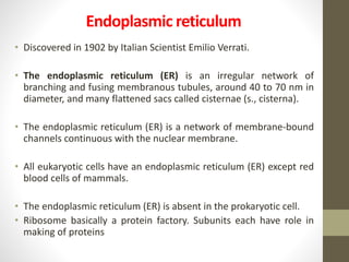
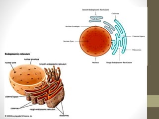
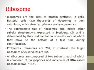
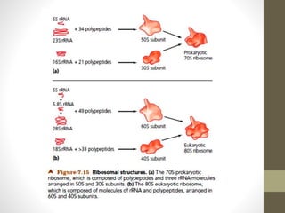
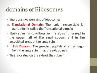
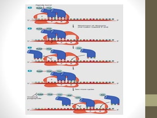
Endoplasmic reticulum,
Ribosome and protien synthesis
Dr. Sarah abdulsalam
1st STAGE / LEC4/ Biology theory/
Radiology and Ulrasonography Techniques dep.
Endoplasmic reticulum
• Discovered in 1902 by Italian Scientist Emilio Verrati.
• The endoplasmic reticulum (ER) is an irregular network of
branching and fusing membranous tubules, around 40 to 70 nm in
diameter, and many flattened sacs called cisternae (s., cisterna).
• The endoplasmic reticulum (ER) is a network of membrane-bound
channels continuous with the nuclear membrane.
• All eukaryotic cells have an endoplasmic reticulum (ER) except red
blood cells of mammals.
• The endoplasmic reticulum (ER) is absent in the prokaryotic cell.
• Ribosome basically a protein factory. Subunits each have role in
making of proteins
Type of ER(smooth and rough)
• It is basically of two functionally distinct types : Smooth Endoplasmic
Reticulum (SER) and Rough Endoplasmic Reticulum (RER) on absence or
presence of ribonucleoprotein granules (Ribosomes).
• The two types of ER are recognized: rough, which contains attached 80S
ribosomes, and smooth, which does not.
• Agranular or Smooth Endoplasmic Reticulum (SER) that does not have
Ribosomes attached to its surface.
• Granular or Rough ER (RER) The endoplasmic reticulum bears ribosomes
on its surface .
• SER and RER change into each other as per the need of the cell.
• Rough ER is a major producer of glycoproteins and produces new
membrane material that is transported throughout the cell; smooth ER
participates in the synthesis of lipids and in some aspects of carbohydrate
metabolism.
FUNCTIONS OF ER
1) Translocation of proteins (such as secretory proteins)
across the ER membrane.
2) Integration of proteins into the plasma membrane.
3) Folding and modification of proteins in the ER lumen.
4) Synthesis of phospholipids and steroids on the cytosolic
side of the ER membrane.
5) Storage of calcium ion in the lumen and their regulated
release into the cytosol.
6) Carbohydrate metabolism.
Ribosome
• Ribosomes are the sites of protein synthesis in cells.
Bacterial cells have thousands of ribosomes in their
cytoplasm, which gives cytoplasm a grainy appearance.
• The approximate size of ribosomes—and indeed other
cellular structures—is expressed in Svedbergs (S), and is
determined by their sedimentation rate—the rate at which
they move to the bottom of a test tube during
centrifugation.
• Prokaryotic ribosomes are 70S; in contrast, the larger
ribosomes of eukaryotes are 80S.
• All ribosomes are composed of two subunits, each of which
is composed of polypeptides and molecules of RNA called
ribosomal RNA (rRNA).
• The subunits of prokaryotic 70S ribosomes are a smaller 30S subunit and
a larger 50S subunit; the 30S subunit contains polypeptides and a single
rRNA molecule, whereas the 50S subunit has polypeptides and two rRNA
molecules.
• Because sedimentation rates depend not only on mass and size but also
on shape, the sedimentation rates of subunits do not add up to the
sedimentation rate of a whole ribosome.
• Each ribosome is composed of two subunits: 50S and 30S .
• The 50S subunit is in turn composed of two rRNA molecules (23S and 5S)
and about 34 different polypeptides, whereas the 30S subunit consists of
one molecule of 16S rRNA and 21 ribosomal polypeptides.
• In contrast, both the cytosol and the rough endoplasmic reticulum (RER)
of eukaryotic cells have 80S ribosomes composed of 60S and 40S
subunits .
Types of ribosomes :
1) Matrix Ribosomes: These synthesize
proteins destined to remain within
the cell.
2) Plasma Membrane Ribosomes:
These make proteins for transport to
the outside.
domains of Ribosomes
• There are two domains of Ribosomes
A. Translational Domain: The region responsible for
translation is called the Translational domain
• Both subunits contribute to this domain, located in
the upper half of the small subunit and in the
associated areas of the large subunit
B. Exit Domain: The growing peptide chain emerges
from the large subunit at the exit domain
• This is located on the side of the subunit.
Types of RNA
1) Messenger RNA (mRNA)—template for the
synthesis of proteins by ribosomes.
2) Transfer RNA (tRNA)—transfers specific amino
acids to growing polypeptide chains at the
ribosomal site of protein synthesis during
translation
3) Ribosomal RNA (rRNA)—a component of
ribosomes
Ribosome and RNA
• mRNA with code for proteins located at 30S subunit.
• • tRNAs responsible for carrying amino acids to mRNA.
• Each tRNA has own nucleotide triplet which binds to
matching triplet on mRNA, ex., tRNA with code AAA
(triple adenine) would match up with mRNA that has
code UUU (triple uracil).
Sites of ribosome
• The ribosome has three
• sites for binding tRNA
• • The Peptidyl or Donor site
• (the P site)
• • The Aminoacyl or Acceptor Site
• (the A site)
• • The Exit Site
• (the E site)
Protein synthesis
• Process starts from DNA
through “transcription”
• • “Translation” is where
ribosome comes in.
• Translation occurs when
protein formed from code
on mRNA
• • Ribosome carries out the
translation of the
nucleotide triplets
• Chart - visual image of transcription
and translation in protein synthesizing
• DNA and RNA have nucleotides that
determine kind of protein
• 3 nucleotides = 1 amino acid of
a protein
• Protein Synthesis is divided into three stages:
• 1. Initiation
• 2. Elongation
• 3. Termination
• Ribosome are made up of 2 subunits, a large one and a smaller
one, each subunit contains ribosomal RNA (rRNA) & proteins.
• Protein synthesis starts when the two subunits bind to mRNA.
• The initiator codon AUG binds to the first anticodon of tRNA,
signaling the start of a protein.
• The anticodon of another tRNA binds to the next mRNA codon,
bringing the 2nd amino acid to be placed in the protein.
• As each anticodon & codon bind together a peptide bond forms
between the two amino acids.
• The protein chain continues to grow until a stop codon reaches
the ribosome, which results in the release of the new protein and
mRNA, completing the process of translation
1. Initiation
• During initiation, the two ribosomal subunits, mRNA, several
protein factors, and tRNAfMet (methionine) form an initiation
complex. Initiation may occur while the cell is still transcribing
mRNA from DNA.
• The smaller ribosomal subunit attaches to mRNA at a ribosome-
binding site so as to position a start codon (AUG) at the ribosomal
subunit’s P site.
• tRNAfMet (whose anticodon, UAC, is complementary to the start
codon, AUG) attaches at the ribosome’s P site.
• The larger ribosomal subunit then attaches to form a complete
initiation complex.
A
E
Large
subunit
P
Small
subunit
fMet
UAC
GAG...CU-AUG--UUC--CUU--AGU--GGU--AGA--GCU--GUA--UGA-AT GCA...TAAAAAA
5’
mRNA
3’
2. Elongation
• Elongation of a polypeptide is a cyclical process that involves the
sequential addition of amino acids to a polypeptide chain growing
at the P site.
• The transfer RNA whose anticodon is complementary to the next
codon—in this example, AAA complementary to the codon
UUU—delivers its amino acid, in this case, phenylalanine (Phe), to
the A site.
• A ribozyme in the larger ribosomal subunit forms a peptide bond
between the terminal amino acid of the growing polypeptide
chain and the newly introduced amino acid. The polypeptide is
now attached to the tRNA occupying the A site.
• Using energy supplied by more GTP, the ribosome moves one
codon down the mRNA. This transfers each tRNA to the
adjacent binding site; that is, the first tRNA moves from the P
site to the E site, and the second tRNA (with the attached
polypeptide) moves to the vacated P site.
• The ribosome releases the “empty” tRNA from the E site. In
the cytosol, the appropriate enzyme recharges it with another
molecule of its specific amino acid.
• The cycle repeats, each time adding another amino acid, at a
rate of about 15 amino acids per second.
A
E
Ribosome
P
Phe
Leu
Met
Ser
Gly
Polypeptide
Arg
Aminoacyl tRNA
UCU
CCA
GAG...CU-AUG--UUC--CUU--AGU--GGU--AGA--GCU--GUA--UGA-AT GCA...TAAAAAA
5’
mRNA
3’
A
E
Ribosome
P
Aminoacyl tRNA
Ala
CCA
Arg
UCU
Phe
Leu
Met
Ser
Gly
Polypeptide
GAG...CU-AUG--UUC--CUU--AGU--GGU--AGA--GCU--GUA--UGA-AT GCA...TAAAAAA
5’
mRNA
3’
A
E
Ribosome
P
Arg
UCU
Phe
Leu
Met
Ser
Gly
Polypeptide
CGA
Ala
GAG...CU-AUG--UUC--CUU--AGU--GGU--AGA--GCU--GUA--UGA-AT GCA...TAAAAAA
5’
mRNA
3’
3. Termination
• Termination does not involve tRNA;
instead, proteins called release factors halt
elongation. It appears that release factors
somehow recognize stop codons and
modify the larger ribosomal subunit in
such a way as to activate another of its
ribozymes, which severs the polypeptide
from the final tRNA (resident at the P site).
The ribosome then dissociates into its
subunits.
3’
5’
5’
3’
Ribosome
Ribosome
5’
mRNA
RNA
Pol.
lec 4 bio اشعة.pptx

lec 4 bio اشعة.pptx

  • 1.
    Endoplasmic reticulum, Ribosome andprotien synthesis Dr. Sarah abdulsalam 1st STAGE / LEC4/ Biology theory/ Radiology and Ulrasonography Techniques dep.
  • 2.
    Endoplasmic reticulum • Discoveredin 1902 by Italian Scientist Emilio Verrati. • The endoplasmic reticulum (ER) is an irregular network of branching and fusing membranous tubules, around 40 to 70 nm in diameter, and many flattened sacs called cisternae (s., cisterna). • The endoplasmic reticulum (ER) is a network of membrane-bound channels continuous with the nuclear membrane. • All eukaryotic cells have an endoplasmic reticulum (ER) except red blood cells of mammals. • The endoplasmic reticulum (ER) is absent in the prokaryotic cell. • Ribosome basically a protein factory. Subunits each have role in making of proteins
  • 3.
    Type of ER(smoothand rough) • It is basically of two functionally distinct types : Smooth Endoplasmic Reticulum (SER) and Rough Endoplasmic Reticulum (RER) on absence or presence of ribonucleoprotein granules (Ribosomes). • The two types of ER are recognized: rough, which contains attached 80S ribosomes, and smooth, which does not. • Agranular or Smooth Endoplasmic Reticulum (SER) that does not have Ribosomes attached to its surface. • Granular or Rough ER (RER) The endoplasmic reticulum bears ribosomes on its surface . • SER and RER change into each other as per the need of the cell. • Rough ER is a major producer of glycoproteins and produces new membrane material that is transported throughout the cell; smooth ER participates in the synthesis of lipids and in some aspects of carbohydrate metabolism.
  • 5.
    FUNCTIONS OF ER 1)Translocation of proteins (such as secretory proteins) across the ER membrane. 2) Integration of proteins into the plasma membrane. 3) Folding and modification of proteins in the ER lumen. 4) Synthesis of phospholipids and steroids on the cytosolic side of the ER membrane. 5) Storage of calcium ion in the lumen and their regulated release into the cytosol. 6) Carbohydrate metabolism.
  • 6.
    Ribosome • Ribosomes arethe sites of protein synthesis in cells. Bacterial cells have thousands of ribosomes in their cytoplasm, which gives cytoplasm a grainy appearance. • The approximate size of ribosomes—and indeed other cellular structures—is expressed in Svedbergs (S), and is determined by their sedimentation rate—the rate at which they move to the bottom of a test tube during centrifugation. • Prokaryotic ribosomes are 70S; in contrast, the larger ribosomes of eukaryotes are 80S. • All ribosomes are composed of two subunits, each of which is composed of polypeptides and molecules of RNA called ribosomal RNA (rRNA).
  • 7.
    • The subunitsof prokaryotic 70S ribosomes are a smaller 30S subunit and a larger 50S subunit; the 30S subunit contains polypeptides and a single rRNA molecule, whereas the 50S subunit has polypeptides and two rRNA molecules. • Because sedimentation rates depend not only on mass and size but also on shape, the sedimentation rates of subunits do not add up to the sedimentation rate of a whole ribosome. • Each ribosome is composed of two subunits: 50S and 30S . • The 50S subunit is in turn composed of two rRNA molecules (23S and 5S) and about 34 different polypeptides, whereas the 30S subunit consists of one molecule of 16S rRNA and 21 ribosomal polypeptides. • In contrast, both the cytosol and the rough endoplasmic reticulum (RER) of eukaryotic cells have 80S ribosomes composed of 60S and 40S subunits .
  • 9.
    Types of ribosomes: 1) Matrix Ribosomes: These synthesize proteins destined to remain within the cell. 2) Plasma Membrane Ribosomes: These make proteins for transport to the outside.
  • 10.
    domains of Ribosomes •There are two domains of Ribosomes A. Translational Domain: The region responsible for translation is called the Translational domain • Both subunits contribute to this domain, located in the upper half of the small subunit and in the associated areas of the large subunit B. Exit Domain: The growing peptide chain emerges from the large subunit at the exit domain • This is located on the side of the subunit.
  • 11.
    Types of RNA 1)Messenger RNA (mRNA)—template for the synthesis of proteins by ribosomes. 2) Transfer RNA (tRNA)—transfers specific amino acids to growing polypeptide chains at the ribosomal site of protein synthesis during translation 3) Ribosomal RNA (rRNA)—a component of ribosomes
  • 12.
    Ribosome and RNA •mRNA with code for proteins located at 30S subunit. • • tRNAs responsible for carrying amino acids to mRNA. • Each tRNA has own nucleotide triplet which binds to matching triplet on mRNA, ex., tRNA with code AAA (triple adenine) would match up with mRNA that has code UUU (triple uracil).
  • 13.
    Sites of ribosome •The ribosome has three • sites for binding tRNA • • The Peptidyl or Donor site • (the P site) • • The Aminoacyl or Acceptor Site • (the A site) • • The Exit Site • (the E site)
  • 14.
    Protein synthesis • Processstarts from DNA through “transcription” • • “Translation” is where ribosome comes in. • Translation occurs when protein formed from code on mRNA • • Ribosome carries out the translation of the nucleotide triplets
  • 15.
    • Chart -visual image of transcription and translation in protein synthesizing • DNA and RNA have nucleotides that determine kind of protein • 3 nucleotides = 1 amino acid of a protein • Protein Synthesis is divided into three stages: • 1. Initiation • 2. Elongation • 3. Termination
  • 16.
    • Ribosome aremade up of 2 subunits, a large one and a smaller one, each subunit contains ribosomal RNA (rRNA) & proteins. • Protein synthesis starts when the two subunits bind to mRNA. • The initiator codon AUG binds to the first anticodon of tRNA, signaling the start of a protein. • The anticodon of another tRNA binds to the next mRNA codon, bringing the 2nd amino acid to be placed in the protein. • As each anticodon & codon bind together a peptide bond forms between the two amino acids. • The protein chain continues to grow until a stop codon reaches the ribosome, which results in the release of the new protein and mRNA, completing the process of translation
  • 17.
    1. Initiation • Duringinitiation, the two ribosomal subunits, mRNA, several protein factors, and tRNAfMet (methionine) form an initiation complex. Initiation may occur while the cell is still transcribing mRNA from DNA. • The smaller ribosomal subunit attaches to mRNA at a ribosome- binding site so as to position a start codon (AUG) at the ribosomal subunit’s P site. • tRNAfMet (whose anticodon, UAC, is complementary to the start codon, AUG) attaches at the ribosome’s P site. • The larger ribosomal subunit then attaches to form a complete initiation complex.
  • 19.
  • 20.
    2. Elongation • Elongationof a polypeptide is a cyclical process that involves the sequential addition of amino acids to a polypeptide chain growing at the P site. • The transfer RNA whose anticodon is complementary to the next codon—in this example, AAA complementary to the codon UUU—delivers its amino acid, in this case, phenylalanine (Phe), to the A site. • A ribozyme in the larger ribosomal subunit forms a peptide bond between the terminal amino acid of the growing polypeptide chain and the newly introduced amino acid. The polypeptide is now attached to the tRNA occupying the A site.
  • 21.
    • Using energysupplied by more GTP, the ribosome moves one codon down the mRNA. This transfers each tRNA to the adjacent binding site; that is, the first tRNA moves from the P site to the E site, and the second tRNA (with the attached polypeptide) moves to the vacated P site. • The ribosome releases the “empty” tRNA from the E site. In the cytosol, the appropriate enzyme recharges it with another molecule of its specific amino acid. • The cycle repeats, each time adding another amino acid, at a rate of about 15 amino acids per second.
  • 22.
  • 23.
  • 24.
  • 26.
    3. Termination • Terminationdoes not involve tRNA; instead, proteins called release factors halt elongation. It appears that release factors somehow recognize stop codons and modify the larger ribosomal subunit in such a way as to activate another of its ribozymes, which severs the polypeptide from the final tRNA (resident at the P site). The ribosome then dissociates into its subunits.
  • 27.