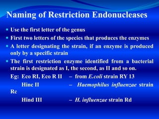
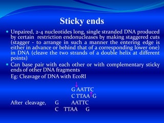
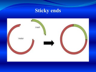
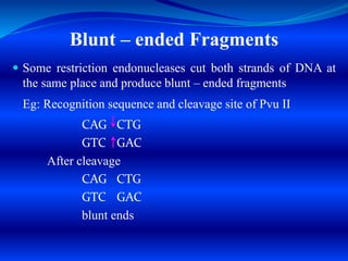
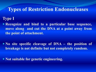
Restriction endonucleases are enzymes found in bacteria that recognize specific nucleotide sequences in DNA and cut the DNA at those sites. This acts as a defense mechanism for bacteria by destroying foreign DNA, such as that from viruses. There are several types of restriction endonucleases, but type II are most useful for genetic engineering as they cut DNA at predictable, site-specific locations. The resulting DNA fragments can be joined back together via DNA ligases. Restriction endonucleases have four- to six-letter recognition sequences that are palindromic, and they produce sticky or blunt ends depending on whether cuts are offset or at the same position.
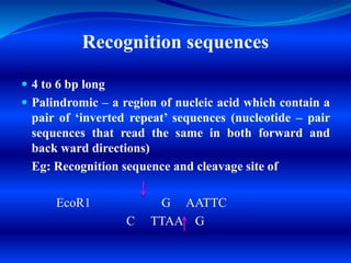
Introduction to ArtificiaI Intelligence in Higher Education
RESTRICTION ENDONUCLEASES & DNA LIGASES SMG
1.
2. Restriction Endonucleases
Pioneer – Werner Arber
Discovered by Hamilton Smith & Daniel
Nathans (1970) (1986 Nobel Prize in
Physiology & Medicine)
Enzymes in Bacteria – protect DNA of
bacteria from “invasion” by foreign DNA (of
another species/bacteriophages).
‘Immune systems of prokaryotes’
3. Cleave foreign DNA into a fixed
number of fragments, depending upon
the number of restriction sites.
Recognize specific nucleotide sequences
–
‘Recognition sequences’ and cut DNA at
these sites – ‘Molecular scalpels’.
4. Recognition sequences
4 to 6 bp long
Palindromic – a region of nucleic acid which contain a
pair of ‘inverted repeat’ sequences (nucleotide – pair
sequences that read the same in both forward and
back ward directions)
Eg: Recognition sequence and cleavage site of
EcoR1 G AATTC
C TTAA G
5. Naming of Restriction Endonucleases
Use the first letter of the genus
First two letters of the species that produces the enzymes
A letter designating the strain, if an enzyme is produced
only by a specific strain
The first restriction enzyme identified from a bacterial
strain is designated as I, the second, as II and so on.
Eg: Eco RI, Eco R II – from E.coli strain RY 13
Hinc II – Haemophilus influenzae strain
Rc
Hind III – H. influenzae strain Rd
6. Sticky ends
Unpaired, 2-4 nucleotides long, single stranded DNA produced
by certain restriction endonucleases by making staggered cuts
(stagger - to arrange in such a manner the entering edge is
either in advance or behind that of a corresponding lower one)
in DNA (cleave the two strands of a double helix at different
points)
Can base pair with each other or with complementary sticky
ends of other DNA fragments
Eg: Cleavage of DNA with EcoRI
G AATTC
C TTAA G
After cleavage, G AATTC
C TTAA G
8. Blunt – ended Fragments
Some restriction endonucleases cut both strands of DNA at
the same place and produce blunt – ended fragments
Eg: Recognition sequence and cleavage site of Pvu II
CAG CTG
GTC GAC
After cleavage
CAG CTG
GTC GAC
blunt ends
9. Types of Restriction Endonucleases
Type I
Recognize and bind to a particular base sequence,
move along and cut the DNA at a point away from
the point of attachment.
No site specific cleavage of DNA – the position of
breakage is not definite but not completely random.
Not suitable for genetic engineering.
10. Type II
Site specific cleavage – recognize a specific
sequence in DNA and cut at the same site.
Used in Genetic Engineering
Type III
No site specific cleavage – cut the DNA at a specific
point that is a measured distance (about 25 bp
[bp= base pair] from the recognition sequence)
Not suitable for Genetic Engineering
11. DNA Ligases
Enzymes used to join DNA fragments.
Mediate the sealing of exposed single stranded breaks or
gaps in the DNA molecule.
Catalyses the formation of phosphodiester bonds.
‘Molecular glue’ – used to join DNA fragments to produce
recombinant DNA
Types of DNA Ligases
1. E.coli ligase – requires NAD as cofactor
2. T4 DNA ligase – uses ATP as cofactor