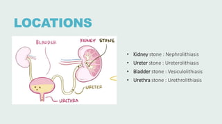
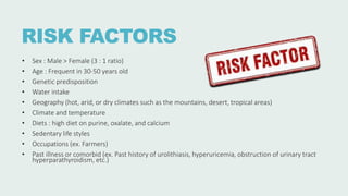
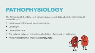
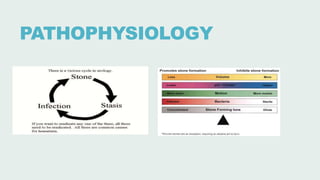
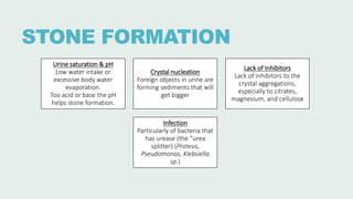
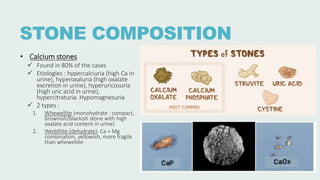
UROLITHIASIS
DEFINITION
A condition where urinary
stones/calculi are formed or
located anywhere in the urinary
system
LOCATIONS
• Kidney stone : Nephrolithiasis
• Ureter stone : Ureterolithiasis
• Bladder stone : Vesiculolithiasis
• Urethra stone : Urethrolithiasis
RISK FACTORS
• Sex : Male > Female (3 : 1 ratio)
• Age : Frequent in 30-50 years old
• Genetic predisposition
• Water intake
• Geography (hot, arid, or dry climates such as the mountains, desert, tropical areas)
• Climate and temperature
• Diets : high diet on purine, oxalate, and calcium
• Sedentary life styles
• Occupations (ex. Farmers)
• Past illness or comorbid (ex. Past history of urolithiasis, hyperuricemia, obstruction of urinary tract
hyperparathyroidism, etc.)
PATHOPHYSIOLOGY
The formation of the stones is a complex process, and depend on the interaction of
several factors:
 Urinary concentration of stone forming ions
 Urinary pH
 Urinary flow rate
 The balance between promoter and inhibitory factors of crystallization
 Anatomic factors that encourage urinary statis
PATHOPHYSIOLOGY
STONE FORMATION
Urine saturation & pH
Low water intake or
excessive body water
evaporation.
Too acid or base the pH
helps stone formation.
Crystal nucleation
Foreign objects in urine are
forming sediments that will
get bigger
Lack of Inhibitors
Lack of inhibitors to the
crystal aggregations,
especially to citrates,
magnesium, and cellulose
Infection
Particularly of bacteria that
has urease (the “urea
splitter) (Proteus,
Pseudomonas, Klebsiella
sp.)
STONE COMPOSITION
• Calcium stones
 Found in 80% of the cases
 Etiologies : hypercalciuria (high Ca in
urine), hyperoxaluria (high oxalate
excretion in urine), hyperuricosuria
(high uric acid in urine),
hypercitraturia. Hypomagnesuria
 2 types :
1. Whewellite (monohydrate : compact,
brownish/blackish stone with high
oxalate acid content in urine)
2. Weddllite (dehydrate): Ca + Mg
combination, yellowish, more fragile
than whewellite
STONE COMPOSITION
• Uric stones
 5-10% of all urinary stone cases
 Risk factors : gout, patients that
received anti-cancer treatment,
uricosuric drugs, obesity, alcohol, high
protein diet
 Varied in size (small to big. The big
one can be shaped like a deer horn
(staghorn stone))
STONE COMPOSITION
• Uric stones
 5-10% of all urinary stone cases
 Risk factors : gout, patients that received anti-cancer treatment, uricosuric drugs,
obesity, alcohol, high protein diet.
Others:
1. Too acid urinary pH (pH < 6)
2. Too low urinary volumes (< 2 L/day or dehydration)
3. Hyperuricosuria or high uric acid level.
 Varied in size (small to big so that shaped like a deer horn (staghorn)
 Radiolucent : filling defect shadow in IVP test
STONE COMPOSITION
• Struvit stones
 Also called as infection stone
 Etiology : urea splitter bacteria (bacteria that can produce urease enzyme and
change urine pH into base through urea hydrolysis to ammonia); such as Proteus
spp, Klebsiella spp, Serratia, Enterobacteria, Pseudomonas, and Staphylococcus
• Cystine stones
 2% of urinary stone cases
 Caused by metabolic defect that causing failure of reabsorption of cystine,
ornithine, lysine, and arginine in renal.
CLINICAL MANIFESTATION
Varied, can be without any symptoms.
Others are:
• Pain in the flank area or radiating. If colic is present : sharp pain
• Pain in urination
• Urinary symptoms: hematuria, suprapubic pain, weak stream, dysuria
• Urine accompanied by stones/sands
• Fever
• Could be accompanied by signs of renal failure
• Others : urgency, various bowel symptoms, pain in the tip of the penis
DIAGNOSIS
 History
‐ Pain (onset, duration, location, characteristics, radiation)
In colic, pain is acute, sharp, and constant. Acute colic
pain can induce nausea and vomiting.
‐ Nausea, vomiting
‐ Infection (fever, chills)
‐ Urinary symptoms: hematuria, dysuria
‐ History of urinary calculi, urinary tract infections, family
history of calculi, history of
hyperuricemia/hyperparathyroidism
 Physical examination
Costovertebral angle tenderness, palpable renal due to
hydronephrosis, sign of renal failure, urine retention, and
fever
DIAGNOSIS
 Laboratory examinations
 Urinalysis : yellow, dark-chocolate, RBC and WBC usually are found, minerals, crystals
 Urine (24 hours) : increased creatinine, uric acid, calcium, phosphate, oxalate, or cystine
 Urine culture : showing urinary tract infection
 Chloride and bicarbonate serum
 Complete blood count : WBC can be increased due to the infection. Hb and Ht can be
abnormal if there is severe dehydration.
 Renal function
DIAGNOSIS
 Radiographic examination
 Abdominal X-ray :
• Evaluating stone in renal, ureter,
and bladder area
• High density stones (radiopaque)
are calcium oxalate and calcium
phosphate stones; low density
stones (radiolucent) are struvite,
cystine, or mixed stones
 Intravenous pyelogram (IVP) :
evaluating renal anatomy and function
 USG : showing the location, the shape
of stone
 Others: Abdominal CT scan, MRI
DIFFERENTIAL DIAGNOSIS
• Acute pyelonephritis
• Renal, ureter, and bladder tumor
• Acute cholecystitis
• Acute appendicitis
TREATMENT
Non – surgical
• Stones that <5 mm in size usually can spontaneously passes
• Diet
• Hydration : drinking water 2 L/day  to reserve precipitation
• Medications:
 Reduce pain : NSAIDs (1st choice), tramadol, pentazocine, etc.
 Reduce stone formation (e.g. potassium citrate)
 Help pass stones (e.g. alpha adrenergic blockers, calcium channel
blockers)
• If there is an infection : antibiotics, such as cotrimoxazole and
amoxicillin
• ESWL (extracorporeal shock wave lithotripsy): non-invasive
treatment; using external shock waves to break down the
stone
• URS (Ureteroscopy)
ESWL illustration
TREATMENT
Surgical
American Urological Association/Endourological Society Guideline (2016) recommends surgery in the
following scenarios:
 Ureteral stones >10 mm
 Uncomplicated distal ureteral stones ≤10 mm that have not passed after 4-6 weeks of observation,
with or without MET
 Symptomatic renal stones in patients without any other etiology for pain
 Pediatric patients with ureteral stones that are unlikely to pass or in whom MET has failed
 Pregnant patients with ureteral or renal stones in whom failed observation has failed
General contraindications to definitive stone manipulation:
• Active, untreated UTI
• Uncorrected bleeding diathesis
• Pregnancy (a relative, but not absolute, contraindication)
TREATMENT
Surgical
• Reserved for larger stones
• Primary indications: pain, infection, and obstruction.
Infection combined with urinary tract obstruction is an extremely dangerous
situation  significant risk of urosepsis and death
• Surgery and stent placement
• Endourology : minimally invasive technique to extract the stone (breaking the stone
and extracting the stone from urinary tract using device that directly inserted to the
urinary tract
PREVENTION
• Water intake – output = 2-3 L/day
• Low-protein diet
• Uric stone : low-purine diet
• Calcium stone : low-calcium diet
• Oxalate stone : low oxalate diet, cut down chocolate and nuts
PROGNOSIS
Good if there is no complication
REFERENCES
• [Guideline] Assimos DG, Krambeck A, Miller NL, et al. Surgical Management of Stones: American Urological Association/Endourological
Society Guideline. American Urological Association. Available at https://www.auanet.org/guidelines-and-quality/guidelines/kidney-
stones-surgical-management-guideline. 2016
• Chirag N Dave, MD. Nephrolithiasis. Medscape. Available at https://emedicine.medscape.com/article/437096-overview. 2023
• Anna Hernández Castillo, MD. Nephrolithiasis. Elsevier. Available at https://www.osmosis.org/answers/nephrolithiasis.
• Gottlieb, M., Long, B., & Koyfman, A. (2018). The evaluation and management of urolithiasis in the ED: A review of the literature. The
American Journal of Emergency Medicine, 36(4): 699–706.
• Han, H., Segal, A. M., Seifter, J. .L, & Dwyer, J. T. (2015). Nutritional Management of Kidney Stones (Nephrolithiasis). Clinical Nutrition
Research, 4(3): 137–152.
• Matlaga, B. R., Shah, O. D., & Assimos, D. G. (2003). Drug-induced urinary calculi. Reviews in Urology, 5(4), 227–231.
• Scales, C. D., Jr, Smith, A. C., Hanley, J. M., & Saigal, C. S. (2012). Prevalence of kidney stones in the United States. European Urology,
62(1): 160–165.
• Semins, M. J. & Matlaga, B. R. (2010). Medical evaluation and management of urolithiasis. Therapeutic Advances in Urology, 2(1):3–9
• Campbell, M.F., Kavoussi, L.R. and Wein, A.J. (2020) Campbell-Walsh Urology. 12th Edition, Elsevier Saunders, Philadelphia, PA..

UROLITHIASIS.pptx

  • 1.
  • 2.
    DEFINITION A condition whereurinary stones/calculi are formed or located anywhere in the urinary system
  • 3.
    LOCATIONS • Kidney stone: Nephrolithiasis • Ureter stone : Ureterolithiasis • Bladder stone : Vesiculolithiasis • Urethra stone : Urethrolithiasis
  • 4.
    RISK FACTORS • Sex: Male > Female (3 : 1 ratio) • Age : Frequent in 30-50 years old • Genetic predisposition • Water intake • Geography (hot, arid, or dry climates such as the mountains, desert, tropical areas) • Climate and temperature • Diets : high diet on purine, oxalate, and calcium • Sedentary life styles • Occupations (ex. Farmers) • Past illness or comorbid (ex. Past history of urolithiasis, hyperuricemia, obstruction of urinary tract hyperparathyroidism, etc.)
  • 5.
    PATHOPHYSIOLOGY The formation ofthe stones is a complex process, and depend on the interaction of several factors:  Urinary concentration of stone forming ions  Urinary pH  Urinary flow rate  The balance between promoter and inhibitory factors of crystallization  Anatomic factors that encourage urinary statis
  • 6.
  • 7.
    STONE FORMATION Urine saturation& pH Low water intake or excessive body water evaporation. Too acid or base the pH helps stone formation. Crystal nucleation Foreign objects in urine are forming sediments that will get bigger Lack of Inhibitors Lack of inhibitors to the crystal aggregations, especially to citrates, magnesium, and cellulose Infection Particularly of bacteria that has urease (the “urea splitter) (Proteus, Pseudomonas, Klebsiella sp.)
  • 8.
    STONE COMPOSITION • Calciumstones  Found in 80% of the cases  Etiologies : hypercalciuria (high Ca in urine), hyperoxaluria (high oxalate excretion in urine), hyperuricosuria (high uric acid in urine), hypercitraturia. Hypomagnesuria  2 types : 1. Whewellite (monohydrate : compact, brownish/blackish stone with high oxalate acid content in urine) 2. Weddllite (dehydrate): Ca + Mg combination, yellowish, more fragile than whewellite
  • 9.
    STONE COMPOSITION • Uricstones  5-10% of all urinary stone cases  Risk factors : gout, patients that received anti-cancer treatment, uricosuric drugs, obesity, alcohol, high protein diet  Varied in size (small to big. The big one can be shaped like a deer horn (staghorn stone))
  • 10.
    STONE COMPOSITION • Uricstones  5-10% of all urinary stone cases  Risk factors : gout, patients that received anti-cancer treatment, uricosuric drugs, obesity, alcohol, high protein diet. Others: 1. Too acid urinary pH (pH < 6) 2. Too low urinary volumes (< 2 L/day or dehydration) 3. Hyperuricosuria or high uric acid level.  Varied in size (small to big so that shaped like a deer horn (staghorn)  Radiolucent : filling defect shadow in IVP test
  • 11.
    STONE COMPOSITION • Struvitstones  Also called as infection stone  Etiology : urea splitter bacteria (bacteria that can produce urease enzyme and change urine pH into base through urea hydrolysis to ammonia); such as Proteus spp, Klebsiella spp, Serratia, Enterobacteria, Pseudomonas, and Staphylococcus • Cystine stones  2% of urinary stone cases  Caused by metabolic defect that causing failure of reabsorption of cystine, ornithine, lysine, and arginine in renal.
  • 12.
    CLINICAL MANIFESTATION Varied, canbe without any symptoms. Others are: • Pain in the flank area or radiating. If colic is present : sharp pain • Pain in urination • Urinary symptoms: hematuria, suprapubic pain, weak stream, dysuria • Urine accompanied by stones/sands • Fever • Could be accompanied by signs of renal failure • Others : urgency, various bowel symptoms, pain in the tip of the penis
  • 13.
    DIAGNOSIS  History ‐ Pain(onset, duration, location, characteristics, radiation) In colic, pain is acute, sharp, and constant. Acute colic pain can induce nausea and vomiting. ‐ Nausea, vomiting ‐ Infection (fever, chills) ‐ Urinary symptoms: hematuria, dysuria ‐ History of urinary calculi, urinary tract infections, family history of calculi, history of hyperuricemia/hyperparathyroidism  Physical examination Costovertebral angle tenderness, palpable renal due to hydronephrosis, sign of renal failure, urine retention, and fever
  • 14.
    DIAGNOSIS  Laboratory examinations Urinalysis : yellow, dark-chocolate, RBC and WBC usually are found, minerals, crystals  Urine (24 hours) : increased creatinine, uric acid, calcium, phosphate, oxalate, or cystine  Urine culture : showing urinary tract infection  Chloride and bicarbonate serum  Complete blood count : WBC can be increased due to the infection. Hb and Ht can be abnormal if there is severe dehydration.  Renal function
  • 15.
    DIAGNOSIS  Radiographic examination Abdominal X-ray : • Evaluating stone in renal, ureter, and bladder area • High density stones (radiopaque) are calcium oxalate and calcium phosphate stones; low density stones (radiolucent) are struvite, cystine, or mixed stones  Intravenous pyelogram (IVP) : evaluating renal anatomy and function  USG : showing the location, the shape of stone  Others: Abdominal CT scan, MRI
  • 16.
    DIFFERENTIAL DIAGNOSIS • Acutepyelonephritis • Renal, ureter, and bladder tumor • Acute cholecystitis • Acute appendicitis
  • 17.
    TREATMENT Non – surgical •Stones that <5 mm in size usually can spontaneously passes • Diet • Hydration : drinking water 2 L/day  to reserve precipitation • Medications:  Reduce pain : NSAIDs (1st choice), tramadol, pentazocine, etc.  Reduce stone formation (e.g. potassium citrate)  Help pass stones (e.g. alpha adrenergic blockers, calcium channel blockers) • If there is an infection : antibiotics, such as cotrimoxazole and amoxicillin • ESWL (extracorporeal shock wave lithotripsy): non-invasive treatment; using external shock waves to break down the stone • URS (Ureteroscopy) ESWL illustration
  • 18.
    TREATMENT Surgical American Urological Association/EndourologicalSociety Guideline (2016) recommends surgery in the following scenarios:  Ureteral stones >10 mm  Uncomplicated distal ureteral stones ≤10 mm that have not passed after 4-6 weeks of observation, with or without MET  Symptomatic renal stones in patients without any other etiology for pain  Pediatric patients with ureteral stones that are unlikely to pass or in whom MET has failed  Pregnant patients with ureteral or renal stones in whom failed observation has failed General contraindications to definitive stone manipulation: • Active, untreated UTI • Uncorrected bleeding diathesis • Pregnancy (a relative, but not absolute, contraindication)
  • 19.
    TREATMENT Surgical • Reserved forlarger stones • Primary indications: pain, infection, and obstruction. Infection combined with urinary tract obstruction is an extremely dangerous situation  significant risk of urosepsis and death • Surgery and stent placement • Endourology : minimally invasive technique to extract the stone (breaking the stone and extracting the stone from urinary tract using device that directly inserted to the urinary tract
  • 20.
    PREVENTION • Water intake– output = 2-3 L/day • Low-protein diet • Uric stone : low-purine diet • Calcium stone : low-calcium diet • Oxalate stone : low oxalate diet, cut down chocolate and nuts
  • 21.
    PROGNOSIS Good if thereis no complication
  • 22.
    REFERENCES • [Guideline] AssimosDG, Krambeck A, Miller NL, et al. Surgical Management of Stones: American Urological Association/Endourological Society Guideline. American Urological Association. Available at https://www.auanet.org/guidelines-and-quality/guidelines/kidney- stones-surgical-management-guideline. 2016 • Chirag N Dave, MD. Nephrolithiasis. Medscape. Available at https://emedicine.medscape.com/article/437096-overview. 2023 • Anna Hernández Castillo, MD. Nephrolithiasis. Elsevier. Available at https://www.osmosis.org/answers/nephrolithiasis. • Gottlieb, M., Long, B., & Koyfman, A. (2018). The evaluation and management of urolithiasis in the ED: A review of the literature. The American Journal of Emergency Medicine, 36(4): 699–706. • Han, H., Segal, A. M., Seifter, J. .L, & Dwyer, J. T. (2015). Nutritional Management of Kidney Stones (Nephrolithiasis). Clinical Nutrition Research, 4(3): 137–152. • Matlaga, B. R., Shah, O. D., & Assimos, D. G. (2003). Drug-induced urinary calculi. Reviews in Urology, 5(4), 227–231. • Scales, C. D., Jr, Smith, A. C., Hanley, J. M., & Saigal, C. S. (2012). Prevalence of kidney stones in the United States. European Urology, 62(1): 160–165. • Semins, M. J. & Matlaga, B. R. (2010). Medical evaluation and management of urolithiasis. Therapeutic Advances in Urology, 2(1):3–9 • Campbell, M.F., Kavoussi, L.R. and Wein, A.J. (2020) Campbell-Walsh Urology. 12th Edition, Elsevier Saunders, Philadelphia, PA..