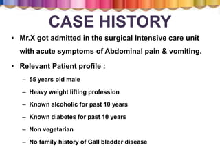
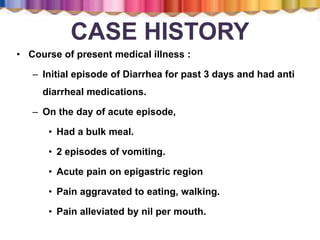
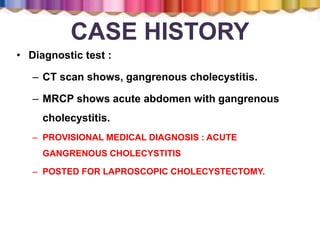
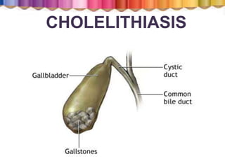
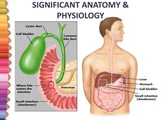
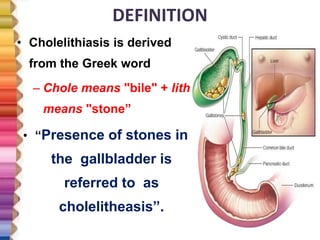
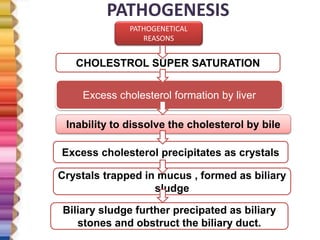
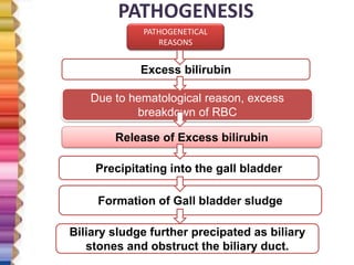
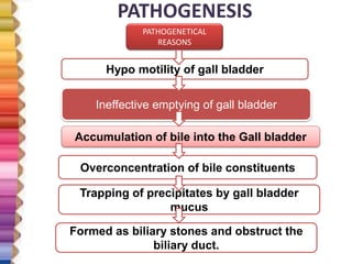
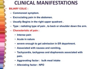
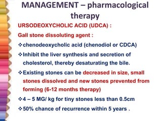
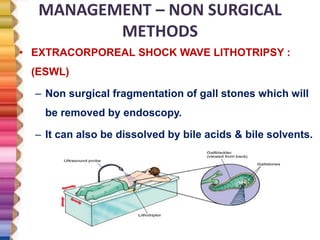
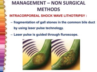
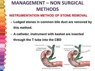
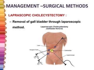
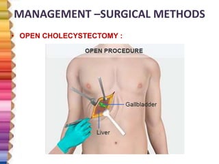
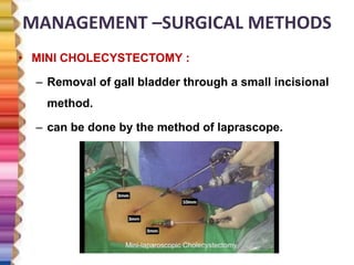
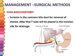
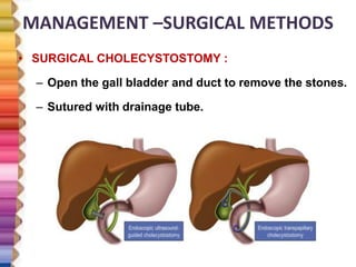
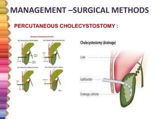
MS.MUTHU RAJATHI, M.SC (N)
ASST., PROFESSOR
DEPARTMENT OF MEDICAL SURGICAL NURSING
GANGA INSTITUTE OF HEALTH SCIENCES
COIMBATORE
CASE HISTORY
• Mr.X got admitted in the surgical Intensive care unit
with acute symptoms of Abdominal pain & vomiting.
• Relevant Patient profile :
– 55 years old male
– Heavy weight lifting profession
– Known alcoholic for past 10 years
– Known diabetes for past 10 years
– Non vegetarian
– No family history of Gall bladder disease
CASE HISTORY
• Course of present medical illness :
– Initial episode of Diarrhea for past 3 days and had anti
diarrheal medications.
– On the day of acute episode,
• Had a bulk meal.
• 2 episodes of vomiting.
• Acute pain on epigastric region
• Pain aggravated to eating, walking.
• Pain alleviated by nil per mouth.
CASE HISTORY
• Diagnostic test :
– CT scan shows, gangrenous cholecystitis.
– MRCP shows acute abdomen with gangrenous
cholecystitis.
– PROVISIONAL MEDICAL DIAGNOSIS : ACUTE
GANGRENOUS CHOLECYSTITIS
– POSTED FOR LAPROSCOPIC CHOLECYSTECTOMY.
CHOLELITHIASIS
INTRODUCTION
• Common disorder of the abdominal cavity.
• Requires emergency surgical intervention
• Disorders of the gall bladder & duct is more common
than gall bladder cancer.
• commonly affects sedentary life style people and
obese community than others.
SIGNIFICANT ANATOMY &
PHYSIOLOGY
SIGNIFICANT ANATOMY &
PHYSIOLOGY
Emulsi
fication
of fat
Storage of
bile
Digestion
of fat
soluble
vitamins
Elimination
of excess
cholesterol.
DEFINITION
• Cholelithiasis is derived
from the Greek word
– Chole means "bile" + lith
means "stone”
• “Presence of stones in
the gallbladder is
referred to as
cholelitheasis”.
DEFINITION
• Gallstones are collections of cholesterol,
bile pigment or a combination of the two,
which can form in the gallbladder or
within the bile ducts of the liver.
DEFINITION
“Cholelithiasis” (calculi or gallstones)
usually form in the gallbladder from the
solid constituents of bile and vary
greatly in size, shape and composition.
The stones in the biliary tract shift into
common bile duct is known as
“Choledochocholelithiasis”
INCIDENCE AND PREVALENCE
• Sex :
– Females more affected than male.
– Multi parous women are more at risk.
• Age :
– 40 years and above are common
• Region :
– Higher incidence in north India.
– Overall Indian prevalence 20 – 30%
CLASSIFICATION OF GALL STONES
Cholesterol
stones
Pigment
stones
Mixed
stones
Cholesterol stones (80% cholesterol by
weight)
Color : vary in color from light-yellow to
dark-green or brown
Size : oval 2 to 3 cm in length, often
having a tiny dark central spot.
Composition : Cholesterol gallstones
usually contain >50% cholesterol
monohydrate plus an admixture of
calcium salts, bile pigments, and
proteins
Pigment stones :
Composed almost entirely of calcium
bilirubinate.
They are mostly small, black and multiple.
Some are hard and coral like stones.
Most of them seems to be soft and really
concretions of sludge rather than stones.
.
Pigment stones :
Two types
Hemolytic and liver disease are associated with the black
stones;
The brown, earthy stones more frequently are formed
outside the gallbladder and often are associated with
bacterial infections of the biliary tract
 Small, dark stones made of bilirubin, calcium salts and
20% cholesterol that are found in bile.
Mixed stones :
• Typically
Contain 20–80% cholesterol.
Other common constituents
Are calcium carbonate, palmitate
Phosphate, bilirubin, and
Other bile pigments.
Because of their calcium content,
they are often radiographically
visible.
COMMON RISK FACTORS- 5Fs
FATTY
FORTY
FEMALE
FERTILE
FAIR
CAUSES – PROLONGED FASTING
PROLONGED
FASTING
FORMATION OF
BILIARY SLUDGE
MICROLITHIASIS
CAUSES AND RISK FACTORS
Obesity
pregnancy
genetics
Rapid weight
loss
Liver and
pancreatic
disorder
Alcoholics
CAUSES AND RISK FACTORS
Oral
contraceptive
pills
Hormonal
therapy
Ileal disease or
resection leads
to malabsorption
of bile acids
Hemolytic
disease
Infection
CAUSES AND RISK FACTORS
Spinal cord injury leads
to delayed gall bladder
emptying
Prolonged total
parenteral nutrition
CAUSES- Lithogenic bile
Increased amount of cholesterol with high amount of
lecithin molecules in bile. Due to,
• Defective bile salt synthesis
• Excessive intestinal loss of bile salts
• Excessive cholesterol secretion
• Abnormal gall bladder function
PATHOGENESIS
PATHOGENETICAL
REASONS
CHOLESTROL SUPER SATURATION
Excess cholesterol formation by liver
Inability to dissolve the cholesterol by bile
Excess cholesterol precipitates as crystals
Crystals trapped in mucus , formed as biliary
sludge
Biliary sludge further precipated as biliary
stones and obstruct the biliary duct.
PATHOGENESIS
PATHOGENETICAL
REASONS
Excess bilirubin
Due to hematological reason, excess
breakdown of RBC
Release of Excess bilirubin
Precipitating into the gall bladder
Formation of Gall bladder sludge
Biliary sludge further precipated as biliary
stones and obstruct the biliary duct.
PATHOGENESIS
PATHOGENETICAL
REASONS
Hypo motility of gall bladder
Ineffective emptying of gall bladder
Accumulation of bile into the Gall bladder
Overconcentration of bile constituents
Trapping of precipitates by gall bladder
mucus
Formed as biliary stones and obstruct the
biliary duct.
CLINICAL MANIFESTATIONS
• BILIARY COLIC :
– Commonest symptom.
– Excruciating pain in the abdomen.
– Usually Begins in the right upper quadrant .
– Type : radiating type of pain , to back or shoulder down the arm.
– Characteristic of pain :
• Intense pain
• Acute in nature
• severe enough to get admission in ER department.
• Associated with nausea and vomiting,
• Tachycardia, tachypnea and diaphoresis associated with
pain.
• Aggravating factor : bulk meal intake
• Alleviating factor : NPO
CLINICAL MANIFESTATIONS
• Jaundice :
– Due to absorption of excess bile into the blood
causing jaundice.
– Yellowish discoloration of eyes and skin
associated with marked itching.
– Dark yellowish urine with pale color stools.
– Steatorrhea.
• Cholecystitis :
– If gall stones obstructs the cystic duct causes
acute cholecystitis.
– Manifested with fever and chillness, palpable
abdominal mass.
• Vitamin deficiency :
– Poor absorption of fat soluble vitamins due to
bile obstruction.
CLINICAL MANIFESTATIONS
• MURPHY SIGN :
– Place the hand below the right costal
margin along with mid clavicular line.
– Ask the patient to take deep breath.
– Patient experiences sharp pain at the end of
inspiration.
– Patient stops breathing in and winces with a
“ catch in breath”.
STAGES OF STONE FORMATION
• Viscous & non homogenous bile.
• Bile sludge with microlitheasisINITIAL
• Localization of stone in site.
• Symptoms appear according
to obstruction.
FORMATION
• Recurrent obstruction
• Prolonged severe symptomsCHRONIC
• Chronic obstruction causes
complications.COMPLICATIONS
DIAGNOSTIC EVALUATION
• History collection
• Physical examination : Murphy sign & biliary colic
• Liver function test
– Shows Increased WBC
– Increased CRP
– Increased bilirubin & alkaline phosphate
– Increased transminase
– Elevated prothrombin time
– Elevated serum amylase and lipase
• Plain X ray abdomen
– Mixed stones visualized due to high content of calcium.
• USG abdomen : posterior acoustic shadowing confirms the
location of stone.
DIAGNOSTIC EVALUATION
Cholescintigraphy (HIDA
scan)
• Used to diagnose
abnormal contraction
of gallbladder or
obstruction of bile
ducts
DIAGNOSTIC EVALUATION
ERCP :
• Used to identify the
exact location of the
gall stones.
DIAGNOSTIC EVALUATION
PERCUTANEOUS
TRANSHEPATIC
CHOLANGIOGRAPHY :
• Used to identify the exact
location of the gall stones
in the biliary tree, with the
help of contrast medium.
DIAGNOSTIC EVALUATION
MAGNETIC RESONANCE
CHOLANGIO
PANCREATOGRAPHY :
To identify the extent of
Disease with involved
Anatomical areas.
MANAGEMENT
To
resolve
the
symptoms.
To
remove
the
stones.
To
prevent
and treat
the
complicat
ions
MANAGEMENT – nutritional therapy
• Low fat diet should be followed.
• Increased protein and carbohydrate supplements to
be included.
• Fatty foods may initiate an acute episodic
symptoms.
• Fried items to be avoided.
MANAGEMENT – pharmacological
therapy
• Administer ,
– Analgesics : eg : Diclofenac
– Anti emetics : eg : emeset
– Antibiotics : inj. ciprofloxacin
– H2 receptor antagonist : tab. rantac
– Fluid replacement : IVF. NS. IVF. DNS
MANAGEMENT – pharmacological
therapy
URSODEOXYCHOLIC ACID (UDCA) :
Gall stone dissoluting agent :
chenodeoxycholic acid (chenodiol or CDCA)
Inhibit the liver synthesis and secretion of
cholesterol, thereby desaturating the bile.
Existing stones can be decreased in size, small
stones dissolved and new stones prevented from
forming (6-12 months therapy)
4 – 5 MG/ kg for tiny stones less than 0.5cm
50% chance of recurrence within 5 years .
MANAGEMENT – NON SURGICAL
METHODS
• EXTRACORPOREAL SHOCK WAVE LITHOTRIPSY :
(ESWL)
– Non surgical fragmentation of gall stones which will
be removed by endoscopy.
– It can also be dissolved by bile acids & bile solvents.
MANAGEMENT – NON SURGICAL
METHODS
• INTRACORPOREAL SHOCK WAVE LITHOTRIPSY :
– fragmentation of gall stones in the common bile duct
by using laser pulse technology.
– Laser pulse is guided through fluroscope.
MANAGEMENT – NON SURGICAL
METHODS
• DISSOLUTING SOLVENT : METHYL TERTIARY BUTYL
ETHER (MTBE)
– Inject into the gallbladder.
– Can be infused through a tube or catheter inserted
percutaneously directly into the gallbladder;
– A tube or drain inserted through a T-tube tract to
dissolve stones.
MANAGEMENT – NON SURGICAL
METHODS
• INSTRUMENTATION METHOD OF STONE REMOVAL
– Lodged stones in common bile duct are removed by
this method.
– A catheter, instrument with basket are inserted
through the T tube into the CBD
MANAGEMENT –SURGICAL METHODS
• LAPRASCOPIC CHOLECYSTECTOMY :
– Removal of gall bladder through laparoscopic
method.
MANAGEMENT –SURGICAL METHODS
• OPEN CHOLECYSTECTOMY :
MANAGEMENT –SURGICAL METHODS
• MINI CHOLECYSTECTOMY :
– Removal of gall bladder through a small incisional
method.
– can be done by the method of laprascope.
MANAGEMENT –SURGICAL METHODS
• CHOLEDOCHOSTOMY:
– Incision in the common bile duct for removal of
stones. After that T tube will be placed in the incision
site for drainage.
MANAGEMENT –SURGICAL METHODS
• SURGICAL CHOLECYSTOSTOMY :
– Open the gall bladder and duct to remove the stones.
– Sutured with drainage tube.
MANAGEMENT –SURGICAL METHODS
• PERCUTANEOUS CHOLECYSTOSTOMY :
COMPLICATIONS
1. Cholecystitis
2. Choledocholelithiasis.
3. Perforated Gallbladder
4. Gallbladder Cancer
5. Cholangitis
6. Pancreatitis
7. Gangrene or Abscesses.
NURSING MANAGEMENT
• Nursing assessment :
– Assess the general condition of the
patient.
– Collect detailed history relevant to present
medical history.
– Identify the possible physical examination
findings.
NURSING MANAGEMENT
• Nursing diagnosis: pre operative
– Acute pain related to inflammation process
secondary to bile obstruction as evidenced by pain
scale score.
– Imbalanced nutrition less than body requirement
related to vomiting as evidenced by verbalization.
– Impaired bowel elimination related to improper
digestion process secondary to bile insufficiency as
evidenced by bowel sounds.
– Risk for peritonitis related to rupture of inflamed gall
bladder.
NURSING MANAGEMENT
• Nursing diagnosis: postoperative
– Acute pain related to surgical procedure as
evidenced by pain scale score.
– Ineffective airway clearance related to increased
tracheobronchial secretion secondary to high
incision abdominal surgery as evidenced by breath
sounds.
– Imbalanced nutrition less than body requirement
related to NPO as evidenced by verbalization.
– Risk for infection related surgical procedure.
NURSING MANAGEMENT
• Nursing intervention: postoperative pain
– Administer analgesic agents as prescribed to relieve
the pain
– Help the patient to turn, cough, breathe deeply, and
ambulate as indicated.
– Use of a pillow or binder over the incision during
these maneuvers
NURSING MANAGEMENT
• Nursing intervention: improving respiratory status
– Remind patients to take deep breaths and cough
every hour to expand the lungs fully and prevent
atelectasis.
– Promote early ambulation. Early ambulation prevents
pulmonary complications as well
– Monitor elderly and obese patients must closely for
respiratory problem. .
NURSING MANAGEMENT
• Nursing intervention: promoting skin care and biliary
drainage:
– observed for indications of infection, leakage of bile
into the peritoneal cavity, and obstruction of bile
drainage, clay colored stool and vital sign.
– note and report right upper quadrant abdominal, pain,
nausea and vomiting. Observe for jaundice.
– changes frequently the outer dressings and protection
of the skin from irritation.
– Maintain a careful record of fluid intake and output
NURSING MANAGEMENT
• Nursing intervention: improving nutritional status
– Encourage the patient to eat a diet low in fats and high
in carbohydrates and proteins immediately after
surgery.
– Monitoring and managing potential complications:
– Closely monitor vital signs and inspects the surgical
incisions and drains, if in place, for evidence of
bleeding. Periodically assesses the patient for
increased tenderness and rigidity of the abdomen and
report to the surgeon. Instructs the patient and family
to report to the surgeon any change in the color of
NURSING MANAGEMENT
Monitoring and managing potential complications:
Closely monitor vital signs and inspects the surgical
incisions and drains, if in place, for evidence of
bleeding.
Periodically assesses the patient for increased
tenderness and rigidity of the abdomen and report to
the surgeon.
Instructs the patient and family to report to the surgeon
any change in the color of stools because this may
indicate complications.
REFERENCES
 Brunner & sudharth's textbook of medical surgical nursing. 13th edition. I
volume .New delhi
 Lewis’s medical surgical nursing. 2 edition. I volume. New delhi: elsevier
publication
 Black mj, hawks hj. Medical surgical nursing. 8th edition. Ii volume .New
delhi: elsevier publications
 Lippincott, williams & wilkins. Manual of nursing practice. 10th
edition.New delhi
 Www.Ncbi.Nlm.Nih.Gov.In
 www. Pubmed.com
 www. Pancreapedia.com
CHOLELITHIASIS

CHOLELITHIASIS

  • 1.
    MS.MUTHU RAJATHI, M.SC(N) ASST., PROFESSOR DEPARTMENT OF MEDICAL SURGICAL NURSING GANGA INSTITUTE OF HEALTH SCIENCES COIMBATORE
  • 2.
    CASE HISTORY • Mr.Xgot admitted in the surgical Intensive care unit with acute symptoms of Abdominal pain & vomiting. • Relevant Patient profile : – 55 years old male – Heavy weight lifting profession – Known alcoholic for past 10 years – Known diabetes for past 10 years – Non vegetarian – No family history of Gall bladder disease
  • 3.
    CASE HISTORY • Courseof present medical illness : – Initial episode of Diarrhea for past 3 days and had anti diarrheal medications. – On the day of acute episode, • Had a bulk meal. • 2 episodes of vomiting. • Acute pain on epigastric region • Pain aggravated to eating, walking. • Pain alleviated by nil per mouth.
  • 4.
    CASE HISTORY • Diagnostictest : – CT scan shows, gangrenous cholecystitis. – MRCP shows acute abdomen with gangrenous cholecystitis. – PROVISIONAL MEDICAL DIAGNOSIS : ACUTE GANGRENOUS CHOLECYSTITIS – POSTED FOR LAPROSCOPIC CHOLECYSTECTOMY.
  • 5.
  • 6.
    INTRODUCTION • Common disorderof the abdominal cavity. • Requires emergency surgical intervention • Disorders of the gall bladder & duct is more common than gall bladder cancer. • commonly affects sedentary life style people and obese community than others.
  • 7.
  • 8.
    SIGNIFICANT ANATOMY & PHYSIOLOGY Emulsi fication offat Storage of bile Digestion of fat soluble vitamins Elimination of excess cholesterol.
  • 9.
    DEFINITION • Cholelithiasis isderived from the Greek word – Chole means "bile" + lith means "stone” • “Presence of stones in the gallbladder is referred to as cholelitheasis”.
  • 10.
    DEFINITION • Gallstones arecollections of cholesterol, bile pigment or a combination of the two, which can form in the gallbladder or within the bile ducts of the liver.
  • 11.
    DEFINITION “Cholelithiasis” (calculi orgallstones) usually form in the gallbladder from the solid constituents of bile and vary greatly in size, shape and composition. The stones in the biliary tract shift into common bile duct is known as “Choledochocholelithiasis”
  • 12.
    INCIDENCE AND PREVALENCE •Sex : – Females more affected than male. – Multi parous women are more at risk. • Age : – 40 years and above are common • Region : – Higher incidence in north India. – Overall Indian prevalence 20 – 30%
  • 13.
    CLASSIFICATION OF GALLSTONES Cholesterol stones Pigment stones Mixed stones
  • 14.
    Cholesterol stones (80%cholesterol by weight) Color : vary in color from light-yellow to dark-green or brown Size : oval 2 to 3 cm in length, often having a tiny dark central spot. Composition : Cholesterol gallstones usually contain >50% cholesterol monohydrate plus an admixture of calcium salts, bile pigments, and proteins
  • 15.
    Pigment stones : Composedalmost entirely of calcium bilirubinate. They are mostly small, black and multiple. Some are hard and coral like stones. Most of them seems to be soft and really concretions of sludge rather than stones. .
  • 16.
    Pigment stones : Twotypes Hemolytic and liver disease are associated with the black stones; The brown, earthy stones more frequently are formed outside the gallbladder and often are associated with bacterial infections of the biliary tract  Small, dark stones made of bilirubin, calcium salts and 20% cholesterol that are found in bile.
  • 17.
    Mixed stones : •Typically Contain 20–80% cholesterol. Other common constituents Are calcium carbonate, palmitate Phosphate, bilirubin, and Other bile pigments. Because of their calcium content, they are often radiographically visible.
  • 18.
    COMMON RISK FACTORS-5Fs FATTY FORTY FEMALE FERTILE FAIR
  • 19.
    CAUSES – PROLONGEDFASTING PROLONGED FASTING FORMATION OF BILIARY SLUDGE MICROLITHIASIS
  • 20.
    CAUSES AND RISKFACTORS Obesity pregnancy genetics Rapid weight loss Liver and pancreatic disorder Alcoholics
  • 21.
    CAUSES AND RISKFACTORS Oral contraceptive pills Hormonal therapy Ileal disease or resection leads to malabsorption of bile acids Hemolytic disease Infection
  • 22.
    CAUSES AND RISKFACTORS Spinal cord injury leads to delayed gall bladder emptying Prolonged total parenteral nutrition
  • 23.
    CAUSES- Lithogenic bile Increasedamount of cholesterol with high amount of lecithin molecules in bile. Due to, • Defective bile salt synthesis • Excessive intestinal loss of bile salts • Excessive cholesterol secretion • Abnormal gall bladder function
  • 24.
    PATHOGENESIS PATHOGENETICAL REASONS CHOLESTROL SUPER SATURATION Excesscholesterol formation by liver Inability to dissolve the cholesterol by bile Excess cholesterol precipitates as crystals Crystals trapped in mucus , formed as biliary sludge Biliary sludge further precipated as biliary stones and obstruct the biliary duct.
  • 25.
    PATHOGENESIS PATHOGENETICAL REASONS Excess bilirubin Due tohematological reason, excess breakdown of RBC Release of Excess bilirubin Precipitating into the gall bladder Formation of Gall bladder sludge Biliary sludge further precipated as biliary stones and obstruct the biliary duct.
  • 26.
    PATHOGENESIS PATHOGENETICAL REASONS Hypo motility ofgall bladder Ineffective emptying of gall bladder Accumulation of bile into the Gall bladder Overconcentration of bile constituents Trapping of precipitates by gall bladder mucus Formed as biliary stones and obstruct the biliary duct.
  • 27.
    CLINICAL MANIFESTATIONS • BILIARYCOLIC : – Commonest symptom. – Excruciating pain in the abdomen. – Usually Begins in the right upper quadrant . – Type : radiating type of pain , to back or shoulder down the arm. – Characteristic of pain : • Intense pain • Acute in nature • severe enough to get admission in ER department. • Associated with nausea and vomiting, • Tachycardia, tachypnea and diaphoresis associated with pain. • Aggravating factor : bulk meal intake • Alleviating factor : NPO
  • 28.
    CLINICAL MANIFESTATIONS • Jaundice: – Due to absorption of excess bile into the blood causing jaundice. – Yellowish discoloration of eyes and skin associated with marked itching. – Dark yellowish urine with pale color stools. – Steatorrhea. • Cholecystitis : – If gall stones obstructs the cystic duct causes acute cholecystitis. – Manifested with fever and chillness, palpable abdominal mass. • Vitamin deficiency : – Poor absorption of fat soluble vitamins due to bile obstruction.
  • 29.
    CLINICAL MANIFESTATIONS • MURPHYSIGN : – Place the hand below the right costal margin along with mid clavicular line. – Ask the patient to take deep breath. – Patient experiences sharp pain at the end of inspiration. – Patient stops breathing in and winces with a “ catch in breath”.
  • 30.
    STAGES OF STONEFORMATION • Viscous & non homogenous bile. • Bile sludge with microlitheasisINITIAL • Localization of stone in site. • Symptoms appear according to obstruction. FORMATION • Recurrent obstruction • Prolonged severe symptomsCHRONIC • Chronic obstruction causes complications.COMPLICATIONS
  • 31.
    DIAGNOSTIC EVALUATION • Historycollection • Physical examination : Murphy sign & biliary colic • Liver function test – Shows Increased WBC – Increased CRP – Increased bilirubin & alkaline phosphate – Increased transminase – Elevated prothrombin time – Elevated serum amylase and lipase • Plain X ray abdomen – Mixed stones visualized due to high content of calcium. • USG abdomen : posterior acoustic shadowing confirms the location of stone.
  • 32.
    DIAGNOSTIC EVALUATION Cholescintigraphy (HIDA scan) •Used to diagnose abnormal contraction of gallbladder or obstruction of bile ducts
  • 33.
    DIAGNOSTIC EVALUATION ERCP : •Used to identify the exact location of the gall stones.
  • 34.
    DIAGNOSTIC EVALUATION PERCUTANEOUS TRANSHEPATIC CHOLANGIOGRAPHY : •Used to identify the exact location of the gall stones in the biliary tree, with the help of contrast medium.
  • 35.
    DIAGNOSTIC EVALUATION MAGNETIC RESONANCE CHOLANGIO PANCREATOGRAPHY: To identify the extent of Disease with involved Anatomical areas.
  • 36.
  • 37.
    MANAGEMENT – nutritionaltherapy • Low fat diet should be followed. • Increased protein and carbohydrate supplements to be included. • Fatty foods may initiate an acute episodic symptoms. • Fried items to be avoided.
  • 38.
    MANAGEMENT – pharmacological therapy •Administer , – Analgesics : eg : Diclofenac – Anti emetics : eg : emeset – Antibiotics : inj. ciprofloxacin – H2 receptor antagonist : tab. rantac – Fluid replacement : IVF. NS. IVF. DNS
  • 39.
    MANAGEMENT – pharmacological therapy URSODEOXYCHOLICACID (UDCA) : Gall stone dissoluting agent : chenodeoxycholic acid (chenodiol or CDCA) Inhibit the liver synthesis and secretion of cholesterol, thereby desaturating the bile. Existing stones can be decreased in size, small stones dissolved and new stones prevented from forming (6-12 months therapy) 4 – 5 MG/ kg for tiny stones less than 0.5cm 50% chance of recurrence within 5 years .
  • 40.
    MANAGEMENT – NONSURGICAL METHODS • EXTRACORPOREAL SHOCK WAVE LITHOTRIPSY : (ESWL) – Non surgical fragmentation of gall stones which will be removed by endoscopy. – It can also be dissolved by bile acids & bile solvents.
  • 41.
    MANAGEMENT – NONSURGICAL METHODS • INTRACORPOREAL SHOCK WAVE LITHOTRIPSY : – fragmentation of gall stones in the common bile duct by using laser pulse technology. – Laser pulse is guided through fluroscope.
  • 42.
    MANAGEMENT – NONSURGICAL METHODS • DISSOLUTING SOLVENT : METHYL TERTIARY BUTYL ETHER (MTBE) – Inject into the gallbladder. – Can be infused through a tube or catheter inserted percutaneously directly into the gallbladder; – A tube or drain inserted through a T-tube tract to dissolve stones.
  • 43.
    MANAGEMENT – NONSURGICAL METHODS • INSTRUMENTATION METHOD OF STONE REMOVAL – Lodged stones in common bile duct are removed by this method. – A catheter, instrument with basket are inserted through the T tube into the CBD
  • 44.
    MANAGEMENT –SURGICAL METHODS •LAPRASCOPIC CHOLECYSTECTOMY : – Removal of gall bladder through laparoscopic method.
  • 45.
    MANAGEMENT –SURGICAL METHODS •OPEN CHOLECYSTECTOMY :
  • 46.
    MANAGEMENT –SURGICAL METHODS •MINI CHOLECYSTECTOMY : – Removal of gall bladder through a small incisional method. – can be done by the method of laprascope.
  • 47.
    MANAGEMENT –SURGICAL METHODS •CHOLEDOCHOSTOMY: – Incision in the common bile duct for removal of stones. After that T tube will be placed in the incision site for drainage.
  • 48.
    MANAGEMENT –SURGICAL METHODS •SURGICAL CHOLECYSTOSTOMY : – Open the gall bladder and duct to remove the stones. – Sutured with drainage tube.
  • 49.
    MANAGEMENT –SURGICAL METHODS •PERCUTANEOUS CHOLECYSTOSTOMY :
  • 50.
    COMPLICATIONS 1. Cholecystitis 2. Choledocholelithiasis. 3.Perforated Gallbladder 4. Gallbladder Cancer 5. Cholangitis 6. Pancreatitis 7. Gangrene or Abscesses.
  • 51.
    NURSING MANAGEMENT • Nursingassessment : – Assess the general condition of the patient. – Collect detailed history relevant to present medical history. – Identify the possible physical examination findings.
  • 52.
    NURSING MANAGEMENT • Nursingdiagnosis: pre operative – Acute pain related to inflammation process secondary to bile obstruction as evidenced by pain scale score. – Imbalanced nutrition less than body requirement related to vomiting as evidenced by verbalization. – Impaired bowel elimination related to improper digestion process secondary to bile insufficiency as evidenced by bowel sounds. – Risk for peritonitis related to rupture of inflamed gall bladder.
  • 53.
    NURSING MANAGEMENT • Nursingdiagnosis: postoperative – Acute pain related to surgical procedure as evidenced by pain scale score. – Ineffective airway clearance related to increased tracheobronchial secretion secondary to high incision abdominal surgery as evidenced by breath sounds. – Imbalanced nutrition less than body requirement related to NPO as evidenced by verbalization. – Risk for infection related surgical procedure.
  • 54.
    NURSING MANAGEMENT • Nursingintervention: postoperative pain – Administer analgesic agents as prescribed to relieve the pain – Help the patient to turn, cough, breathe deeply, and ambulate as indicated. – Use of a pillow or binder over the incision during these maneuvers
  • 55.
    NURSING MANAGEMENT • Nursingintervention: improving respiratory status – Remind patients to take deep breaths and cough every hour to expand the lungs fully and prevent atelectasis. – Promote early ambulation. Early ambulation prevents pulmonary complications as well – Monitor elderly and obese patients must closely for respiratory problem. .
  • 56.
    NURSING MANAGEMENT • Nursingintervention: promoting skin care and biliary drainage: – observed for indications of infection, leakage of bile into the peritoneal cavity, and obstruction of bile drainage, clay colored stool and vital sign. – note and report right upper quadrant abdominal, pain, nausea and vomiting. Observe for jaundice. – changes frequently the outer dressings and protection of the skin from irritation. – Maintain a careful record of fluid intake and output
  • 57.
    NURSING MANAGEMENT • Nursingintervention: improving nutritional status – Encourage the patient to eat a diet low in fats and high in carbohydrates and proteins immediately after surgery. – Monitoring and managing potential complications: – Closely monitor vital signs and inspects the surgical incisions and drains, if in place, for evidence of bleeding. Periodically assesses the patient for increased tenderness and rigidity of the abdomen and report to the surgeon. Instructs the patient and family to report to the surgeon any change in the color of
  • 58.
    NURSING MANAGEMENT Monitoring andmanaging potential complications: Closely monitor vital signs and inspects the surgical incisions and drains, if in place, for evidence of bleeding. Periodically assesses the patient for increased tenderness and rigidity of the abdomen and report to the surgeon. Instructs the patient and family to report to the surgeon any change in the color of stools because this may indicate complications.
  • 59.
    REFERENCES  Brunner &sudharth's textbook of medical surgical nursing. 13th edition. I volume .New delhi  Lewis’s medical surgical nursing. 2 edition. I volume. New delhi: elsevier publication  Black mj, hawks hj. Medical surgical nursing. 8th edition. Ii volume .New delhi: elsevier publications  Lippincott, williams & wilkins. Manual of nursing practice. 10th edition.New delhi  Www.Ncbi.Nlm.Nih.Gov.In  www. Pubmed.com  www. Pancreapedia.com