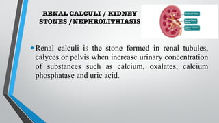
This document discusses renal calculi (kidney stones), including their incidence, causes, risk factors, types, clinical manifestations, diagnosis, and management. It provides an overview of the different types of kidney stones such as calcium, struvite, uric acid and cystine stones. Diagnostic tests including imaging, blood tests and urine analysis are used to identify stones and determine their composition. Treatment involves pain relief, increasing fluid intake, preventing infections, and sometimes surgical procedures if stones do not pass spontaneously. Nursing care focuses on relieving pain, ensuring adequate hydration and output, and educating patients on preventing future stone recurrences.
Kidney stone disease, also known as urolithiasis, is when a solid piece of material (kidney stone) occurs in the urinary tract. Kidney stones typically form in the kidney and leave the body in the urine stream. A small stone may pass without causing symptoms.
Benign prostatic hyperplasia is an enlargement of the prostate gland resulting from an increase in the number of epithelial cells and stromal tissue and developing upward into the bladder and obstructing the outflow of urine.
Kidney stones (also called renal calculi, nephrolithiasis or urolithiasis) are hard deposits made of minerals and salts that form inside your kidneys. Diet, excess body weight, some medical conditions, and certain supplements and medications are among the many causes of kidney stones.
Benign prostatic hyperplasia (BPH), also called prostate enlargement, is a noncancerous increase in size of the prostate gland. Symptoms may include frequent urination, trouble starting to urinate, weak stream, inability to urinate, or loss of bladder control.
definition of hydronephrosis,
causes and types of hydronephrosis
pathophysiology of hydronephrosis
clinical manifestation and diagnostic test for hydronephrosis
management
continuation on the urinary tract disorders. congenital and acquired disorders well covered. pyelonephritis also forms part of the text. thanks for reading. remeber to like and follow
Nephrolithiasis is the term employed for kidney stones, also known as renal calculi, and they are crystal concretions formed typically in the kidney. Calculi typically form in the kidneys and ideally leave the body via the urethra without pain. Larger stones are painful and may need surgical intervention
Urolithiasis Is the process of forming stones in the kidney, bladder or urethra
Kidney stones(calculi) are formed of minerals deposits commonly calcium oxalate and calcium phosphate; however uric acid, struvite and cystine are also calculus formers.
Nephrolithiasis refers to stones (calculi) in kidney when urinary concentration of substances such as calcium oxalate, calcium phosphate and uric acid increases, but they can form in or migrate to the lower urinary system. They are typically asymptomatic until they pass into the lower urinary tract.
Kidney Stones Causes, Symptoms and treatment options.pptxSaket Narnoli
Kidney stones, often caused by diet and dehydration, lead to intense pain, blood in urine, and urinary issues. Treatment varies from hydration and pain relief to surgical procedures.
Nephrolithiasis, commonly known as kidney stones, refers to the formation of hard mineral and salt deposits within the kidneys or urinary tract. These stones can vary in size, ranging from tiny grains to larger, more substantial formations. Nephrolithiasis is a relatively common condition and can affect people of all ages, although it is more prevalent in adults.
RENAL CALCULUS AETIOLOGY
Males- radio-opaque gall stones
Females - Radiolucent gall stones
Diet:Vitamin A deficiency
it causes desquamation of epithelium
which acts as a nidus for stone formation.
Climate:
In hot climate urinary solutes will increase with decrease in colloids,
Navigating the Health Insurance Market_ Understanding Trends and Options.pdfEnterprise Wired
From navigating policy options to staying informed about industry trends, this comprehensive guide explores everything you need to know about the health insurance market.
R3 Stem Cells and Kidney Repair A New Horizon in Nephrology.pptxR3 Stem Cell
R3 Stem Cells and Kidney Repair: A New Horizon in Nephrology" explores groundbreaking advancements in the use of R3 stem cells for kidney disease treatment. This insightful piece delves into the potential of these cells to regenerate damaged kidney tissue, offering new hope for patients and reshaping the future of nephrology.
Defecation
Normal defecation begins with movement in the left colon, moving stool toward the anus. When stool reaches the rectum, the distention causes relaxation of the internal sphincter and an awareness of the need to defecate. At the time of defecation, the external sphincter relaxes, and abdominal muscles contract, increasing intrarectal pressure and forcing the stool out
The Valsalva maneuver exerts pressure to expel faeces through a voluntary contraction of the abdominal muscles while maintaining forced expiration against a closed airway. Patients with cardiovascular disease, glaucoma, increased intracranial pressure, or a new surgical wound are at greater risk for cardiac dysrhythmias and elevated blood pressure with the Valsalva maneuver and need to avoid straining to pass the stool.
Normal defecation is painless, resulting in passage of soft, formed stool
CONSTIPATION
Constipation is a symptom, not a disease. Improper diet, reduced fluid intake, lack of exercise, and certain medications can cause constipation. For example, patients receiving opiates for pain after surgery often require a stool softener or laxative to prevent constipation. The signs of constipation include infrequent bowel movements (less than every 3 days), difficulty passing stools, excessive straining, inability to defecate at will, and hard feaces
IMPACTION
Fecal impaction results from unrelieved constipation. It is a collection of hardened feces wedged in the rectum that a person cannot expel. In cases of severe impaction the mass extends up into the sigmoid colon.
DIARRHEA
Diarrhea is an increase in the number of stools and the passage of liquid, unformed feces. It is associated with disorders affecting digestion, absorption, and secretion in the GI tract. Intestinal contents pass through the small and large intestine too quickly to allow for the usual absorption of fluid and nutrients. Irritation within the colon results in increased mucus secretion. As a result, feces become watery, and the patient is unable to control the urge to defecate. Normally an anal bag is safe and effective in long-term treatment of patients with fecal incontinence at home, in hospice, or in the hospital. Fecal incontinence is expensive and a potentially dangerous condition in terms of contamination and risk of skin ulceration
HEMORRHOIDS
Hemorrhoids are dilated, engorged veins in the lining of the rectum. They are either external or internal.
FLATULENCE
As gas accumulates in the lumen of the intestines, the bowel wall stretches and distends (flatulence). It is a common cause of abdominal fullness, pain, and cramping. Normally intestinal gas escapes through the mouth (belching) or the anus (passing of flatus)
FECAL INCONTINENCE
Fecal incontinence is the inability to control passage of feces and gas from the anus. Incontinence harms a patient’s body image
PREPARATION AND GIVING OF LAXATIVESACCORDING TO POTTER AND PERRY,
An enema is the instillation of a solution into the rectum and sig
CHAPTER 1 SEMESTER V PREVENTIVE-PEDIATRICS.pdfSachin Sharma
This content provides an overview of preventive pediatrics. It defines preventive pediatrics as preventing disease and promoting children's physical, mental, and social well-being to achieve positive health. It discusses antenatal, postnatal, and social preventive pediatrics. It also covers various child health programs like immunization, breastfeeding, ICDS, and the roles of organizations like WHO, UNICEF, and nurses in preventive pediatrics.
How many patients does case series should have In comparison to case reports.pdfpubrica101
Pubrica’s team of researchers and writers create scientific and medical research articles, which may be important resources for authors and practitioners. Pubrica medical writers assist you in creating and revising the introduction by alerting the reader to gaps in the chosen study subject. Our professionals understand the order in which the hypothesis topic is followed by the broad subject, the issue, and the backdrop.
https://pubrica.com/academy/case-study-or-series/how-many-patients-does-case-series-should-have-in-comparison-to-case-reports/
CHAPTER 1 SEMESTER V - ROLE OF PEADIATRIC NURSE.pdfSachin Sharma
Pediatric nurses play a vital role in the health and well-being of children. Their responsibilities are wide-ranging, and their objectives can be categorized into several key areas:
1. Direct Patient Care:
Objective: Provide comprehensive and compassionate care to infants, children, and adolescents in various healthcare settings (hospitals, clinics, etc.).
This includes tasks like:
Monitoring vital signs and physical condition.
Administering medications and treatments.
Performing procedures as directed by doctors.
Assisting with daily living activities (bathing, feeding).
Providing emotional support and pain management.
2. Health Promotion and Education:
Objective: Promote healthy behaviors and educate children, families, and communities about preventive healthcare.
This includes tasks like:
Administering vaccinations.
Providing education on nutrition, hygiene, and development.
Offering breastfeeding and childbirth support.
Counseling families on safety and injury prevention.
3. Collaboration and Advocacy:
Objective: Collaborate effectively with doctors, social workers, therapists, and other healthcare professionals to ensure coordinated care for children.
Objective: Advocate for the rights and best interests of their patients, especially when children cannot speak for themselves.
This includes tasks like:
Communicating effectively with healthcare teams.
Identifying and addressing potential risks to child welfare.
Educating families about their child's condition and treatment options.
4. Professional Development and Research:
Objective: Stay up-to-date on the latest advancements in pediatric healthcare through continuing education and research.
Objective: Contribute to improving the quality of care for children by participating in research initiatives.
This includes tasks like:
Attending workshops and conferences on pediatric nursing.
Participating in clinical trials related to child health.
Implementing evidence-based practices into their daily routines.
By fulfilling these objectives, pediatric nurses play a crucial role in ensuring the optimal health and well-being of children throughout all stages of their development.
QA Paediatric dentistry department, Hospital Melaka 2020Azreen Aj
QA study - To improve the 6th monthly recall rate post-comprehensive dental treatment under general anaesthesia in paediatric dentistry department, Hospital Melaka
Global launch of the Healthy Ageing and Prevention Index 2nd wave – alongside...ILC- UK
The Healthy Ageing and Prevention Index is an online tool created by ILC that ranks countries on six metrics including, life span, health span, work span, income, environmental performance, and happiness. The Index helps us understand how well countries have adapted to longevity and inform decision makers on what must be done to maximise the economic benefits that comes with living well for longer.
Alongside the 77th World Health Assembly in Geneva on 28 May 2024, we launched the second version of our Index, allowing us to track progress and give new insights into what needs to be done to keep populations healthier for longer.
The speakers included:
Professor Orazio Schillaci, Minister of Health, Italy
Dr Hans Groth, Chairman of the Board, World Demographic & Ageing Forum
Professor Ilona Kickbusch, Founder and Chair, Global Health Centre, Geneva Graduate Institute and co-chair, World Health Summit Council
Dr Natasha Azzopardi Muscat, Director, Country Health Policies and Systems Division, World Health Organisation EURO
Dr Marta Lomazzi, Executive Manager, World Federation of Public Health Associations
Dr Shyam Bishen, Head, Centre for Health and Healthcare and Member of the Executive Committee, World Economic Forum
Dr Karin Tegmark Wisell, Director General, Public Health Agency of Sweden
1. RENAL CALCULI AND ITS
MANAGEMENT
Presented by
Karan choudhary
CON AIIMS JODHPUR
2. INTRODUCTION
•A kidney stone, is a solid concentration or crystal
aggregation formed in the kidneys from dietary
minerals in the urine.
•A calculus often called a stone, is a concretion of
material, usually mineral salts, that forms in an organ
or duct of the body.
•Formation of calculi is known as lithiasis which can
cause a number of medical conditions.
•Urinary stones are typically classified by their location
in three types.
3. RENAL CALCULI / KIDNEY
STONES /NEPHROLITHIASIS
•Renal calculi is the stone formed in renal tubules,
calyces or pelvis when increase urinary concentration
of substances such as calcium, oxalates, calcium
phosphatase and uric acid.
4. INCIDENCE AND PREVALENCE
•More common in men than in women.
•In India it most commonly occurs in the ‘stone belt’.
(Maharashtra, Gujarat, Rajasthan, Punjab, Haryana,
Delhi, Madhya Pradesh, Bihar, and West Bengal)
•The incidence is higher in persons with family history
of stone formation.
•Most common in summer months
•The incidence is higher in persons with family history
of stone formation.
5. ETIOLOGY AND RISK FACTORS
•Infection
•Urinary stasis
•Periods of immobility
•With inflammatory bowel disease and with an
ileostomy or bowel resection.
•Some medications also cause stones e.g., antacids,
acetazolamide vitamin D.
•Urine PH and solute load affect the formation of
stones.
6. Continued…
•Increased calcium concentrations in blood and urine
promote precipitation of calcium and formation of
stones (about 75% of all renal stones are calcium-
based).
•Hyperparathyroidism
•Tumour of parathyroid gland
•Renal tubular acidosis
•Cancers (multiple myeloma)
•Excessive intake of milk
7. Continued…
•Granulomatous diseases (sarcoidosis, tuberculosis),
which may cause increased vitamin D production by
the granulomatous tissue.
•Fracture and prolonged immobilization due to
resorption of bone
•Large intake of dietary protein and calcium
•Excessive amount of tea and fruit juices that elevate
the urinary oxalate levels
•Prolong catheterisation
8. TYPES OF URINARY STONES
•There are five major categories of stones:
1. Calcium stone
2. Struvite stone (Triple phosphate stone):-
3. Uric acid stones
4. Cystine stones
5. Other e.g., Xanthine etc.
9. CALCIUM STONES
•It is the most common 80% .
•Alkaline in nature so urine ph is
alkaline and risk of UTI.
•Irregular in shape.
•Covered with sharp projections,
which cause bleeding.
10. URIC ACID STONES
•About 8% of renal stones
contain uric acid.
•Caused by excessive dietary
purine or gout patient.
•Red to yellow colour
•Acidic in nature so urine ph
is acidic in nature
11. STRUVITE STONE
•About 10% of all the renal stones
contain magnesium ammonium
phosphate
•Associated with chronic urinary tract
infection.
•Caused by urea splitting bacteria
(Proteus, Pseudomonas,
Staphylococcus)
•Alkaline in nature.
•more common women then the man
12. CYSTEINE STONES
• A rare type of kidney stone.
•Stones made up of chemical called cysteine.
•Caused by excessive consumption of methionine
(essential amino acid )and cystine (non essential AA)
•Acidic in nature.
14. CLINICAL MANIFESTATIONS
•Pain (pain pattern depend on location of obstruction ) -
MC symptom in 75% of people.
•It may be worse on movement, particularly on
climbing stairs.
•sharp pain in the flank area, back or lower abdomen.
•Pyuria
•Nausea
•vomiting
15. CONTD,,,
•Sleep disturbance and discomfort.
•Cloudy or foul smelling urine
•Fever or chills if infection present
•Abdominal discomfort
•Haematuria
•Urinating small amount of urine
16. ASSESSMENT AND DIAGNOSTIC FINDINGS
History and Physical examination
•Dietary, medication and family history.
•patient drinking enough?
•Profession, enquire about UTI
•Long illness bedridden patient
•Abdomen tense and rigid
•Tenderness present
17. Diagnosis is confirmed by
•X-ray films of the kidneys, ureter, and bladder (KUB)
•Non contrast spiral CT called CT/KUB commonly
used in renal colic patient.
•Ultrasonography
•Intravenous urography or pyelogram (IVP)
•Retrograde pyelography.
18. Conted,,
•Blood chemistries - serum calcium, phosphorus,
sodium, potassium, bicarbonate, uric acid, BUN, and
creatinine levels.
•24-hour urine test - for measurement of calcium,
uric acid, creatinine, sodium, pH, and total volume.
•Chemical analysis is carried out to determine their
composition when stones are recovered.
19. MEDICAL MANAGEMENT
The basic goals of management are:
•The immediate objective of treatment of renal or
ureteral colic is to relieve the pain until its cause can
be eliminated.
•To eradicate the stone
•To determine the stone type
•to prevent nephron destruction
•To control infection
•To relieve any obstruction that may be present.
20. PHARMACOLOGICAL MANAGEMENT
1. Analgesic:
• Narcotic analgesic: Morphine sulphate is DOC for parenteral use.
• Opioid analgesics: to prevent shock and syncope (resulting from the
excruciating pain).
⎯Note: Most stones are 4mm or less in size and will probably pass
spontaneously.
2. Alpha-adrenergic blockers: by relaxing the smooth muscles in the
ureter, can be used to facilitate stone passage. E.g., Tamsulosin or
terazosin
3. NSAIDs - specific pain relief by inhibiting the synthesis of
prostaglandin E.
5. Antiemetic agent -(metoclopramide)
21. Dietary Recommendations for Prevention of
Kidney Stones
•Restricting protein to 60 g/day is recommended to
decrease urinary excretion of calcium and uric acid.
•Oxalate-containing foods (spinach, strawberries, tea,
peanuts, and wheat bran) may be restricted.
•Restrict calcium in the diet (when stones are clearly
due to excess dietary calcium)
•Liberal fluid intake along with dietary restriction of
protein and sodium.
•Increase fluid intake.
22. SURGICAL MANAGEMENT
•Indications:
•Stones too large for spontaneous passage (usually
greater than 7 mm).
•Stones associated with symptomatic infection.
•Stones causing impaired renal function.
•Stones causing persistent pain or worsening of
symptoms.
•Patient with only one kidney.
23. Ureteroscopy
•It involves first visualizing the
stone and then destroying it.
•A stent may be inserted and
left in place for 48 hours or
more after the procedure to
keep the ureter patent.
•Hospital stays are generally
brief, and some patients can be
treated as outpatients.
24. ESWL(extracorporeal shockwave lithotripsy)
•It is a non-invasive procedure to break up stones in the
calyx of the kidney.
•After the stones are fragmented to the size of grains of
sand, the remnants of the stones are spontaneously
voided.
•An average treatment comprises between 1,000 and
3,000 shocks.
•The patient is observed for obstruction and infection
resulting from blockage of the urinary tract by stone
fragments.
25.
26. Percutaneous nephrostomy or a percutaneous
nephrolithotomy
•A nephroscope is introduced through the dilated
percutaneous tract into the renal parenchyma and the
stone may be extracted with forceps or by a stone
retrieval basket.
•Small stone fragments and stone dust are irrigated and
suctioned out of collecting system.
•Larger stones may be further reduced by ultrasonic
disintegration and then removed with forceps or a
stone retrieval basket.
29. NURSING MANAGEMENT
•Nursing Assessment
•Assess for pain and discomfort as well as nausea,
vomiting, diarrhoea, and abdominal distention.
•Severity, location and radiation of pain are determined.
•Symptoms of UTI (chills, fever, dysuria, frequency, and
hesitancy) and obstruction (frequent urination of small
amounts, oliguria, or anuria).
•See for Haematuria
•Precipitating factors: episodes of dehydration, prolonged
immobilization, and infection.
30. NURSING DIAGNOSES
•Acute pain related to inflammation, obstruction, and abrasion of the
urinary tract.
•Impaired Urinary Elimination related to stimulation of the bladder
by calculi, renal or ureteral irritation, mechanical obstruction and
inflammation as evidenced by urgency and frequency; oliguria
(retention); haematuria.
•Disturbed sleep pattern related to acute pain as manifested by
decreased sleep duration.
•Risk of deficient fluid volume related to nausea and vomiting as
manifested by decreased appetite and fatigue.
•Deficient knowledge regarding prevention of recurrence of renal
stones.
31. •Relieving pain
•Opioid analgesic agents (intravenous or intramuscular
administration may be prescribed to provide rapid
relief) or NSAIDs for immediate relief.
•The patient is encouraged and assisted to assume a
position of comfort.
•If activity brings some pain relief, the patient is assisted
to ambulate.
•The pain level is monitored closely, and increases in
severity are reported promptly to the physician so that
relief can be provided and additional treatment initiated.
•
32. PREVENTING RECURRENT KIDNEY STONE
•Follow prescribed diet closely.
•During the day, drink fluids enough to excrete greater
than 2,000 mL of urine every 24 hours (preferably 3,000
to 4,000 mL) to assist in the passage of stone fragments.
•Avoid sudden increases in environmental temperatures,
which may cause excessive sweating, dehydration and
fall in urinary volume.
•Avoid activities that cause excessive sweating and
dehydration.
33. SUMMARY
•Today we have discussed introduction, definition of
urolithiasis and nephrolithiasis, their incidences and
causes /risk factors. Classification/ Types of urinary
stones, its pathophysiology, clinical manifestations,
assessment and diagnostic findings, medical,
surgical and nursing management, potential
complications and prevention of kidney stones.
34. CONCLUSION
•A calculus (plural calculi), often called a stone, is a
concretion of material, usually mineral salts, that
forms in an organ or duct of the body. These are
common and recur frequently. Nurse should educate
the preventive strategies to the patient such as drinking
adequate water and avoiding dehydration and high
temperature areas.
35. Reference
•Brunner & Suddarths. Textbook of Medical Surgical Nursing, 13th Edition
Volume -2 .New Delhi: Wolters Kluwer; 2015;
•Black.M.J, Haawks.J.H. Medical Surgical Nursing. VoII. 7th Edition. New
delhi Saunders publication.
•Lewis. S. Heitkemper M. Medical surgical Nursing. 6th edition. Missouri:
Elseveir’s publications.
•Li DF, Gao YL, Liu HC, Huang XC, Zhu RF, Zhu CT. Use of thiazide
diuretics for the prevention of recurrent kidney calculi: a systematic
review and meta-analysis. J Transl Med. 2020 Feb 28;18(1):106. doi:
10.1186/s12967-020-02270-7.
•Rodgers AL, Siener R. The Efficacy of Polyunsaturated Fatty Acids as
Protectors against Calcium Oxalate Renal Stone Formation: A Review.
Nutrients. 2020 Apr 12;12(4). pii: E1069. doi: 10.3390/nu12041069.