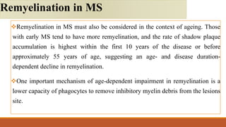
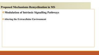
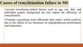
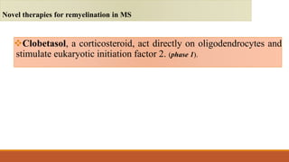
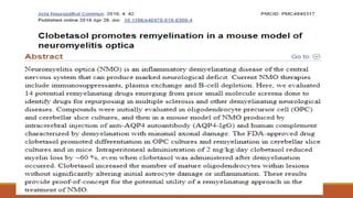
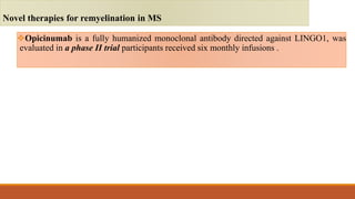
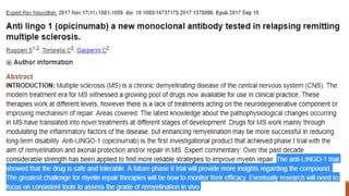
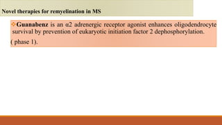
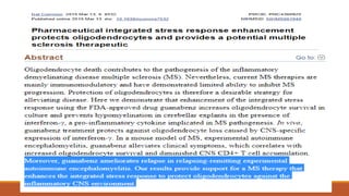
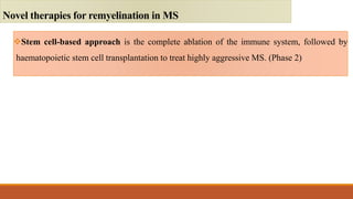
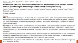
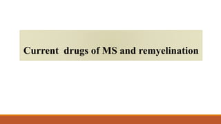
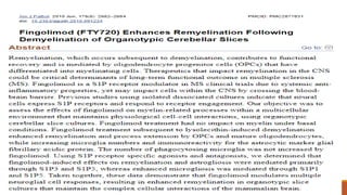
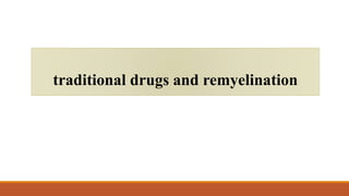
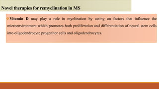
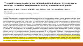
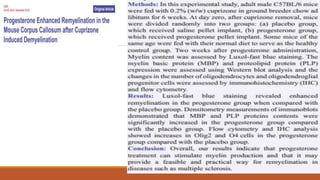
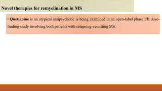
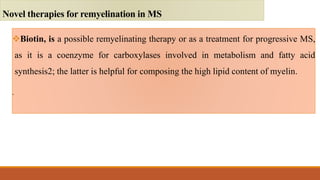
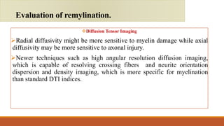
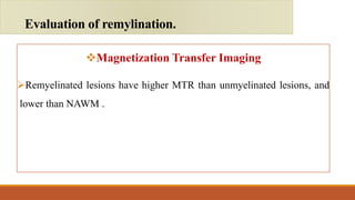
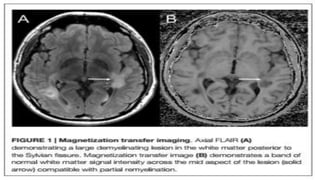
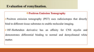
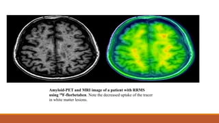
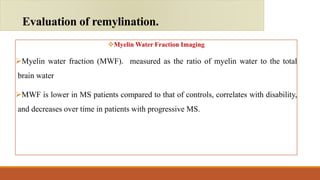
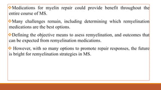
3. Multiple sclerosis (MS) is an immune-mediated disorder of
the central nervous system that results in destruction of the
myelin sheath that surrounds axons.
Introduction
4. Remyelination can restore neuronal function and prevent further
neuronal loss and clinical disability.
Molecular and cellular mechanisms regulating myelination, resulted in
identification of agents that enhance myelination, and therapies used in
preclinical and early clinical development.
Introduction
5. There are many important factors must be considered to effectively translate
remyelination therapies into clinical reality:
Developing validated biomarkers to measure myelin status.
Selecting of the most appropriate form of MS to test remyelination therapies.
Determining the most effective period to use a therapeutic for remyelination
(for example, early after a relapse or during stable disease in relapsing–
remitting MS).
Introduction
7. Myelination begins with oligodendrocyte progenitor cells (OPCs)
differentiating into oligodendrocytes, followed by their maturation
into myelinating oligodendrocytes.
Following white matter injury, neighboring OPCs proliferate and
migrate towards the site of injury or demyelination.
The process of myelination is subject to both positive and negative
regulation
Development of myelin
10. As oligodendrocytes are metabolically coupled to axons, so after
demyelination this loss of support might prime axons to degenerate.
The increase in the number of Na+ channels along the length of
the axon that occurs upon demyelination elevates the energy
demands on the axon, and reverse the Na+/Ca2+ exchanger,
resulting in toxic levels of Ca+ in the axon.
Consequence of demyelination
12. On the basis of histological assessments in patients with MS,
remyelination in humans is highly variable: considerable in some
cases, and absent in others.
In tissue sections, the hallmark of remyelination in MS is a shadow plaque,
because these lesions possess an amount of lipid staining that is intermediate
between the normal white matter and a demyelinated plaque.
Remyelination in MS
13.
14.
15.
16. Remyelination in MS must also be considered in the context of ageing. Those
with early MS tend to have more remyelination, and the rate of shadow plaque
accumulation is highest within the first 10 years of the disease or before
approximately 55 years of age, suggesting an age- and disease duration-
dependent decline in remyelination.
One important mechanism of age-dependent impairment in remyelination is a
lower capacity of phagocytes to remove inhibitory myelin debris from the lesions
site.
Remyelination in MS
21. Number of adult OPCs available for remyelination is depleted over
time.
Disruptions to the blood–brain barrier, leads to aberrant deposition
of extracellular matrix (ECM) components, including fibronectin,
hyaluronic acid (HA), and chondroitin sulfate proteoglycans
(CSPGs), which can block the differentiation of OPCs and
premyelinating oligodendrocytes
Causes of remyeleination failure in MS
22. Demyelination can expose OPCs inhibitory cues, including
components of damaged myelin such as the proteins MAG (myelin-
associated glycoprotein), OMgp (oligodendrocyte myelin
glycoprotein), and and LINGO-1 (leucine-rich repeat- and Ig
domain-containing Nogo receptor-interacting protein 1) to inhibit
oligodendrocyte differentiation and remyelination.
Causes of remyileination failure in MS
23. several non-disease-related factors such as age, sex, diet, and
individual genetic background can also impact the efficiency of
remyelination.
Females remyelinate more efficiently than males, which could be
due to the effects of sex hormones on oligodendrocyte proliferation
and maturation .
Causes of remyileination failure in MS
25. Clobetasol, a corticosteroid, act directly on oligodendrocytes and
stimulate eukaryotic initiation factor 2. (phase 1).
Novel therapies for remyelination in MS
26.
27. Opicinumab is a fully humanized monoclonal antibody directed against LINGO1, was
evaluated in a phase II trial participants received six monthly infusions .
Novel therapies for remyelination in MS
28.
29. Guanabenz is an α2 adrenergic receptor agonist enhances oligodendrocyte
survival by prevention of eukaryotic initiation factor 2 dephosphorylation.
( phase 1).
Novel therapies for remyelination in MS
30.
31. Olesoxime, is a cholesterol-oxime compound and mitochondrial pore
modulator, accelerates oligodendrocyte maturation and enhanced myelination
in vitro and in vivo. (Phase 1)
Novel therapies for remyelination in MS
32.
33. Blockade of ASIC1 through amiloride, a potassium-sparing diuretic that
showed neuroprotective and myeloprotective effects in experimental models
of MS ( clinical trials)
Novel therapies for remyelination in MS
34.
35. Stem cell-based approach is the complete ablation of the immune system, followed by
haematopoietic stem cell transplantation to treat highly aggressive MS. (Phase 2)
Novel therapies for remyelination in MS
41. Vitamin D may play a role in myelination by acting on factors that influence the
microenvironment which promotes both proliferation and differentiation of neural stem cells
into oligodendrocyte progenitor cells and oligodendrocytes.
Novel therapies for remyelination in MS
42.
43. Thyroid hormone can induce more OPCs from neural stem cells (NSCs), and promote the
differentiation and myelination of OPCs
Novel therapies for remyelination in MS
44.
45.
46. Tamoxifen, improved oligodendrocyte maturation and accelerated remyelination even in
the presence of inhibitory myelin debris.
Novel therapies for remyelination in MS
47.
48. Quetiapine is an atypical antipsychotic is being examined in an open-label phase I/II dose-
finding study involving both patients with relapsing–remitting MS.
Novel therapies for remyelination in MS
49.
50. Biotin, is a possible remyelinating therapy or as a treatment for progressive MS,
as it is a coenzyme for carboxylases involved in metabolism and fatty acid
synthesis2; the latter is helpful for composing the high lipid content of myelin.
.
Novel therapies for remyelination in MS
54. Diffusion Tensor Imaging
Radial diffusivity might be more sensitive to myelin damage while axial
diffusivity may be more sensitive to axonal injury.
Newer techniques such as high angular resolution diffusion imaging,
which is capable of resolving crossing fibers and neurite orientation
dispersion and density imaging, which is more specific for myelination
than standard DTI indices.
Evaluation of remylination.
55. Images in 35 years old female RRMS patient; (a) axial view FLAIR MRI shows bilateral peri-ventricular WM lesions (the
largest one on left side shows black holes). Suggestive of multifocal white matter disease. (b) ROIs for the evaluation of
tracts in MS white matter lesions. (c and d) Fiber tractography; shows educed number of fibers when they traverse white
matter lesions and cross-sectional area of the CST (green) on the affected side (FA at fibers measures 0.43 while MD 0.90).
58. Positron Emission Tomography
Positron emission tomography (PET) uses radioisotopes that directly
bind to different tissue substrates to enable molecular imaging.
18F-florbetaben derivative has an affinity for CNS myelin and
demonstrates differential binding to normal and demyelinated white
matter.
Evaluation of remylination.
59. Amyloid-PET and MRI image of a patient with RRMS
using 18F-florbetaben. Note the decreased uptake of the tracer
in white matter lesions.
60. Myelin Water Fraction Imaging
Myelin water fraction (MWF). measured as the ratio of myelin water to the total
brain water
MWF is lower in MS patients compared to that of controls, correlates with disability,
and decreases over time in patients with progressive MS.
Evaluation of remylination.
61. T2-weighted image (left), axial and sagittal Z-score map of
MWF values (middle) and histogram of MWF values (right)
for three PPMS patients.
PPMS Patient A had an EDSS of 1.5, Patient B had an
EDSS of 5.5 and Patient C had an EDSS of 6.5
63. Medications for myelin repair could provide benefit throughout the
entire course of MS.
Many challenges remain, including determining which remyelination
medications are the best options.
Defining the objective means to asess remyelination, and outcomes that
can be expected from remyelination medications.
However, with so many options to promote repair responses, the future
is bright for remyelination strategies in MS.