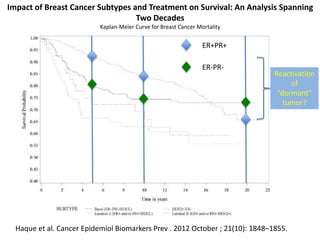
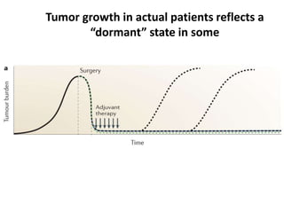
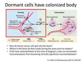
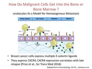
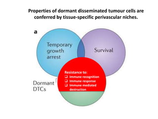
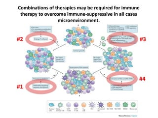
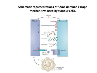
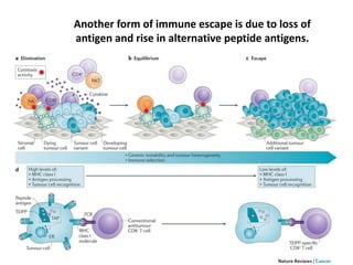
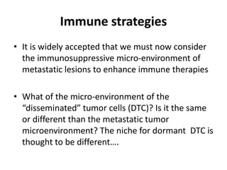
Tumor Dormancy
H. Kim Lyerly, M.D., F.A.C.S.
Duke University
Impact of Breast Cancer Subtypes and Treatment on Survival: An Analysis Spanning
Two Decades
Kaplan-Meier Curve for Breast Cancer Mortality
Haque et al. Cancer Epidemiol Biomarkers Prev . 2012 October ; 21(10): 1848–1855.
ER+PR+
ER-PR-
Reactivation
of
“dormant”
tumor?
Tumor growth in actual patients reflects a
“dormant” state in some
C L Chaffer, R A Weinberg Science 2011;331:1559-1564
Dormant cells have colonized body
• How do breast cancer cells get into the bone?
• Where in the bone do they reside during the years prior to relapse?
• If the have colonized bone at the time of diagnosis, how can we prevent
micrometastases from becoming clinically significant macrometastases?
How Do Malignant Cells Get Into the Bone or
Bone Marrow ?
Leukocytes As a Model for Hematogenous Metastasis
• Breast cancer cells express multiple E-selectin ligands
• They express CXCR4; CXCR4 expression correlates with late
relapse (Price et al., Sci Trans Med 2016)
Adapted from Immunobiology, 5th Ed., Janeway et al.
Models of dormancy control
Tumor cells evade
the immune system
and expand:
 Not recognized
 Immune system
is modulated
 Tumor cells
resistant to
immune killing
GROWTH BALANCED
BY APOPTOSIS
GROWTH LIMITED
BY ANGIOGENISIS
GROWTH LIMITED
BY IMMUNE SYSTEM
Disseminated cells had two types of
“dormancy”- “cellular” and “population”
Goss and Chambers, Nat Rev Cancer 2010
CELLULAR
DORMANCY
POPULATION
(of cells)
DORMANCY
DISSEMINATED METASTATIC
Parallels between stem cell regulation & regulation of dormant
disseminated tumour cells by the perivascular niche.
Properties of dormant disseminated tumour cells are
conferred by tissue-specific perivascular niches.
E-Selectin and SDF-1 are Expressed by the
Bone Marrow Vasculature in Discrete,
Sinusoidal and Peri-Sinusoidal Regions
Sipkins, et al. Nature 2005
Blood pool
(LMW
Dextrans)
E-selectin
BCCs Enter Bone Through the BM
Sinusoidal Vasculature
Patient Bone Marrow Biopsies Show
Micrometastatic Disease is “Dormant” and
Located in SDF-1+ Sinusoidal Niches
Properties of dormant disseminated tumour cells are
conferred by tissue-specific perivascular niches.
Resistance to:
 Immune recognition
 Immune response
 Immune mediated
destruction
Antigens (what is recognized by the immune system) are generated from
degraded proteins and presented on cell surface to be recognized by T cells
Tumor cell or
cell infected
with a virus
Antigen
Presenting Cell,
(A Normal
Component Of
The Immune
System)
Cytotoxic T Cell Helper T Cell
Both antigen and co-stimulation are required to
elicit an immune response
Molecular Mechanisms of Down Modulation
Combinations of therapies may be required for immune
therapy to overcome immune-suppressive in all cases
microenvironment.
#1
#2 #3
#4
Schematic representations of some immune escape
mechanisms used by tumour cells.
Another form of immune escape is due to loss of
antigen and rise in alternative peptide antigens.
Immune strategies
• It is widely accepted that we must now consider
the immunosuppressive micro-environment of
metastatic lesions to enhance immune therapies
• What of the micro-environment of the
“disseminated” tumor cells (DTC)? Is it the same
or different than the metastatic tumor
microenvironment? The niche for dormant DTC is
thought to be different….
E0771 mammary tumor cells in C57/BL6 mice model to
probe DTC interactions with engineered T cells in vivo.
• A model of breast cancer that allows expression a
defined antigen in tumor cells, implantation of
tumor cells in immune competent mice, and
analysis of antigen specific T cells recognizing,
interacting, and eliminating DTCs derived from
the tumor.
• OVA (a model antigen) and all associated
reagents have been developed in C57/BL6 mice,
so we determined that the C57/BL6-derived
mammary cancer line E0771 generates
detectable DTCs and could be used as a model.
The E0771 Model of dormant DTC
B. BM whole mounts generated from femurs of these mice, stained, and imaged to identify
dormant (vimentin-positive, Ki67-negative) DTCs (ii, iii). Vimentin-positive cells are not
present in the marrow of uninoculated, control mice (i; scale bar = 20μm).
C. DTCs present in BM of inoculated mice can reconstitute tumors, as they form luciferase-
positive colonies once isolated from BM and expanded in 3D culture.
A. Hundreds of GFP-luc E0771 cells detected in BM flushed from femur of C57/BL6 mice
inoculated with E0771 cells in the mammary fat pad (MFP) 4-weeks prior.
OVA is detected by qPCR specifically in
E0771 mammary tumor cells engineered
to specifically express the antigen
ovalbumin (OVA) and not in wild-type
control cells.
OT1 cells eliminate OVA-expressing DTCs
isolated from bone marrow of mice
inoculated with E0771-OVA cells in
culture. Increasing the ratio of OT1 cells:E0771
DTCs isolated from the bone marrow of E0771-OVA
inoculated C57/BL6 mice results in enhanced
cytotoxicity. This demonstrates the feasibility of using
the OVA system to functionally probe DTC-T cell
interactions.
The E0771 OVA Model of Immune
Recognition
Future Objectives
• Metastatic cancer remains a clinical challenge.
• Can we understand how the DTC escape
immune recognition and elimination?
• Can we develop a non-toxic strategy to
eliminate DTC from residing in the body?
• Can we develop a non-toxic therapy to
prevent DTC from developing into clinically
relevant metastasis?
Thanks
Zach Hartman, Ph.D.
Duke University
Dorothy Sipkins, M.D., Ph.D.
Duke University
Cyrus M. Ghajar, Ph.D.
Fred Hutchinson Cancer Research Center

Tumor Dormancy

  • 1.
    Tumor Dormancy H. KimLyerly, M.D., F.A.C.S. Duke University
  • 2.
    Impact of BreastCancer Subtypes and Treatment on Survival: An Analysis Spanning Two Decades Kaplan-Meier Curve for Breast Cancer Mortality Haque et al. Cancer Epidemiol Biomarkers Prev . 2012 October ; 21(10): 1848–1855. ER+PR+ ER-PR- Reactivation of “dormant” tumor?
  • 3.
    Tumor growth inactual patients reflects a “dormant” state in some
  • 4.
    C L Chaffer,R A Weinberg Science 2011;331:1559-1564 Dormant cells have colonized body • How do breast cancer cells get into the bone? • Where in the bone do they reside during the years prior to relapse? • If the have colonized bone at the time of diagnosis, how can we prevent micrometastases from becoming clinically significant macrometastases?
  • 5.
    How Do MalignantCells Get Into the Bone or Bone Marrow ? Leukocytes As a Model for Hematogenous Metastasis • Breast cancer cells express multiple E-selectin ligands • They express CXCR4; CXCR4 expression correlates with late relapse (Price et al., Sci Trans Med 2016) Adapted from Immunobiology, 5th Ed., Janeway et al.
  • 6.
    Models of dormancycontrol Tumor cells evade the immune system and expand:  Not recognized  Immune system is modulated  Tumor cells resistant to immune killing GROWTH BALANCED BY APOPTOSIS GROWTH LIMITED BY ANGIOGENISIS GROWTH LIMITED BY IMMUNE SYSTEM
  • 7.
    Disseminated cells hadtwo types of “dormancy”- “cellular” and “population” Goss and Chambers, Nat Rev Cancer 2010 CELLULAR DORMANCY POPULATION (of cells) DORMANCY DISSEMINATED METASTATIC
  • 8.
    Parallels between stemcell regulation & regulation of dormant disseminated tumour cells by the perivascular niche.
  • 9.
    Properties of dormantdisseminated tumour cells are conferred by tissue-specific perivascular niches.
  • 11.
    E-Selectin and SDF-1are Expressed by the Bone Marrow Vasculature in Discrete, Sinusoidal and Peri-Sinusoidal Regions Sipkins, et al. Nature 2005 Blood pool (LMW Dextrans) E-selectin
  • 12.
    BCCs Enter BoneThrough the BM Sinusoidal Vasculature
  • 13.
    Patient Bone MarrowBiopsies Show Micrometastatic Disease is “Dormant” and Located in SDF-1+ Sinusoidal Niches
  • 14.
    Properties of dormantdisseminated tumour cells are conferred by tissue-specific perivascular niches. Resistance to:  Immune recognition  Immune response  Immune mediated destruction
  • 15.
    Antigens (what isrecognized by the immune system) are generated from degraded proteins and presented on cell surface to be recognized by T cells Tumor cell or cell infected with a virus Antigen Presenting Cell, (A Normal Component Of The Immune System) Cytotoxic T Cell Helper T Cell
  • 16.
    Both antigen andco-stimulation are required to elicit an immune response
  • 17.
    Molecular Mechanisms ofDown Modulation
  • 18.
    Combinations of therapiesmay be required for immune therapy to overcome immune-suppressive in all cases microenvironment. #1 #2 #3 #4
  • 19.
    Schematic representations ofsome immune escape mechanisms used by tumour cells.
  • 20.
    Another form ofimmune escape is due to loss of antigen and rise in alternative peptide antigens.
  • 21.
    Immune strategies • Itis widely accepted that we must now consider the immunosuppressive micro-environment of metastatic lesions to enhance immune therapies • What of the micro-environment of the “disseminated” tumor cells (DTC)? Is it the same or different than the metastatic tumor microenvironment? The niche for dormant DTC is thought to be different….
  • 22.
    E0771 mammary tumorcells in C57/BL6 mice model to probe DTC interactions with engineered T cells in vivo. • A model of breast cancer that allows expression a defined antigen in tumor cells, implantation of tumor cells in immune competent mice, and analysis of antigen specific T cells recognizing, interacting, and eliminating DTCs derived from the tumor. • OVA (a model antigen) and all associated reagents have been developed in C57/BL6 mice, so we determined that the C57/BL6-derived mammary cancer line E0771 generates detectable DTCs and could be used as a model.
  • 23.
    The E0771 Modelof dormant DTC B. BM whole mounts generated from femurs of these mice, stained, and imaged to identify dormant (vimentin-positive, Ki67-negative) DTCs (ii, iii). Vimentin-positive cells are not present in the marrow of uninoculated, control mice (i; scale bar = 20μm). C. DTCs present in BM of inoculated mice can reconstitute tumors, as they form luciferase- positive colonies once isolated from BM and expanded in 3D culture. A. Hundreds of GFP-luc E0771 cells detected in BM flushed from femur of C57/BL6 mice inoculated with E0771 cells in the mammary fat pad (MFP) 4-weeks prior.
  • 24.
    OVA is detectedby qPCR specifically in E0771 mammary tumor cells engineered to specifically express the antigen ovalbumin (OVA) and not in wild-type control cells. OT1 cells eliminate OVA-expressing DTCs isolated from bone marrow of mice inoculated with E0771-OVA cells in culture. Increasing the ratio of OT1 cells:E0771 DTCs isolated from the bone marrow of E0771-OVA inoculated C57/BL6 mice results in enhanced cytotoxicity. This demonstrates the feasibility of using the OVA system to functionally probe DTC-T cell interactions. The E0771 OVA Model of Immune Recognition
  • 25.
    Future Objectives • Metastaticcancer remains a clinical challenge. • Can we understand how the DTC escape immune recognition and elimination? • Can we develop a non-toxic strategy to eliminate DTC from residing in the body? • Can we develop a non-toxic therapy to prevent DTC from developing into clinically relevant metastasis?
  • 26.
    Thanks Zach Hartman, Ph.D. DukeUniversity Dorothy Sipkins, M.D., Ph.D. Duke University Cyrus M. Ghajar, Ph.D. Fred Hutchinson Cancer Research Center

Editor's Notes

  • #5 So, spread to the metastatic niche is a very early event in breast cancer, and micrometastases may develop very slowly over many years before overt clinical relapse in the bone occurs- a period of clinical tumor dormancy. BCCs are getting into and out of these metastatic niche from the peripheral circulation. How do they do this and where are they residing during those years?
  • #6 How could this specialized vasculature be playing a role in BCC bone metastasis? Well, BCCs express multiple E-selectin ligands as well as CXCR4. . .
  • #8 There is evidence that tumor cells can exist in distant organs in 2 other states Angiogenic dormancy (Folkman 1971) It was taken for granted that tumors progressed from single surviving cells to this state But as imaging improved and other approaches were taken, it became apparent that tumor cells could persist as single cells or small cell clusters in these tissues The fact that they persist at these distant sites for decades implies 3 things:
  • #12 Our lab has previously characterized this unique BM vasculature. It is
  • #13 What else did we observe? BCCs homed to or entered the BM in very discrete anatomic locations surrounding sinusoidal vasculature