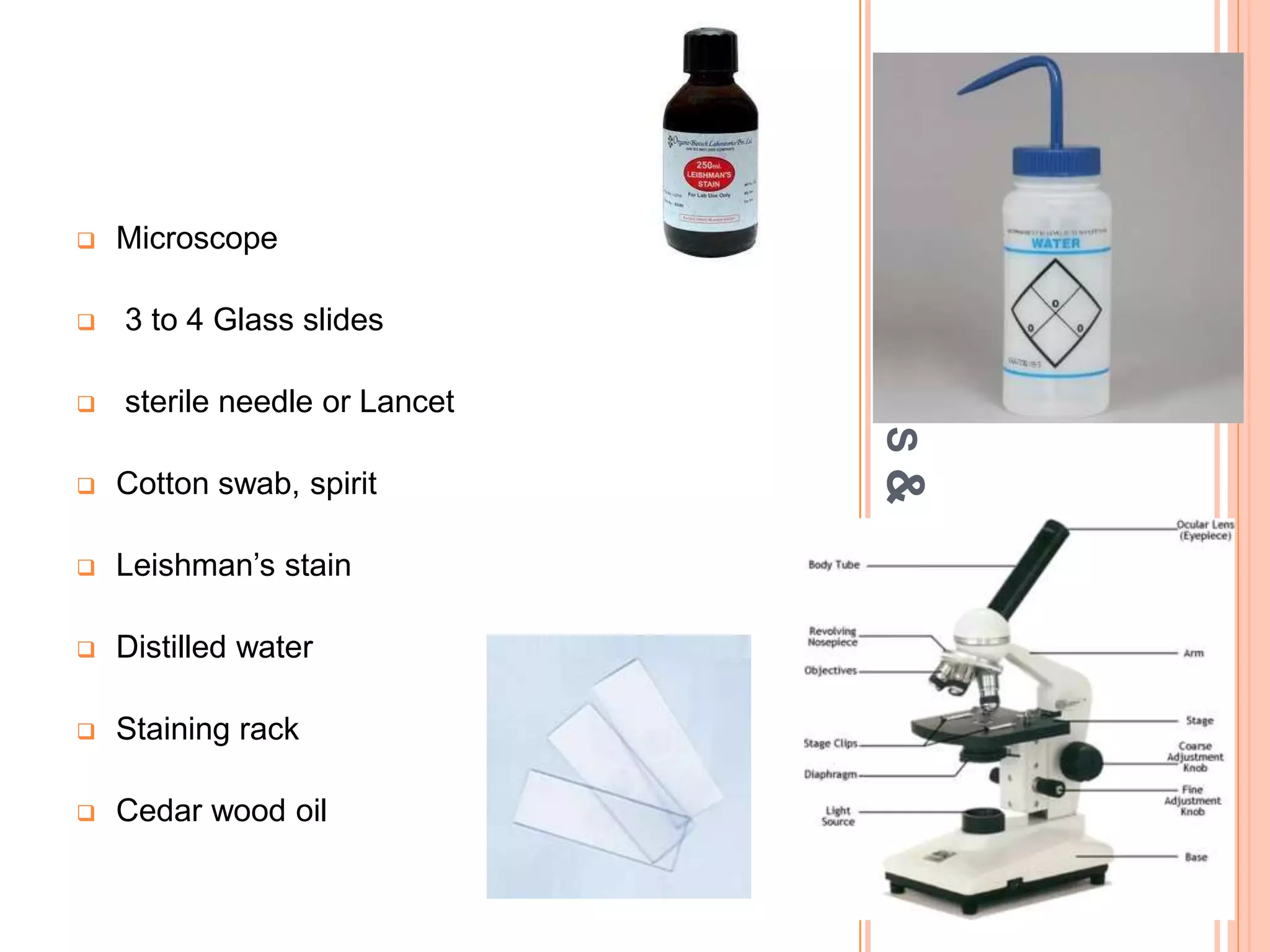
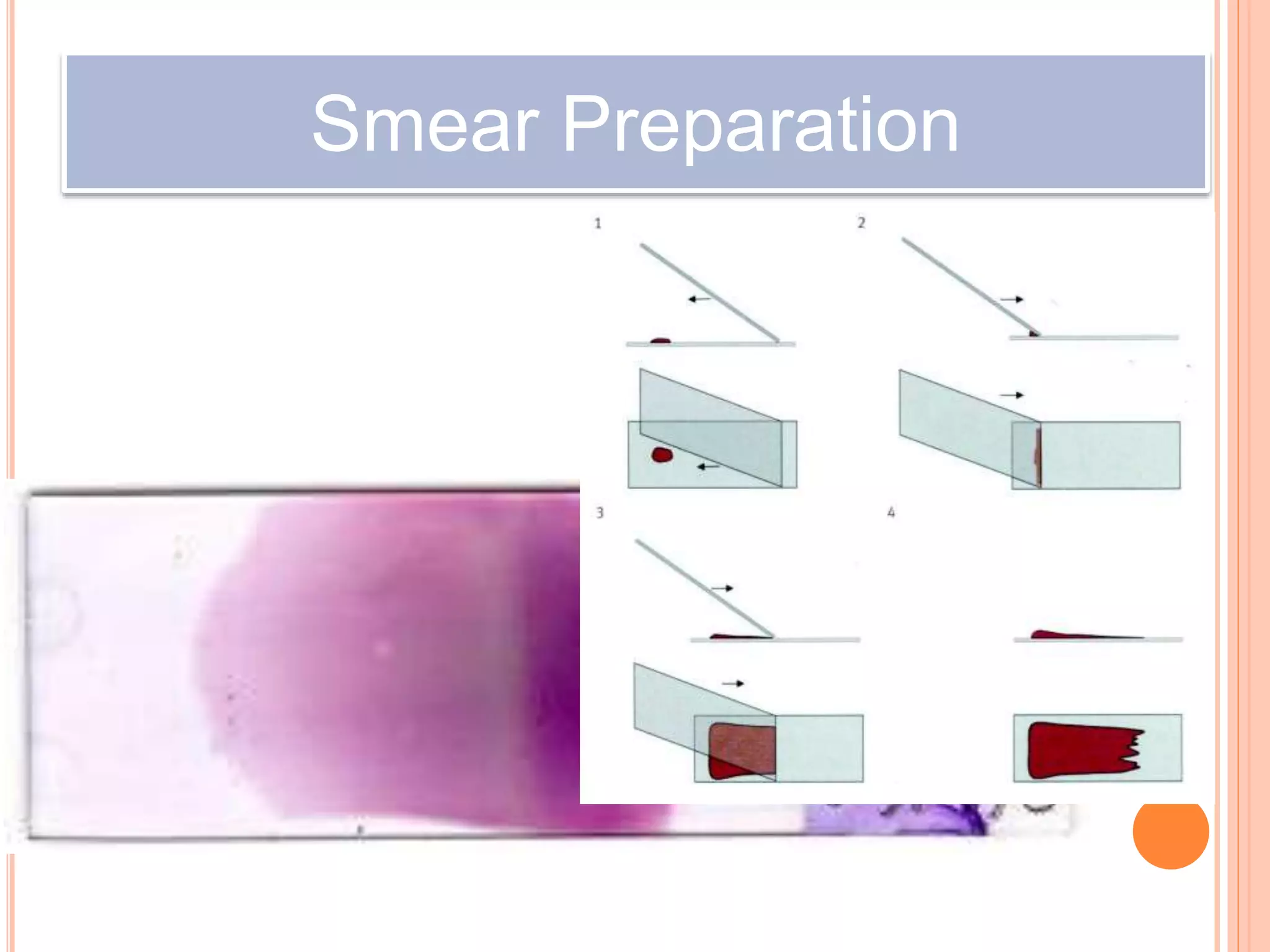
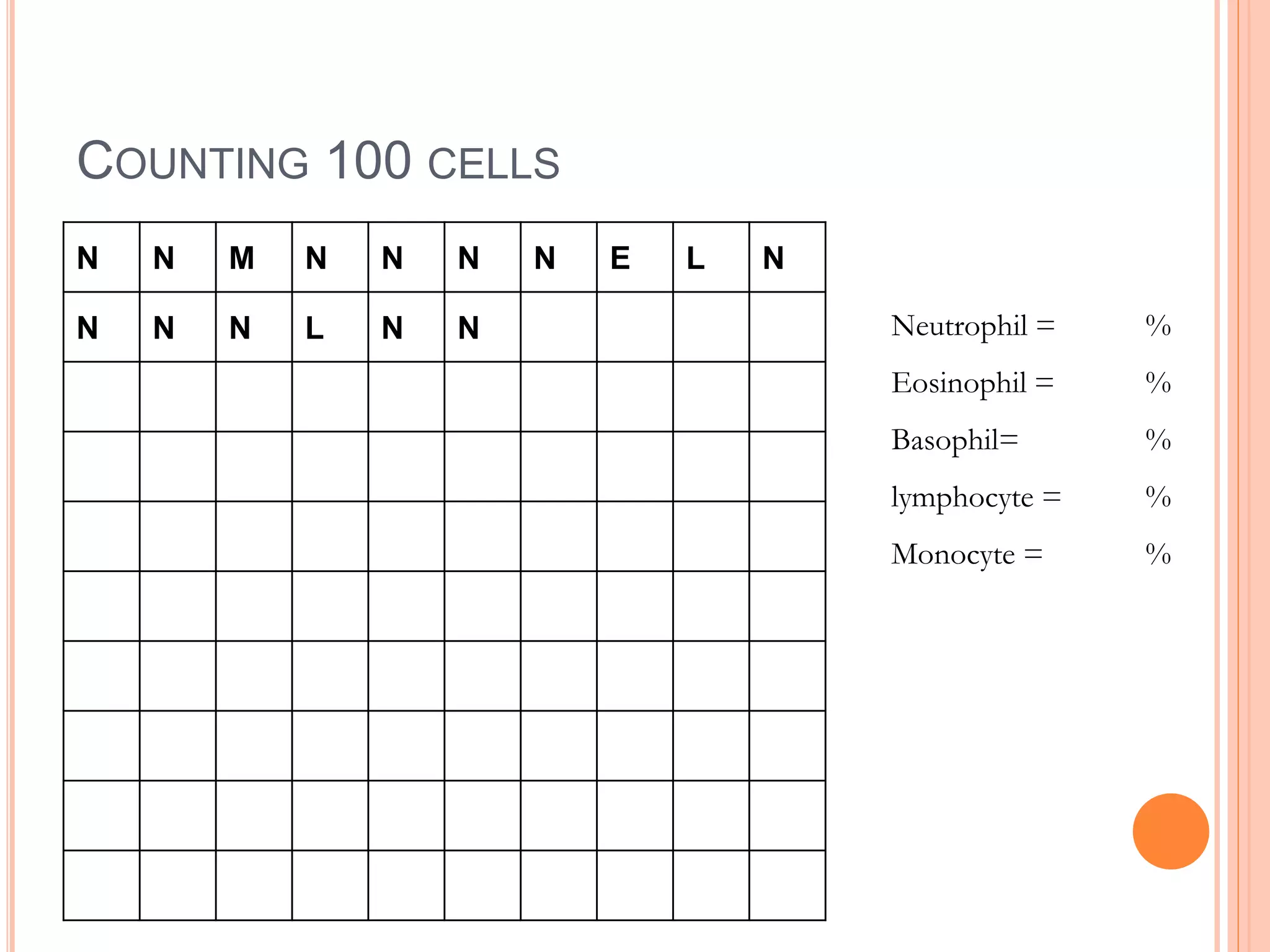
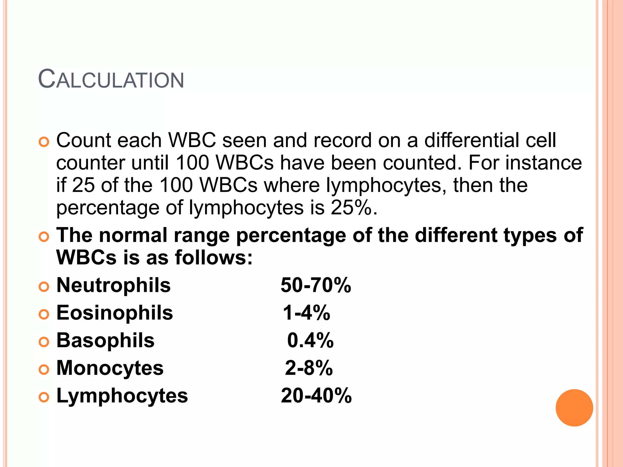
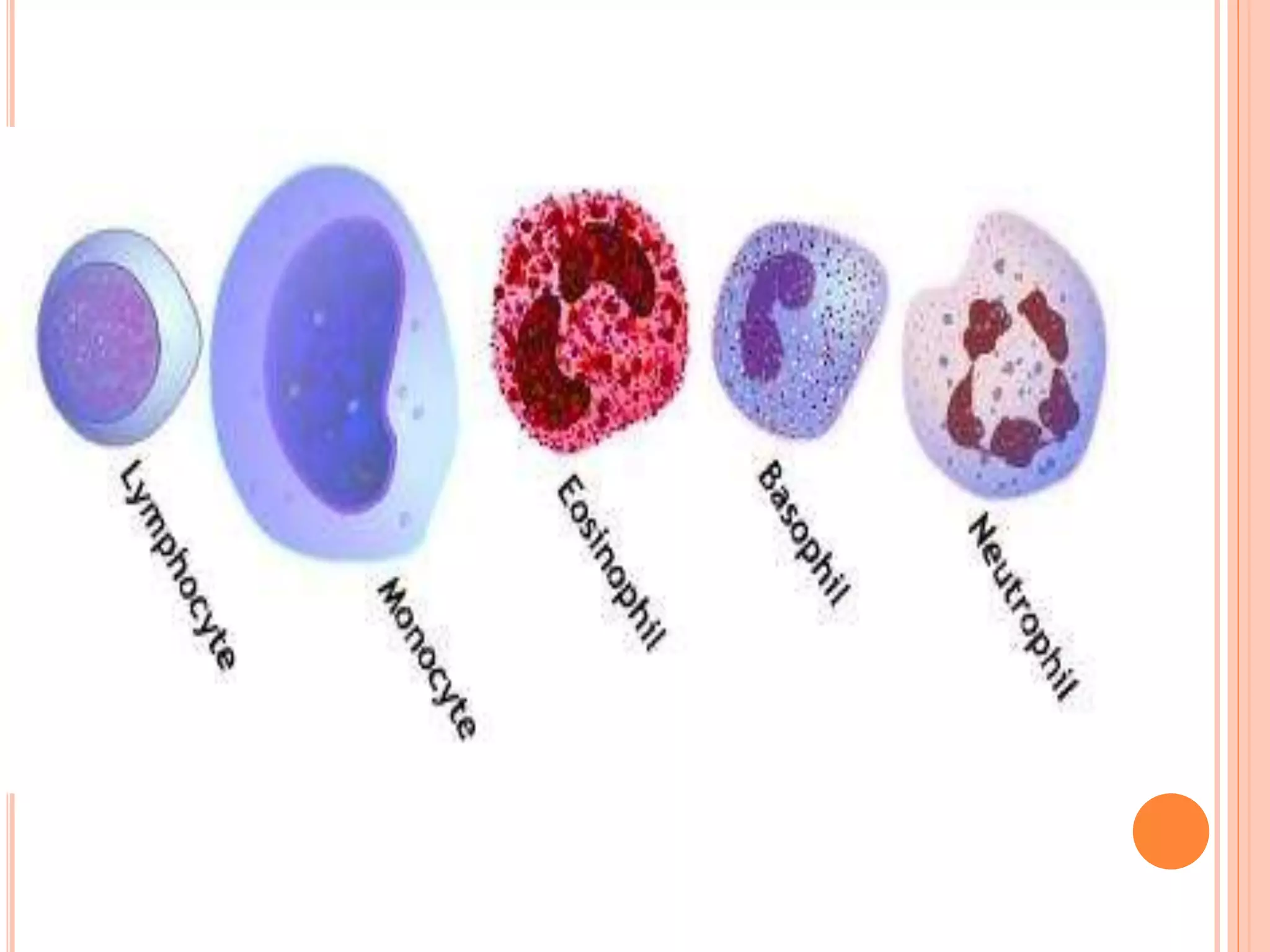
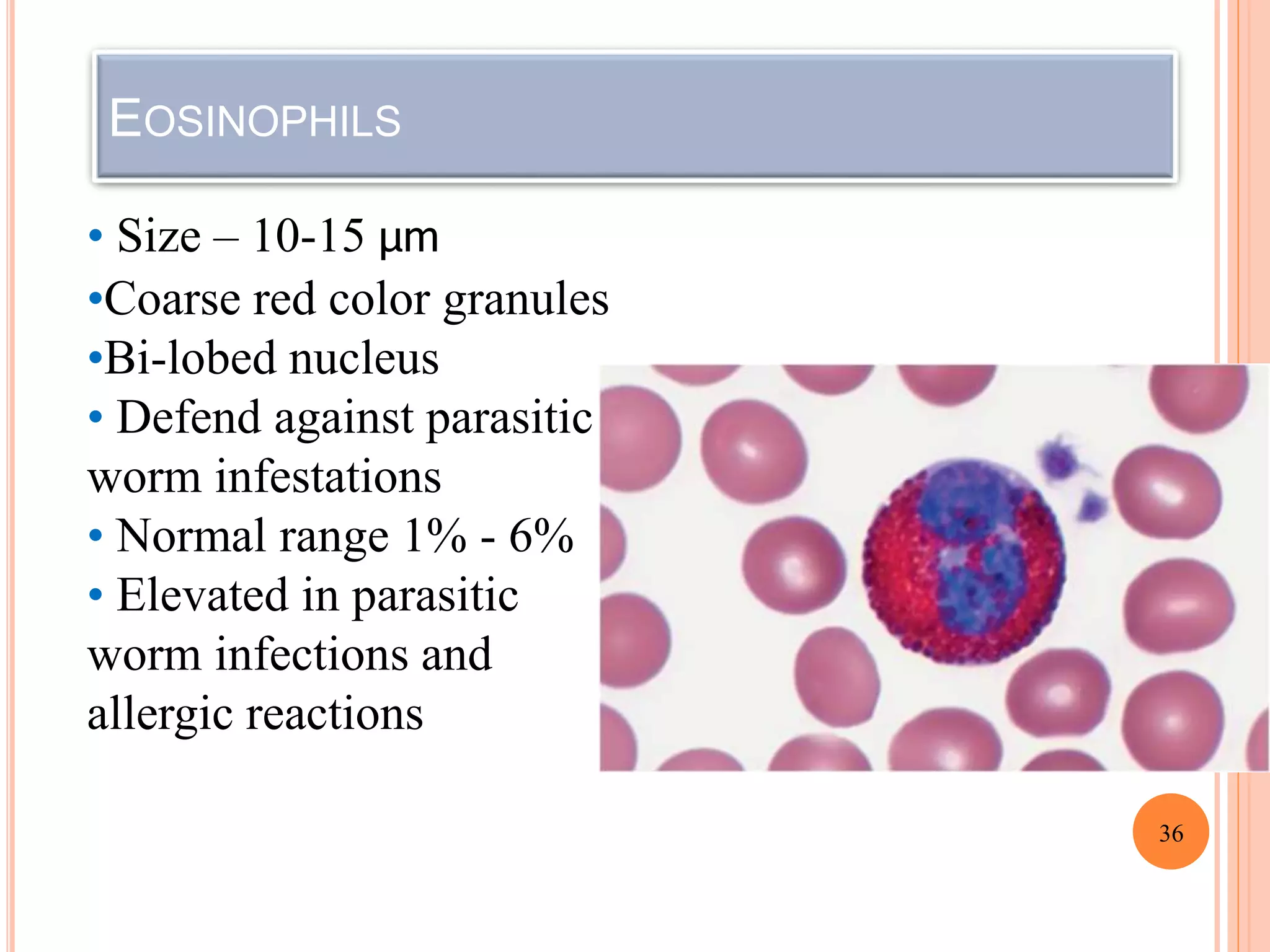
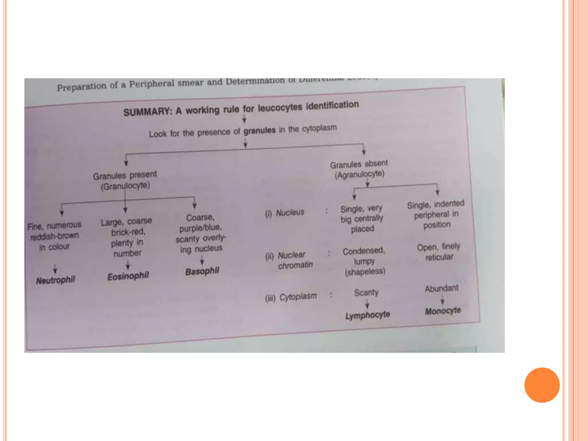
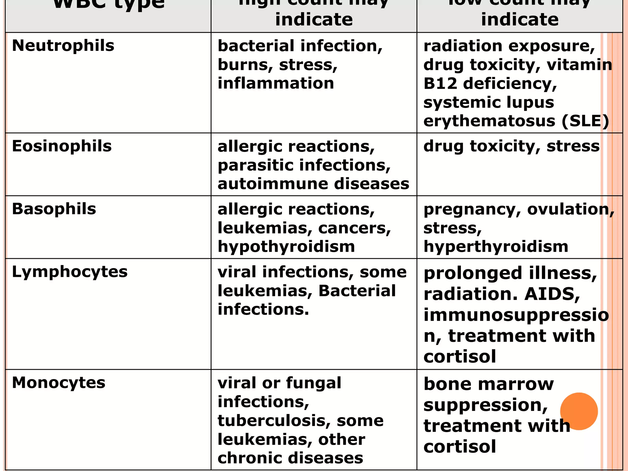
SlideShare a Scribd company logo
DIFFERENTIAL LEUCOCYTE COUNT
INTRODUCTION
 The differential WBC count is performed to
determine the relative number of each type of WBC
in the blood. In disease states a particular white
blood cell type shows increase in number in the
blood.
OBJECTIVES
 1. Describe the relevance and special importance ofpreparing and staining a
blood smear and doing the differential leukocyte count.
 2. Name the components of Leishman’s stain and explain the function of each.
 3. Prepare satisfactory blood films, fix and stain them, and describe the
features of a well-stained film.
 4. Identify different blood cells in a film, and indicate the identifying features of
each type of leukocyte.
 5. Differentiate between neutrophils, eosinophils, and basophils and between a
large lymphocyte and a monocyte.
 6. Carry out the differential count and express your results in their percentages
and absolute numbers.
 7. Describe the functions of each type of leukocyte
 8. List the conditions in which their numbers increase and decrease.
 AIM
 APPARATUS
 PRINCIPLE
 PROCEDURE
 OBSERVATION and CALCULATION
 RESULT
 COMMENT
 PRECAUTIONS
 DISCUSSION
A
PPARATUS
&
MATERIALS
 Microscope
 3 to 4 Glass slides
 sterile needle or Lancet
 Cotton swab, spirit
 Leishman’s stain
 Distilled water
 Staining rack
 Cedar wood oil
Leishman’s stain: It is probably one of the simplest and
most precise methods of staining blood for diagnostic
purposes.
Romanowsky stain, contains acidic and basic dye.
 i. Eosin. It is an acidic dye (negatively charged) and stains
basic (positive) particles—granules of eosinophils, and
RBCs a pink color.
 ii. Methylene-blue. It is a basic dye (positively charged)
and stains acidic (negatively charged) granules in the
cytoplasm, nuclei of leukocytes, especially the granules of
basophils, a blue-violet color.
 iii. Acetone-free and water-free absolute methyl alcohol.
The methyl alcohol is a fixative and must be free from
acetone and water. It serves two functions:
 a. It fixes the blood smear to the glass slide.
 The alcohol precipitates the plasma proteins, which then act
as a ‘glue’ which attaches (fixes) the blood cells to the slide so
that they are not washed away during staining.
 b. The alcohol preserves the morphology and chemical
status of the cells.
PRINCIPLE
 A blood film is stained with Leishman’s stain and
scanned under oil immersion, from one end to the
other. As each WBC is encountered, it is identified
until 100 leukocytes have been examined. The
percentage distribution of each type of WBC is then
calculated.
 Knowing the TLC and the differential count, it is
easy to determine the number of each type of cell
per mm3 of blood.
PROCEDURE
Prepare the blood smear
 Clean the slides, 2 to be covered with the
blood film and one to be used as spreader.
 Clean the finger with alcohol, allow it to dry
and then prick it with a disposable lancet to
obtain a drop of blood.
 Make a fine touch of one end of a slide with
the drop of blood (only a small amount is
required).
 Place the edge of the other slide on the
surface of the first slide just in front of the
drop of blood and at an angle of 45˚.
 Draw the spreader back until it makes contact with
the drop of blood.
 Push the spreader slowly and smoothly to the other
end of the slide.
 Allow the film to dry at room temperature i.e. the
blood smears should be air-dried
Smear Preparation
STAINING THE BLOOD SMEAR BY:-
 Put the dried slide on a staining rack.
 The blood smear should be stained as soon as
possible.
 Carefully drop Leishman's stain onto the blood film ( 8
to 10 drops )until the film is covered.
 Allow the stain to act for two minutes- fixation time.
 Add distilled water ( 20 drops) to the stain and mix by
blowing, this gives dilution of or 1:2
 The diluted stain should act for 10 minutes ( staining
time) -greenish scum
 Then wash it off with distilled water, continue washing
until the film has a pink color.
 Shake off excess water and allow it to dry at room temperature
EXAMINATION OF THE STAINED SMEAR
 For examining the blood smear a microscope with a
low-power objective (10 x) and an oil immersion
objective (100 x) is necessary.
 Place the slide (smear side up) ?? on the microscope
stage.
 Examine the blood smear using the low power (10 x)
objective. Choose an area where there are plenty of
WBCs. This area is usually located near the wedge
shaped end of blood smear.
 Place a drop of immersion oil on the selected site and
carefully change to the oil immersion objective (100x)
 Perform the differential cell count and, at the same
time examine the morphology of the WBCs.
Blood smear under microscope
Counting directions
COUNTING 100 CELLS
N N M N N N N E L N
N N N L N N Neutrophil = %
Eosinophil = %
Basophil= %
lymphocyte = %
Monocyte = %
CALCULATION
 Count each WBC seen and record on a differential cell
counter until 100 WBCs have been counted. For instance
if 25 of the 100 WBCs where lymphocytes, then the
percentage of lymphocytes is 25%.
 The normal range percentage of the different types of
WBCs is as follows:
 Neutrophils 50-70%
 Eosinophils 1-4%
 Basophils 0.4%
 Monocytes 2-8%
 Lymphocytes 20-40%
 Result
 comment
PRECAUTIONS
 1. The slides should be absolutely free from dust and
grease, because blood will not stick to areas where oils from
your fingers have been left.
 2. The edge of the spreader should be smooth and not
chipped, otherwise the slide would leave striations along or
across the smear. Leukocytes may also be caught in chipped
places and be carried towards the tail.
 3. When applying the slide to the blood drop from a finger-
prick, do not touch the skin with the slide, but only the
periphery (top) of the blood drop. This is to avoid taking up
epidermal squames or sweat.
 4. The blood should be spread immediately after taking it
on the slide. Any delay will cause clumping of cells due to
partial coagulation. This will give a ‘granular’ appearance to
the blood film, which is visible to the naked eye.
 5. The angle of the spreader should be 35° to 40°. The
more the angle of the spreader approaches the vertical, the
thinner the film, and the lesser the angle, the thicker the film.
 6. The pressure of the spreader on the slide should be
slight and even and the pushing should be fairly quick
while maintaining a uniform pressure throughout.
 7. The film should be dried by waving it in the
air immediately after spreading it. A delay can
cause not only clumping, but also crenation and
distortion of red cells in a damp atmosphere (if
water is allowed to slowly evaporate from the blood
plasma on the slide, crenation occurs due to
gradual increase in the concentration of salts).
 8. By turning the spreader over, you can use it to
make 4 blood films.
 11. Do not allow Leishman stain to become
syrupy (thick) or dry up on the slide as this is
likely to cause precipitation of the stain.
 12. When counting the cells, use the “battlement”
method. This is to avoid counting a cell twice.
 13. Avoid counting the leukocytes in the extreme
ends of the head or tail, and along the edges of
 blood film
DISCUSSION
 Magnification
 Why oil is placed while using this objective.
 Why cedar wood oil is preferred.
 Features of an ideal blood film
 1. The blood film should occupy the middle two-thirds (about 5 cm)
of the slide, with a clear margin of about 2 mm on either side.
 2. It should be tongue-shaped, i.e. broad at the head (starting point),
and taper towards the other end, but without any ‘tails’.
 3. It should be translucent, uniformly thick throughout, with no
vacant areas, striations (longitudinal or transverse), or
‘granular’ areas.
 4. It should be neither very thick nor very thin (this can be learned
only with practice). A thin film looks faintly pink against a white
surface, while
 a thick smear appears red. An ideal film appears ‘buff’
colored.
 5. The red cells, as seen under the microscope, should lie
separately from each other, just touching here and there,
but without any crowding, or rouleaux formation.
 Method used for identification and counting the
cells
 How to indicate the presence of the cell when
projected
 Relevance
 The differential count is done to find out if there is an
increase or decrease of a particular type of WBC. Knowing
the TLC, the absolute number of each type can be calculated.
This information is important in detecting infection or
inflammation, allergic and parasitic infections, and effects
of chemotherapy and radiation therapy.
 Why acetone free and water free
 Fixation time
 Staining time
 Absolute leucocyte count
 Any other cells seen in the smear
 Buffered water/ distilled water
 Tap water used?
 Approximate size of wbcs
 Dlc of child
 Physiological variations in count of leucocytes
 Absolute leukocyte count.
 The absolute values are more significant than the
DLC values alone. The reason is that the DLC may
show only a relative increase or decrease of a
particular type of cell with a corresponding change
in the other cell types.
 For example, a neutrophil count of 85% may suggest
neutrophilia, but if the TLC is, say, 8000/mm3, then the
absolute neutrophil count of 6800/mm3 (8000 × 85/100 =
6800) would be within the normal range.
TYPES OF WHITE BLOOD CELLS
33
• White blood cells:
• Are leukocytes
•There are five types of WBCs in two categories:
• Granulocytes
• Neutrophils
• Eosinophils
• Basophils
• Agranulocytes
• Lymphocytes
• Monocytes
NEUTROPHILS
35
•Size – 10-14 µm.
•Multilobed nucleus
•Light purple (neutral)
granules
•Other names is
Polymorphonuclear leukocyte
• Phagocytic in function
• Normal range = 50% - 70%
• Elevated in acute bacterial
infections
EOSINOPHILS
36
• Size – 10-15 µm
•Coarse red color granules
•Bi-lobed nucleus
• Defend against parasitic
worm infestations
• Normal range 1% - 6%
• Elevated in parasitic
worm infections and
allergic reactions
BASOPHILS
37
•Size – 10 – 15 µm
•Bilobed nucleus
•Deep blue granules
covering the whole nucleus
• Release histamine
• Release heparin
• Normal range- Less than
1%
MONOCYTES
38
• Largest of all blood cells
•Size – 12 – 20 µm
•Spherical, kidney-shaped,
oval or lobed nuclei
•No visible granules
• May leave bloodstream to
become macrophages
• Normal range - 2% - 10%
• Phagocytize bacteria, dead
cells, and other debris
•Increase count – in chronic
infections eg. Tuberculosis
LYMPHOCYTES
39
•Size – 7-9 or 10-15 µm
•Slightly larger than RBC
• Large spherical nucleus
surrounded by thin rim of
cytoplasm
•No visible granules
•Normal range -20% - 40%
•T cells and B cells
• Both important in
immunity
• B cells produce antibodies
•T cells involved in cell
mediated immunity
• Increased count is seen
in viral infections,
chronic infections
APPLICATIONS OF BLOOD SMEAR:
 to detect infection or inflammation,
 determine the effects of possible poisoning by
chemicals, drugs, chemotherapy, radiation, etc.
 DLC is also done to monitor blood diseases
like leukemia,
 or to detect allergic and parasitic infections.
 The determination of each type of WBC helps
in diagnosing the condition because a particular
type may show an increase or decrease.
 Can know the gender of the person
 Normal values. The normal values for
differential and absolute counts are given
below:
 Differential count (percent) Absolute
count (per mm3)
 Neutrophils 40–75 2000–7500
 Eosinophils 1–6 4–440
 Basophils 0–1 0–100
 Monocytes 2–10 500–800
 Lymphocytes (both) 20–45 1300–3500
WBC type high count may
indicate
low count may
indicate
Neutrophils bacterial infection,
burns, stress,
inflammation
radiation exposure,
drug toxicity, vitamin
B12 deficiency,
systemic lupus
erythematosus (SLE)
Eosinophils allergic reactions,
parasitic infections,
autoimmune diseases
drug toxicity, stress
Basophils allergic reactions,
leukemias, cancers,
hypothyroidism
pregnancy, ovulation,
stress,
hyperthyroidism
Lymphocytes viral infections, some
leukemias, Bacterial
infections.
prolonged illness,
radiation. AIDS,
immunosuppressio
n, treatment with
cortisol
Monocytes viral or fungal
infections,
tuberculosis, some
leukemias, other
chronic diseases
bone marrow
suppression,
treatment with
cortisol
Thank you

More Related Content

What's hot

Anticoagulant
AnticoagulantAnticoagulant
Anticoagulant
chinoo281
 

What's hot (20)

Peripheral Smear Using Leishman Stain
Peripheral Smear Using Leishman StainPeripheral Smear Using Leishman Stain
Peripheral Smear Using Leishman Stain
 
White blood cells
White blood cellsWhite blood cells
White blood cells
 
White Blood Cell count (WBC)
White Blood Cell count (WBC)White Blood Cell count (WBC)
White Blood Cell count (WBC)
 
Total leukocyte count
Total leukocyte countTotal leukocyte count
Total leukocyte count
 
Platelet
PlateletPlatelet
Platelet
 
stains
stainsstains
stains
 
Red blood cell count
Red blood cell countRed blood cell count
Red blood cell count
 
Pcv
PcvPcv
Pcv
 
RBC count and WBC count
RBC count and WBC countRBC count and WBC count
RBC count and WBC count
 
Preparation and staining of peripheral blood smear
Preparation and staining of peripheral blood smearPreparation and staining of peripheral blood smear
Preparation and staining of peripheral blood smear
 
Anticoagulants used in haematology
Anticoagulants used in haematologyAnticoagulants used in haematology
Anticoagulants used in haematology
 
White blood cells
White blood cells White blood cells
White blood cells
 
Anticoagulant
AnticoagulantAnticoagulant
Anticoagulant
 
Hemoglobin estimation
Hemoglobin estimationHemoglobin estimation
Hemoglobin estimation
 
AEC COUNT
AEC COUNTAEC COUNT
AEC COUNT
 
Total Erythrocyte Count By Hemocytometer
Total Erythrocyte Count By HemocytometerTotal Erythrocyte Count By Hemocytometer
Total Erythrocyte Count By Hemocytometer
 
Erythrocyte sedimentation rate
Erythrocyte sedimentation rateErythrocyte sedimentation rate
Erythrocyte sedimentation rate
 
RBC Indices- MCV, MCH, MCHC II Blood Physiology
RBC Indices- MCV, MCH, MCHC II Blood PhysiologyRBC Indices- MCV, MCH, MCHC II Blood Physiology
RBC Indices- MCV, MCH, MCHC II Blood Physiology
 
Total rbc count
Total rbc countTotal rbc count
Total rbc count
 
Erythrocyte indices
Erythrocyte  indicesErythrocyte  indices
Erythrocyte indices
 

Similar to DLC practical ppt

Differential leucocyte count & eosinophilia
Differential leucocyte count & eosinophiliaDifferential leucocyte count & eosinophilia
Differential leucocyte count & eosinophilia
sandeep singh
 
pbs-fields staining-converted"""""""...pdf
pbs-fields staining-converted"""""""...pdfpbs-fields staining-converted"""""""...pdf
pbs-fields staining-converted"""""""...pdf
r8fdq7w2m9
 

Similar to DLC practical ppt (20)

1 blood-smear
1 blood-smear1 blood-smear
1 blood-smear
 
1-Blood-smear.ppt
1-Blood-smear.ppt1-Blood-smear.ppt
1-Blood-smear.ppt
 
PERIPHERAL SMEAR EXAMINATION
PERIPHERAL SMEAR EXAMINATIONPERIPHERAL SMEAR EXAMINATION
PERIPHERAL SMEAR EXAMINATION
 
Differential leucocyte count & eosinophilia
Differential leucocyte count & eosinophiliaDifferential leucocyte count & eosinophilia
Differential leucocyte count & eosinophilia
 
Peripheral blood smear [autosaved]
Peripheral blood smear [autosaved]Peripheral blood smear [autosaved]
Peripheral blood smear [autosaved]
 
differentialleucocytecount.pptx
differentialleucocytecount.pptxdifferentialleucocytecount.pptx
differentialleucocytecount.pptx
 
Tlc dlc
Tlc dlcTlc dlc
Tlc dlc
 
Hema I Chapter 7_hemocytometry.ppt
Hema I Chapter 7_hemocytometry.pptHema I Chapter 7_hemocytometry.ppt
Hema I Chapter 7_hemocytometry.ppt
 
Artefacts in hemat part 2
Artefacts in hemat part 2Artefacts in hemat part 2
Artefacts in hemat part 2
 
Prepration and staining of blood film
Prepration and staining of blood filmPrepration and staining of blood film
Prepration and staining of blood film
 
Practical 1 To Determine Differential Leukocytes Count DLC.pptx
Practical 1 To Determine Differential Leukocytes Count DLC.pptxPractical 1 To Determine Differential Leukocytes Count DLC.pptx
Practical 1 To Determine Differential Leukocytes Count DLC.pptx
 
Differential WBC Count
Differential WBC CountDifferential WBC Count
Differential WBC Count
 
Blood smear prep
Blood smear prepBlood smear prep
Blood smear prep
 
Peripheral blood Smear Preparation
Peripheral blood Smear PreparationPeripheral blood Smear Preparation
Peripheral blood Smear Preparation
 
Dc
DcDc
Dc
 
دكتور عبد الامير عملي Reticulucyte stain
 دكتور عبد الامير عملي Reticulucyte stain دكتور عبد الامير عملي Reticulucyte stain
دكتور عبد الامير عملي Reticulucyte stain
 
Total Leukocyte Count by Hemocytometer
Total Leukocyte Count by HemocytometerTotal Leukocyte Count by Hemocytometer
Total Leukocyte Count by Hemocytometer
 
Total leukocyte count by hemocytometer
Total leukocyte count by hemocytometerTotal leukocyte count by hemocytometer
Total leukocyte count by hemocytometer
 
Hemocytometer manual cell counting (1)
Hemocytometer manual cell counting (1)Hemocytometer manual cell counting (1)
Hemocytometer manual cell counting (1)
 
pbs-fields staining-converted"""""""...pdf
pbs-fields staining-converted"""""""...pdfpbs-fields staining-converted"""""""...pdf
pbs-fields staining-converted"""""""...pdf
 

More from Reena Gollapalli

More from Reena Gollapalli (20)

Mechanism of drug action,drug receptor phrmacology
Mechanism of drug action,drug receptor phrmacologyMechanism of drug action,drug receptor phrmacology
Mechanism of drug action,drug receptor phrmacology
 
coronarycirculation. and peculiarities,regulationpptx
coronarycirculation. and peculiarities,regulationpptxcoronarycirculation. and peculiarities,regulationpptx
coronarycirculation. and peculiarities,regulationpptx
 
PROPERTIES OF NERVE FIBRES,and graphs.pptx
PROPERTIES OF NERVE FIBRES,and graphs.pptxPROPERTIES OF NERVE FIBRES,and graphs.pptx
PROPERTIES OF NERVE FIBRES,and graphs.pptx
 
intdofsensorysystemf-150922031359-lva1-app6892.pptx
intdofsensorysystemf-150922031359-lva1-app6892.pptxintdofsensorysystemf-150922031359-lva1-app6892.pptx
intdofsensorysystemf-150922031359-lva1-app6892.pptx
 
hemoglobin and its functions,forms,structure
hemoglobin and its functions,forms,structurehemoglobin and its functions,forms,structure
hemoglobin and its functions,forms,structure
 
Anaemia,jaundice.pptx
Anaemia,jaundice.pptxAnaemia,jaundice.pptx
Anaemia,jaundice.pptx
 
lung function tests.pptx
lung function tests.pptxlung function tests.pptx
lung function tests.pptx
 
anemia mlt.pptx
anemia mlt.pptxanemia mlt.pptx
anemia mlt.pptx
 
wob.pptx
wob.pptxwob.pptx
wob.pptx
 
reflexes ppt.pptx
reflexes ppt.pptxreflexes ppt.pptx
reflexes ppt.pptx
 
GFR MLT.pptx
GFR MLT.pptxGFR MLT.pptx
GFR MLT.pptx
 
rmp new mbbs.pptx
rmp new mbbs.pptxrmp new mbbs.pptx
rmp new mbbs.pptx
 
Tublar transport & Osmolality.ppt
Tublar transport & Osmolality.pptTublar transport & Osmolality.ppt
Tublar transport & Osmolality.ppt
 
physioanatomy of ear.pptx
physioanatomy of ear.pptxphysioanatomy of ear.pptx
physioanatomy of ear.pptx
 
ventilation.pptx
ventilation.pptxventilation.pptx
ventilation.pptx
 
cerebellum MBBS.pptx
cerebellum MBBS.pptxcerebellum MBBS.pptx
cerebellum MBBS.pptx
 
C&D OF URINE.pptx
C&D OF URINE.pptxC&D OF URINE.pptx
C&D OF URINE.pptx
 
chem.reg of resp.pptx
chem.reg of resp.pptxchem.reg of resp.pptx
chem.reg of resp.pptx
 
ovarian hormones bds.pptx
ovarian hormones bds.pptxovarian hormones bds.pptx
ovarian hormones bds.pptx
 
spirometry.pptx
spirometry.pptxspirometry.pptx
spirometry.pptx
 

Recently uploaded

Demystifying-Gene-Editing-The-Promise-and-Peril-of-CRISPR.pdf
Demystifying-Gene-Editing-The-Promise-and-Peril-of-CRISPR.pdfDemystifying-Gene-Editing-The-Promise-and-Peril-of-CRISPR.pdf
Demystifying-Gene-Editing-The-Promise-and-Peril-of-CRISPR.pdf
SasikiranMarri
 
Urinary Elimination BY ANUSHRI SRIVASTAVA.pptx
Urinary Elimination BY ANUSHRI SRIVASTAVA.pptxUrinary Elimination BY ANUSHRI SRIVASTAVA.pptx
Urinary Elimination BY ANUSHRI SRIVASTAVA.pptx
AnushriSrivastav
 
ASSISTING WITH THE USE OF BED PAN BY ANUSHRI SRIVASTAVA.pptx
ASSISTING WITH THE USE OF BED PAN BY ANUSHRI SRIVASTAVA.pptxASSISTING WITH THE USE OF BED PAN BY ANUSHRI SRIVASTAVA.pptx
ASSISTING WITH THE USE OF BED PAN BY ANUSHRI SRIVASTAVA.pptx
AnushriSrivastav
 

Recently uploaded (20)

Master the Art of Yoga with Joga Yoga Training
Master the Art of Yoga with Joga Yoga TrainingMaster the Art of Yoga with Joga Yoga Training
Master the Art of Yoga with Joga Yoga Training
 
The Docs PPG - 30.05.2024.pptx..........
The Docs PPG - 30.05.2024.pptx..........The Docs PPG - 30.05.2024.pptx..........
The Docs PPG - 30.05.2024.pptx..........
 
Deepfake Detection_Using Machine Learning .pptx
Deepfake Detection_Using Machine Learning .pptxDeepfake Detection_Using Machine Learning .pptx
Deepfake Detection_Using Machine Learning .pptx
 
Myopia Management & Control Strategies.pptx
Myopia Management & Control Strategies.pptxMyopia Management & Control Strategies.pptx
Myopia Management & Control Strategies.pptx
 
Demystifying-Gene-Editing-The-Promise-and-Peril-of-CRISPR.pdf
Demystifying-Gene-Editing-The-Promise-and-Peril-of-CRISPR.pdfDemystifying-Gene-Editing-The-Promise-and-Peril-of-CRISPR.pdf
Demystifying-Gene-Editing-The-Promise-and-Peril-of-CRISPR.pdf
 
CHAPTER- 1 SEMESTER - V NATIONAL HEALTH PROGRAMME RELATED TO CHILD.pdf
CHAPTER- 1 SEMESTER - V NATIONAL HEALTH PROGRAMME RELATED TO CHILD.pdfCHAPTER- 1 SEMESTER - V NATIONAL HEALTH PROGRAMME RELATED TO CHILD.pdf
CHAPTER- 1 SEMESTER - V NATIONAL HEALTH PROGRAMME RELATED TO CHILD.pdf
 
Jesse Jhaj: Building Relationships with Patients as a Doctor or Healthcare Wo...
Jesse Jhaj: Building Relationships with Patients as a Doctor or Healthcare Wo...Jesse Jhaj: Building Relationships with Patients as a Doctor or Healthcare Wo...
Jesse Jhaj: Building Relationships with Patients as a Doctor or Healthcare Wo...
 
R3 Stem Cells and Kidney Repair A New Horizon in Nephrology.pptx
R3 Stem Cells and Kidney Repair A New Horizon in Nephrology.pptxR3 Stem Cells and Kidney Repair A New Horizon in Nephrology.pptx
R3 Stem Cells and Kidney Repair A New Horizon in Nephrology.pptx
 
Management of psoriasis.pptx (Recent advances)
Management of psoriasis.pptx (Recent advances)Management of psoriasis.pptx (Recent advances)
Management of psoriasis.pptx (Recent advances)
 
PhRMA Vaccines Deck_05-15_2024_FINAL.pptx
PhRMA Vaccines Deck_05-15_2024_FINAL.pptxPhRMA Vaccines Deck_05-15_2024_FINAL.pptx
PhRMA Vaccines Deck_05-15_2024_FINAL.pptx
 
VVIP Dehradun Girls 9719300533 Heat-bake { Dehradun } Genteel ℂall Serviℂe By...
VVIP Dehradun Girls 9719300533 Heat-bake { Dehradun } Genteel ℂall Serviℂe By...VVIP Dehradun Girls 9719300533 Heat-bake { Dehradun } Genteel ℂall Serviℂe By...
VVIP Dehradun Girls 9719300533 Heat-bake { Dehradun } Genteel ℂall Serviℂe By...
 
Digital Healthcare: The Future of Medical Consultations
Digital Healthcare: The Future of Medical ConsultationsDigital Healthcare: The Future of Medical Consultations
Digital Healthcare: The Future of Medical Consultations
 
Importance of Diet on Dental Health.docx
Importance of Diet on Dental Health.docxImportance of Diet on Dental Health.docx
Importance of Diet on Dental Health.docx
 
Call Girls in Jaipur (Rajasthan) call me [🔝89011-83002🔝] Escort In Jaipur ℂal...
Call Girls in Jaipur (Rajasthan) call me [🔝89011-83002🔝] Escort In Jaipur ℂal...Call Girls in Jaipur (Rajasthan) call me [🔝89011-83002🔝] Escort In Jaipur ℂal...
Call Girls in Jaipur (Rajasthan) call me [🔝89011-83002🔝] Escort In Jaipur ℂal...
 
Urinary Elimination BY ANUSHRI SRIVASTAVA.pptx
Urinary Elimination BY ANUSHRI SRIVASTAVA.pptxUrinary Elimination BY ANUSHRI SRIVASTAVA.pptx
Urinary Elimination BY ANUSHRI SRIVASTAVA.pptx
 
ASSISTING WITH THE USE OF BED PAN BY ANUSHRI SRIVASTAVA.pptx
ASSISTING WITH THE USE OF BED PAN BY ANUSHRI SRIVASTAVA.pptxASSISTING WITH THE USE OF BED PAN BY ANUSHRI SRIVASTAVA.pptx
ASSISTING WITH THE USE OF BED PAN BY ANUSHRI SRIVASTAVA.pptx
 
Valle Egypt Illustrates Consequences of Financial Elder Abuse
Valle Egypt Illustrates Consequences of Financial Elder AbuseValle Egypt Illustrates Consequences of Financial Elder Abuse
Valle Egypt Illustrates Consequences of Financial Elder Abuse
 
CHAPTER- 1 SEMESTER V NATIONAL-POLICIES-AND-LEGISLATION.pdf
CHAPTER- 1 SEMESTER V NATIONAL-POLICIES-AND-LEGISLATION.pdfCHAPTER- 1 SEMESTER V NATIONAL-POLICIES-AND-LEGISLATION.pdf
CHAPTER- 1 SEMESTER V NATIONAL-POLICIES-AND-LEGISLATION.pdf
 
Navigating Healthcare with Telemedicine
Navigating Healthcare with  TelemedicineNavigating Healthcare with  Telemedicine
Navigating Healthcare with Telemedicine
 
What can we really do to give meaning and momentum to equality, diversity and...
What can we really do to give meaning and momentum to equality, diversity and...What can we really do to give meaning and momentum to equality, diversity and...
What can we really do to give meaning and momentum to equality, diversity and...
 

DLC practical ppt

  • 2. INTRODUCTION  The differential WBC count is performed to determine the relative number of each type of WBC in the blood. In disease states a particular white blood cell type shows increase in number in the blood.
  • 3. OBJECTIVES  1. Describe the relevance and special importance ofpreparing and staining a blood smear and doing the differential leukocyte count.  2. Name the components of Leishman’s stain and explain the function of each.  3. Prepare satisfactory blood films, fix and stain them, and describe the features of a well-stained film.  4. Identify different blood cells in a film, and indicate the identifying features of each type of leukocyte.  5. Differentiate between neutrophils, eosinophils, and basophils and between a large lymphocyte and a monocyte.  6. Carry out the differential count and express your results in their percentages and absolute numbers.  7. Describe the functions of each type of leukocyte  8. List the conditions in which their numbers increase and decrease.
  • 4.  AIM  APPARATUS  PRINCIPLE  PROCEDURE  OBSERVATION and CALCULATION  RESULT  COMMENT  PRECAUTIONS  DISCUSSION
  • 5. A PPARATUS & MATERIALS  Microscope  3 to 4 Glass slides  sterile needle or Lancet  Cotton swab, spirit  Leishman’s stain  Distilled water  Staining rack  Cedar wood oil
  • 6. Leishman’s stain: It is probably one of the simplest and most precise methods of staining blood for diagnostic purposes. Romanowsky stain, contains acidic and basic dye.  i. Eosin. It is an acidic dye (negatively charged) and stains basic (positive) particles—granules of eosinophils, and RBCs a pink color.  ii. Methylene-blue. It is a basic dye (positively charged) and stains acidic (negatively charged) granules in the cytoplasm, nuclei of leukocytes, especially the granules of basophils, a blue-violet color.
  • 7.  iii. Acetone-free and water-free absolute methyl alcohol. The methyl alcohol is a fixative and must be free from acetone and water. It serves two functions:  a. It fixes the blood smear to the glass slide.  The alcohol precipitates the plasma proteins, which then act as a ‘glue’ which attaches (fixes) the blood cells to the slide so that they are not washed away during staining.  b. The alcohol preserves the morphology and chemical status of the cells.
  • 8. PRINCIPLE  A blood film is stained with Leishman’s stain and scanned under oil immersion, from one end to the other. As each WBC is encountered, it is identified until 100 leukocytes have been examined. The percentage distribution of each type of WBC is then calculated.  Knowing the TLC and the differential count, it is easy to determine the number of each type of cell per mm3 of blood.
  • 9. PROCEDURE Prepare the blood smear  Clean the slides, 2 to be covered with the blood film and one to be used as spreader.  Clean the finger with alcohol, allow it to dry and then prick it with a disposable lancet to obtain a drop of blood.  Make a fine touch of one end of a slide with the drop of blood (only a small amount is required).  Place the edge of the other slide on the surface of the first slide just in front of the drop of blood and at an angle of 45˚.
  • 10.  Draw the spreader back until it makes contact with the drop of blood.  Push the spreader slowly and smoothly to the other end of the slide.  Allow the film to dry at room temperature i.e. the blood smears should be air-dried
  • 11.
  • 12.
  • 13.
  • 14.
  • 15.
  • 17. STAINING THE BLOOD SMEAR BY:-  Put the dried slide on a staining rack.  The blood smear should be stained as soon as possible.  Carefully drop Leishman's stain onto the blood film ( 8 to 10 drops )until the film is covered.  Allow the stain to act for two minutes- fixation time.  Add distilled water ( 20 drops) to the stain and mix by blowing, this gives dilution of or 1:2  The diluted stain should act for 10 minutes ( staining time) -greenish scum  Then wash it off with distilled water, continue washing until the film has a pink color.  Shake off excess water and allow it to dry at room temperature
  • 18. EXAMINATION OF THE STAINED SMEAR  For examining the blood smear a microscope with a low-power objective (10 x) and an oil immersion objective (100 x) is necessary.  Place the slide (smear side up) ?? on the microscope stage.  Examine the blood smear using the low power (10 x) objective. Choose an area where there are plenty of WBCs. This area is usually located near the wedge shaped end of blood smear.  Place a drop of immersion oil on the selected site and carefully change to the oil immersion objective (100x)  Perform the differential cell count and, at the same time examine the morphology of the WBCs.
  • 19. Blood smear under microscope
  • 21. COUNTING 100 CELLS N N M N N N N E L N N N N L N N Neutrophil = % Eosinophil = % Basophil= % lymphocyte = % Monocyte = %
  • 22. CALCULATION  Count each WBC seen and record on a differential cell counter until 100 WBCs have been counted. For instance if 25 of the 100 WBCs where lymphocytes, then the percentage of lymphocytes is 25%.  The normal range percentage of the different types of WBCs is as follows:  Neutrophils 50-70%  Eosinophils 1-4%  Basophils 0.4%  Monocytes 2-8%  Lymphocytes 20-40%
  • 24. PRECAUTIONS  1. The slides should be absolutely free from dust and grease, because blood will not stick to areas where oils from your fingers have been left.  2. The edge of the spreader should be smooth and not chipped, otherwise the slide would leave striations along or across the smear. Leukocytes may also be caught in chipped places and be carried towards the tail.  3. When applying the slide to the blood drop from a finger- prick, do not touch the skin with the slide, but only the periphery (top) of the blood drop. This is to avoid taking up epidermal squames or sweat.
  • 25.  4. The blood should be spread immediately after taking it on the slide. Any delay will cause clumping of cells due to partial coagulation. This will give a ‘granular’ appearance to the blood film, which is visible to the naked eye.  5. The angle of the spreader should be 35° to 40°. The more the angle of the spreader approaches the vertical, the thinner the film, and the lesser the angle, the thicker the film.  6. The pressure of the spreader on the slide should be slight and even and the pushing should be fairly quick while maintaining a uniform pressure throughout.
  • 26.  7. The film should be dried by waving it in the air immediately after spreading it. A delay can cause not only clumping, but also crenation and distortion of red cells in a damp atmosphere (if water is allowed to slowly evaporate from the blood plasma on the slide, crenation occurs due to gradual increase in the concentration of salts).  8. By turning the spreader over, you can use it to make 4 blood films.
  • 27.  11. Do not allow Leishman stain to become syrupy (thick) or dry up on the slide as this is likely to cause precipitation of the stain.  12. When counting the cells, use the “battlement” method. This is to avoid counting a cell twice.  13. Avoid counting the leukocytes in the extreme ends of the head or tail, and along the edges of  blood film
  • 28. DISCUSSION  Magnification  Why oil is placed while using this objective.  Why cedar wood oil is preferred.  Features of an ideal blood film  1. The blood film should occupy the middle two-thirds (about 5 cm) of the slide, with a clear margin of about 2 mm on either side.  2. It should be tongue-shaped, i.e. broad at the head (starting point), and taper towards the other end, but without any ‘tails’.  3. It should be translucent, uniformly thick throughout, with no vacant areas, striations (longitudinal or transverse), or ‘granular’ areas.  4. It should be neither very thick nor very thin (this can be learned only with practice). A thin film looks faintly pink against a white surface, while
  • 29.  a thick smear appears red. An ideal film appears ‘buff’ colored.  5. The red cells, as seen under the microscope, should lie separately from each other, just touching here and there, but without any crowding, or rouleaux formation.
  • 30.  Method used for identification and counting the cells  How to indicate the presence of the cell when projected  Relevance  The differential count is done to find out if there is an increase or decrease of a particular type of WBC. Knowing the TLC, the absolute number of each type can be calculated. This information is important in detecting infection or inflammation, allergic and parasitic infections, and effects of chemotherapy and radiation therapy.
  • 31.  Why acetone free and water free  Fixation time  Staining time  Absolute leucocyte count  Any other cells seen in the smear  Buffered water/ distilled water  Tap water used?  Approximate size of wbcs  Dlc of child  Physiological variations in count of leucocytes
  • 32.  Absolute leukocyte count.  The absolute values are more significant than the DLC values alone. The reason is that the DLC may show only a relative increase or decrease of a particular type of cell with a corresponding change in the other cell types.  For example, a neutrophil count of 85% may suggest neutrophilia, but if the TLC is, say, 8000/mm3, then the absolute neutrophil count of 6800/mm3 (8000 × 85/100 = 6800) would be within the normal range.
  • 33. TYPES OF WHITE BLOOD CELLS 33 • White blood cells: • Are leukocytes •There are five types of WBCs in two categories: • Granulocytes • Neutrophils • Eosinophils • Basophils • Agranulocytes • Lymphocytes • Monocytes
  • 34.
  • 35. NEUTROPHILS 35 •Size – 10-14 µm. •Multilobed nucleus •Light purple (neutral) granules •Other names is Polymorphonuclear leukocyte • Phagocytic in function • Normal range = 50% - 70% • Elevated in acute bacterial infections
  • 36. EOSINOPHILS 36 • Size – 10-15 µm •Coarse red color granules •Bi-lobed nucleus • Defend against parasitic worm infestations • Normal range 1% - 6% • Elevated in parasitic worm infections and allergic reactions
  • 37. BASOPHILS 37 •Size – 10 – 15 µm •Bilobed nucleus •Deep blue granules covering the whole nucleus • Release histamine • Release heparin • Normal range- Less than 1%
  • 38. MONOCYTES 38 • Largest of all blood cells •Size – 12 – 20 µm •Spherical, kidney-shaped, oval or lobed nuclei •No visible granules • May leave bloodstream to become macrophages • Normal range - 2% - 10% • Phagocytize bacteria, dead cells, and other debris •Increase count – in chronic infections eg. Tuberculosis
  • 39. LYMPHOCYTES 39 •Size – 7-9 or 10-15 µm •Slightly larger than RBC • Large spherical nucleus surrounded by thin rim of cytoplasm •No visible granules •Normal range -20% - 40% •T cells and B cells • Both important in immunity • B cells produce antibodies •T cells involved in cell mediated immunity • Increased count is seen in viral infections, chronic infections
  • 40.
  • 41. APPLICATIONS OF BLOOD SMEAR:  to detect infection or inflammation,  determine the effects of possible poisoning by chemicals, drugs, chemotherapy, radiation, etc.  DLC is also done to monitor blood diseases like leukemia,  or to detect allergic and parasitic infections.  The determination of each type of WBC helps in diagnosing the condition because a particular type may show an increase or decrease.  Can know the gender of the person
  • 42.  Normal values. The normal values for differential and absolute counts are given below:  Differential count (percent) Absolute count (per mm3)  Neutrophils 40–75 2000–7500  Eosinophils 1–6 4–440  Basophils 0–1 0–100  Monocytes 2–10 500–800  Lymphocytes (both) 20–45 1300–3500
  • 43. WBC type high count may indicate low count may indicate Neutrophils bacterial infection, burns, stress, inflammation radiation exposure, drug toxicity, vitamin B12 deficiency, systemic lupus erythematosus (SLE) Eosinophils allergic reactions, parasitic infections, autoimmune diseases drug toxicity, stress Basophils allergic reactions, leukemias, cancers, hypothyroidism pregnancy, ovulation, stress, hyperthyroidism Lymphocytes viral infections, some leukemias, Bacterial infections. prolonged illness, radiation. AIDS, immunosuppressio n, treatment with cortisol Monocytes viral or fungal infections, tuberculosis, some leukemias, other chronic diseases bone marrow suppression, treatment with cortisol