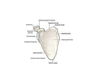
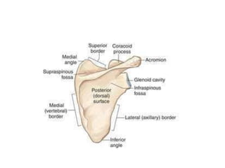
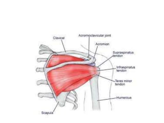
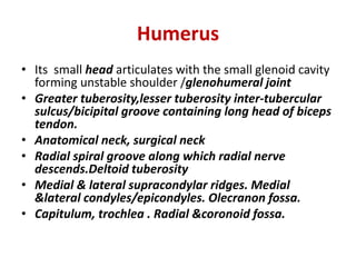
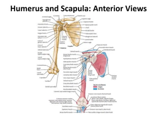
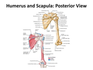
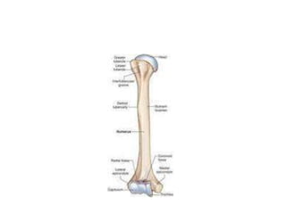
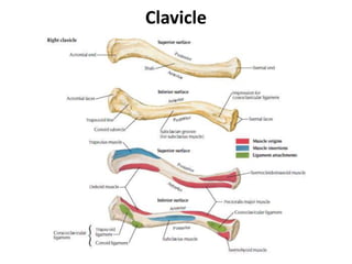
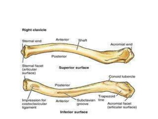
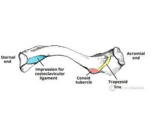
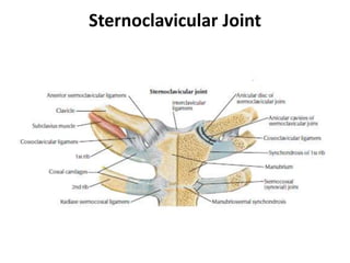
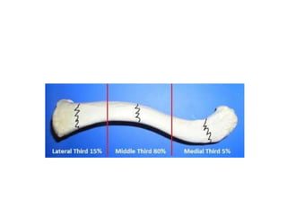
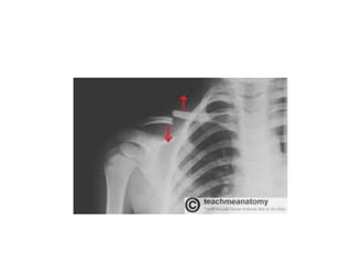
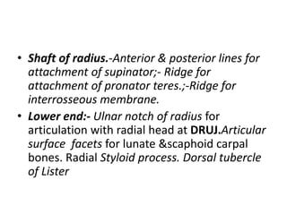
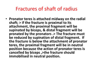
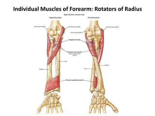
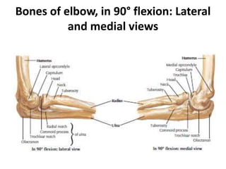
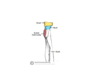
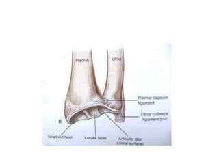
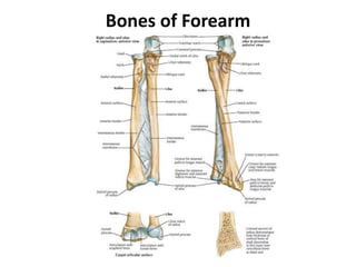
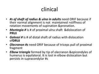
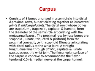
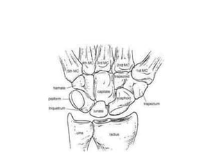
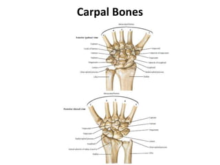
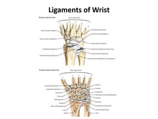
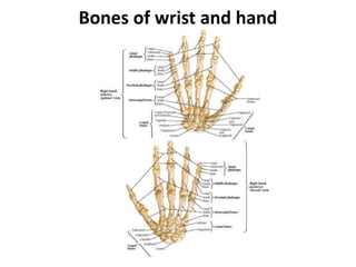
OSTEOLOGY UPPER LIMB
Scapula (shoulder blade)
• Triangular flat bone on the postero-lateral aspect of the thorax
over-lying 2nd to 7th ribs.It has lateral/axillary border ,
medial/vertebral border (extending from the superior angle to
inferior angle) & superior border (which has a suprascapular
notch transmitting suprascapular nerve; It is covered by superior
transverse scapular ligament thus converting it into suprascapular
foramen above which are suprascapular vessels.
-It has the body with:-Posterior/dorsal surface divided by the spine of
scapula into smaller supraspinous fossa containing supraspinatus
muscle & larger Infraspinous fossa containing infraspinatus
muscle. The spine of scapula extends laterally as the expanded
acromium. The costal surface is concave, from which subscapularis
muscle arise.
• Neck of scapula
• Glenoid (head) with shallow glenoid
cavity/fossa .Long head of biceps is attached
to supraglenoid tubercle.Long head of triceps
is attached to infraglenoid tubercle
• Coracoid process projecting from the glenoid
& is palpable anteriorly.
Coracobrachialis,P.minor & short head of
biceps are attached to it.
Humerus
• Its small head articulates with the small glenoid cavity
forming unstable shoulder /glenohumeral joint
• Greater tuberosity,lesser tuberosity inter-tubercular
sulcus/bicipital groove containing long head of biceps
tendon.
• Anatomical neck, surgical neck
• Radial spiral groove along which radial nerve
descends.Deltoid tuberosity
• Medial & lateral supracondylar ridges. Medial
&lateral condyles/epicondyles. Olecranon fossa.
• Capitulum, trochlea . Radial &coronoid fossa.
Humerus and Scapula: Anterior Views
Humerus and Scapula: Posterior View
• Fractures of surgical neck with involvement of
tuberosities are very common especially in
elderly.
• Anterior shoulder dislocation are commonest as
the joint is the most unstable
• Fractures of shaft of humerus-Always check for
wrist drop from damage of radial nerve especially
at spiral groove.
• Supracondylar #s are very common especially in
children.
Clavicle (collar bone)
• Its primary ossification is membranous without
cartilage stage at 5th -6th week.
• Transmits force from upper limb to axial skeleton.
• As a strut holding the arm free from the trunk.
• Protection of neurovascular bundle as it passes
through ‘cervical-axillary canal’.
• Muscle attachment- P.major, Trapezius,
sternocleidomastoid, deltoid,
subclavius(Subclavian groove)
• Long bone but has no medullary cavity.
• Medial 2/3 is rounded & convex forwards for
clearance of neurovascular bundle through ‘cervical-
axillary canal’ from the neck to axilla. The medial
sternal end forms sternoclavicular joint (has a dic)
with the manubrium of the sternum. It also articulates
with 1st costal cartilage.
• The lateral acromial end forms acromialclavicular joint
(has incomplete disc) with acromium of spine of
scapula at the shoulder.
• Coracoclavicular ligaments (conoid &trapezoid)
joining it to coracoid process & conoid &trapezoid
tubercles of clavicle.
• Fractures of clavicle are very common at the
junction between middle &lateral 1/3 often from
indirect force.The medial fragment becomes
elevated by sternocleidomastoid. The lateral
fragment sags down from the weight of the arm ,
but also adducted especially by P.Major.
• Acromial-clavicular disruptions are also common
with torn acromial clavicular &coracoclavicular
ligaments >diastasis of the joint.
Clavicle
Sternoclavicular Joint
Radius
• Head- cylindrical.Hollow superior surface to
articlulate with capitullum of humerus.Also
articulates with radial notch of ulna to form
PRUJ for pronation & supination.
• Radial neck which is clasped by annular
ligament.
• Radial tuberosity for attachment of biceps
tendon the most powerful supinator.
• Shaft of radius.-Anterior & posterior lines for
attachment of supinator;- Ridge for
attachment of pronator teres.;-Ridge for
interrosseous membrane.
• Lower end:- Ulnar notch of radius for
articulation with radial head at DRUJ.Articular
surface facets for lunate &scaphoid carpal
bones. Radial Styloid process. Dorsal tubercle
of Lister
Fractures of shaft of radius
• Pronator teres is attached midway on the radial
shaft; > if the fracture is proximal to its
attachment, the proximal fragment will be
supinated by biceps, & distal fragment will be
pronated by the pronators .> The fracture must
be reduced by supination of distal fragment. If
the fracture is below the attachment of pronator
teres, the proximal fragment will be in neutral
position because the action of pronator teres is
cancelled by biceps ,>the fracture should
immobilised in neutral position.
Individual Muscles of Forearm: Rotators of Radius
Bones of elbow: Anterior and
Posterior views
Bones of elbow: Lateral and medial
views
Bones of elbow, in 90° flexion: Lateral
and medial views
Elbow: Radiographs
• ‘Pulled elbow ‘ may occur in children by
sudden jerk of the radial head inferiorly out of
annular ligament.
• Colles’ fracture very common due to
osteoporosis especially in postmenopausal
women presenting as ‘dinner folk’ deformity
Ulna
• Medial &longer.Angled laterally >10-15 degrees of ‘elbow
carrying angle’>15 in women.Tapers distally (opposite to
radius which tapers proximally).*Distally,the ulna does not
articulate directly with the carpus from which it is
separated by ‘triangular fibro-cartilage articular disc’
• Proximal end;-Olecranon process; coronoid process;
trochlear notch (articulating with trochlea of humerus);
radial notch (Articulating with radial head to form PRUJ);
supinator crest &supinator fossa.
• Shaft of ulna:- interrosseous border
• Head of ulna.
• Styloid process of ulna
Bones of Forearm
clinical
• #s of shaft of radius & ulna in adults need ORIF because if
their normal alignment is not maintained >stiffness of
rotation movements of supination &pronation.
• Montegia # is # of proximal ulna shaft &dislocation of
PRUJ
• Galeazi # is # of distal shaft of radius with dislocation
ofDRUJ
• Olecranon #s need ORIF because of triceps pull of proximal
fragment
• Elbow triangle formed by tip of olecranon &epicondyles of
humerus is equilateral. It is lost in elbow dislocation but
persists in supracondylar #s
Carpus
• Consists of 8 bones arranged in a semicircle into distal
&proximal rows, but articulating together at intercarpal
joints & midcarpal joints.The distal row( whose bones
are trapezium , trapezoid , capitate & hamate, form
the diameter of the semicircle articulating with the
metacarpal bases . The proximal row (whose bones are
scaphoid , lunate, triquetral & pisiform) form the
proximal convexity ,with scaphoid &lunate articulating
with distal radius at the wrist joint. A straight
longitudinal line through 3rd MC, capitate & lunate
passes across the wrist joint.The flexor volar surface of
the carpus is concave to accommodate the flexor
tendons(>10) & median nerve at the carpal tunnel .
• Scaphoid;-Tuberosity of scaphoid; Waist of scaphoid
can be felt at the anatomical snuffbox where
tenderness may be due to # at waist of scaphoid.The
waist has vascular foramina more numerous distally >in
#s waist of scaphoid AVN may occur of proximal
fragment.
• Lunate semilunar .AVN occassionally
• Capitate(keystone)largest carpal bone
• Trapezium forms 1st trapezial metacarpal saddle joint.
• Hamate.Hook of hamate.
• Pisiform is like sesamoid bone
Metacarpus (palm) & phalangese
(Fingers)
• 5 metacarpals; head, neck, shaft ,base
articulating with the bases of proximal phalanges
at MPjoints.1st metacarpal is short &thick.
• The thumb has only two phalanges proximal &
distal articulating at IP joint enabling the thumb
which is shorter & independent mobility across
the palm.The other fingers have got three
phalanges ,proximal, middle &distal articulating
at PIP &DIP joints
Carpal Bones
Ligaments of Wrist
Bones of wrist and hand
Wrist and hand: Radiographs

Osteology upper limb by Dr G Kamau

  • 1.
  • 2.
    Scapula (shoulder blade) •Triangular flat bone on the postero-lateral aspect of the thorax over-lying 2nd to 7th ribs.It has lateral/axillary border , medial/vertebral border (extending from the superior angle to inferior angle) & superior border (which has a suprascapular notch transmitting suprascapular nerve; It is covered by superior transverse scapular ligament thus converting it into suprascapular foramen above which are suprascapular vessels. -It has the body with:-Posterior/dorsal surface divided by the spine of scapula into smaller supraspinous fossa containing supraspinatus muscle & larger Infraspinous fossa containing infraspinatus muscle. The spine of scapula extends laterally as the expanded acromium. The costal surface is concave, from which subscapularis muscle arise.
  • 3.
    • Neck ofscapula • Glenoid (head) with shallow glenoid cavity/fossa .Long head of biceps is attached to supraglenoid tubercle.Long head of triceps is attached to infraglenoid tubercle • Coracoid process projecting from the glenoid & is palpable anteriorly. Coracobrachialis,P.minor & short head of biceps are attached to it.
  • 7.
    Humerus • Its smallhead articulates with the small glenoid cavity forming unstable shoulder /glenohumeral joint • Greater tuberosity,lesser tuberosity inter-tubercular sulcus/bicipital groove containing long head of biceps tendon. • Anatomical neck, surgical neck • Radial spiral groove along which radial nerve descends.Deltoid tuberosity • Medial & lateral supracondylar ridges. Medial &lateral condyles/epicondyles. Olecranon fossa. • Capitulum, trochlea . Radial &coronoid fossa.
  • 8.
    Humerus and Scapula:Anterior Views
  • 9.
    Humerus and Scapula:Posterior View
  • 11.
    • Fractures ofsurgical neck with involvement of tuberosities are very common especially in elderly. • Anterior shoulder dislocation are commonest as the joint is the most unstable • Fractures of shaft of humerus-Always check for wrist drop from damage of radial nerve especially at spiral groove. • Supracondylar #s are very common especially in children.
  • 12.
    Clavicle (collar bone) •Its primary ossification is membranous without cartilage stage at 5th -6th week. • Transmits force from upper limb to axial skeleton. • As a strut holding the arm free from the trunk. • Protection of neurovascular bundle as it passes through ‘cervical-axillary canal’. • Muscle attachment- P.major, Trapezius, sternocleidomastoid, deltoid, subclavius(Subclavian groove) • Long bone but has no medullary cavity.
  • 13.
    • Medial 2/3is rounded & convex forwards for clearance of neurovascular bundle through ‘cervical- axillary canal’ from the neck to axilla. The medial sternal end forms sternoclavicular joint (has a dic) with the manubrium of the sternum. It also articulates with 1st costal cartilage. • The lateral acromial end forms acromialclavicular joint (has incomplete disc) with acromium of spine of scapula at the shoulder. • Coracoclavicular ligaments (conoid &trapezoid) joining it to coracoid process & conoid &trapezoid tubercles of clavicle.
  • 14.
    • Fractures ofclavicle are very common at the junction between middle &lateral 1/3 often from indirect force.The medial fragment becomes elevated by sternocleidomastoid. The lateral fragment sags down from the weight of the arm , but also adducted especially by P.Major. • Acromial-clavicular disruptions are also common with torn acromial clavicular &coracoclavicular ligaments >diastasis of the joint.
  • 15.
  • 18.
  • 21.
    Radius • Head- cylindrical.Hollowsuperior surface to articlulate with capitullum of humerus.Also articulates with radial notch of ulna to form PRUJ for pronation & supination. • Radial neck which is clasped by annular ligament. • Radial tuberosity for attachment of biceps tendon the most powerful supinator.
  • 22.
    • Shaft ofradius.-Anterior & posterior lines for attachment of supinator;- Ridge for attachment of pronator teres.;-Ridge for interrosseous membrane. • Lower end:- Ulnar notch of radius for articulation with radial head at DRUJ.Articular surface facets for lunate &scaphoid carpal bones. Radial Styloid process. Dorsal tubercle of Lister
  • 23.
    Fractures of shaftof radius • Pronator teres is attached midway on the radial shaft; > if the fracture is proximal to its attachment, the proximal fragment will be supinated by biceps, & distal fragment will be pronated by the pronators .> The fracture must be reduced by supination of distal fragment. If the fracture is below the attachment of pronator teres, the proximal fragment will be in neutral position because the action of pronator teres is cancelled by biceps ,>the fracture should immobilised in neutral position.
  • 24.
    Individual Muscles ofForearm: Rotators of Radius
  • 25.
    Bones of elbow:Anterior and Posterior views
  • 26.
    Bones of elbow:Lateral and medial views
  • 27.
    Bones of elbow,in 90° flexion: Lateral and medial views
  • 28.
  • 31.
    • ‘Pulled elbow‘ may occur in children by sudden jerk of the radial head inferiorly out of annular ligament. • Colles’ fracture very common due to osteoporosis especially in postmenopausal women presenting as ‘dinner folk’ deformity
  • 32.
    Ulna • Medial &longer.Angledlaterally >10-15 degrees of ‘elbow carrying angle’>15 in women.Tapers distally (opposite to radius which tapers proximally).*Distally,the ulna does not articulate directly with the carpus from which it is separated by ‘triangular fibro-cartilage articular disc’ • Proximal end;-Olecranon process; coronoid process; trochlear notch (articulating with trochlea of humerus); radial notch (Articulating with radial head to form PRUJ); supinator crest &supinator fossa. • Shaft of ulna:- interrosseous border • Head of ulna. • Styloid process of ulna
  • 33.
  • 34.
    clinical • #s ofshaft of radius & ulna in adults need ORIF because if their normal alignment is not maintained >stiffness of rotation movements of supination &pronation. • Montegia # is # of proximal ulna shaft &dislocation of PRUJ • Galeazi # is # of distal shaft of radius with dislocation ofDRUJ • Olecranon #s need ORIF because of triceps pull of proximal fragment • Elbow triangle formed by tip of olecranon &epicondyles of humerus is equilateral. It is lost in elbow dislocation but persists in supracondylar #s
  • 36.
    Carpus • Consists of8 bones arranged in a semicircle into distal &proximal rows, but articulating together at intercarpal joints & midcarpal joints.The distal row( whose bones are trapezium , trapezoid , capitate & hamate, form the diameter of the semicircle articulating with the metacarpal bases . The proximal row (whose bones are scaphoid , lunate, triquetral & pisiform) form the proximal convexity ,with scaphoid &lunate articulating with distal radius at the wrist joint. A straight longitudinal line through 3rd MC, capitate & lunate passes across the wrist joint.The flexor volar surface of the carpus is concave to accommodate the flexor tendons(>10) & median nerve at the carpal tunnel .
  • 37.
    • Scaphoid;-Tuberosity ofscaphoid; Waist of scaphoid can be felt at the anatomical snuffbox where tenderness may be due to # at waist of scaphoid.The waist has vascular foramina more numerous distally >in #s waist of scaphoid AVN may occur of proximal fragment. • Lunate semilunar .AVN occassionally • Capitate(keystone)largest carpal bone • Trapezium forms 1st trapezial metacarpal saddle joint. • Hamate.Hook of hamate. • Pisiform is like sesamoid bone
  • 38.
    Metacarpus (palm) &phalangese (Fingers) • 5 metacarpals; head, neck, shaft ,base articulating with the bases of proximal phalanges at MPjoints.1st metacarpal is short &thick. • The thumb has only two phalanges proximal & distal articulating at IP joint enabling the thumb which is shorter & independent mobility across the palm.The other fingers have got three phalanges ,proximal, middle &distal articulating at PIP &DIP joints
  • 42.
  • 43.
  • 44.
  • 45.
    Wrist and hand:Radiographs