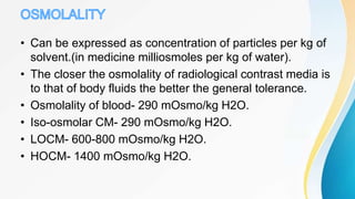
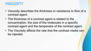
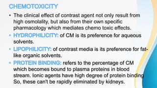
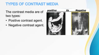
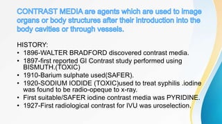
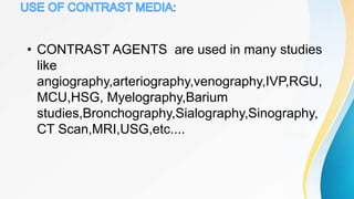
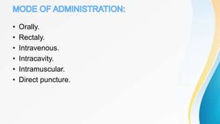
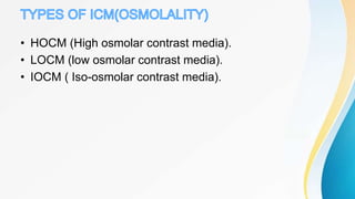
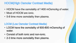
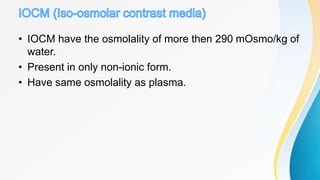
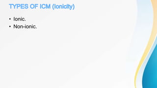
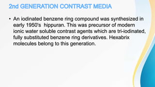
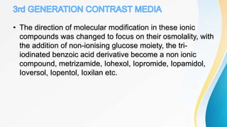
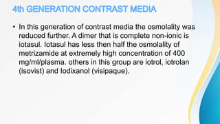
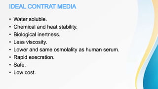
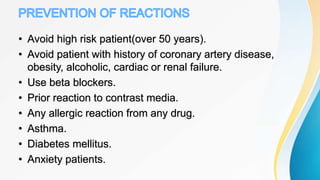
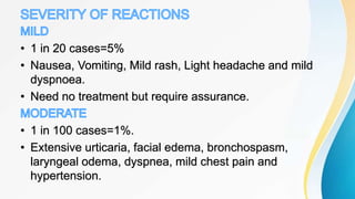
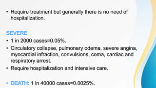
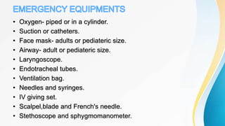
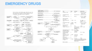
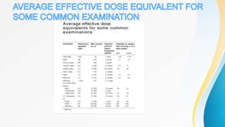
This document discusses the history and use of contrast media and emergency drugs for radiology procedures. It begins by outlining the early discoveries of contrast agents from the 1890s to the 1920s. It then describes how contrast agents are administered and their properties like osmolality and viscosity. The document discusses common contrast agents like barium, iodine-based agents, and categorizes them as high vs low osmolar. It outlines risks of contrast media and expected adverse reaction rates. The document concludes by listing emergency equipment needed to treat potential severe reactions.
Various forms of contrast media have been used to improve medical imaging.
• Their value has long been recognized, as attested to by their common daily use
in imaging departments worldwide.
• Like all other pharmaceuticals, however, these agents are not completely devoid
of risk.
• Adverse side effects from the administration of contrast media vary from minor
physiological disturbances to rare severe life-threatening situations.
• Preparation for prompt treatment of contrast media reactions must include
preparation for the entire spectrum of potential adverse events and include
prearranged response planning with availability of appropriately trained
personnel, equipment, and medications.
• Thorough familiarity with the presentation and emergency treatment of
contrast media reactions must be part of the environment in which all
intravascular contrast media are administered.
VERY BASICS OF CONTRAST MEDIA IN RADIOLOGY.
CLASSIFICATION OF CONTRAST MEDIA.
APPLICATION OF CONTRAST MEDIA.
XRAY, CT, ULTRASOUND AND MRI CONTRAST AGENTS.
Digital image processing in recent year has shown a tremendous potential for application to medical sciences and one of them is “Computed Tomography scanning machine“. Conventional x-ray has a disadvantage while examining the internal structure of the body that it superimposes the 3D image of our body into single plane which makes diagnosis often difficult and confusing. But Computed tomography (CT) imaging systems generate three-dimensional (3-D) images of internal body structures using complex x-ray and computer-aided tomographic imaging techniques. Digital processing geometry are widely used in three-dimensional (3-D) reconstruction of bone geometry and density features for 3-D modelling purposes. For this C.t scanning is widely used in industrial sector and in medical sector. This report represents an overview of “C.T scanning” technique, its advantages and application related to different sector.
this slide sharer contents are basic principle of CT fluoroscopy , software and hardware parts of equipment and image aqua cation and radiation dose comparison and videos related to equipment .
Radiographic Contrast Agents And Contrast-induced Nephropathy
All contrast agents have a basic structure of a benzene ring, which is composed of 6 joined carbon atoms, each of which has an attached hydrogen atom.
Contrast media consist of triiodinated benzene rings, whereby 3 hydrogen atoms are replaced with attached iodine atoms.
Monomers contain 1 triiodinated benzene ring, and dimers contain 2 triiodinated benzene rings
Stain/certified fixed orthodontic courses by Indian dental academyIndian dental academy
The Indian Dental Academy is the Leader in continuing dental education , training dentists in all aspects of dentistry and
offering a wide range of dental certified courses in different formats.for more details please visit
www.indiandentalacademy.com
A brief of contrast media used in various modalities of radiodiagnosis including barium, USG, CT, and MRI with their advantages and disadvantages and ADR.
MAGNETIC RESONANCE ANGIOGRAPHY (MRA).pptxRohit Bansal
MAGNETIC RESONANCE ANGIOGRAPHY (MRA) AND MAGNETIC RESONANCE SPECTROSCOPY (MRS) ARE DESCRIBED IN DETAILIN THIS PPT. CONTENT TAKEN FROM MUTIPLE BOOKS AND GENERALS.
Prix Galien International 2024 Forum ProgramLevi Shapiro
June 20, 2024, Prix Galien International and Jerusalem Ethics Forum in ROME. Detailed agenda including panels:
- ADVANCES IN CARDIOLOGY: A NEW PARADIGM IS COMING
- WOMEN’S HEALTH: FERTILITY PRESERVATION
- WHAT’S NEW IN THE TREATMENT OF INFECTIOUS,
ONCOLOGICAL AND INFLAMMATORY SKIN DISEASES?
- ARTIFICIAL INTELLIGENCE AND ETHICS
- GENE THERAPY
- BEYOND BORDERS: GLOBAL INITIATIVES FOR DEMOCRATIZING LIFE SCIENCE TECHNOLOGIES AND PROMOTING ACCESS TO HEALTHCARE
- ETHICAL CHALLENGES IN LIFE SCIENCES
- Prix Galien International Awards Ceremony
The prostate is an exocrine gland of the male mammalian reproductive system
It is a walnut-sized gland that forms part of the male reproductive system and is located in front of the rectum and just below the urinary bladder
Function is to store and secrete a clear, slightly alkaline fluid that constitutes 10-30% of the volume of the seminal fluid that along with the spermatozoa, constitutes semen
A healthy human prostate measures (4cm-vertical, by 3cm-horizontal, 2cm ant-post ).
It surrounds the urethra just below the urinary bladder. It has anterior, median, posterior and two lateral lobes
It’s work is regulated by androgens which are responsible for male sex characteristics
Generalised disease of the prostate due to hormonal derangement which leads to non malignant enlargement of the gland (increase in the number of epithelial cells and stromal tissue)to cause compression of the urethra leading to symptoms (LUTS
These simplified slides by Dr. Sidra Arshad present an overview of the non-respiratory functions of the respiratory tract.
Learning objectives:
1. Enlist the non-respiratory functions of the respiratory tract
2. Briefly explain how these functions are carried out
3. Discuss the significance of dead space
4. Differentiate between minute ventilation and alveolar ventilation
5. Describe the cough and sneeze reflexes
Study Resources:
1. Chapter 39, Guyton and Hall Textbook of Medical Physiology, 14th edition
2. Chapter 34, Ganong’s Review of Medical Physiology, 26th edition
3. Chapter 17, Human Physiology by Lauralee Sherwood, 9th edition
4. Non-respiratory functions of the lungs https://academic.oup.com/bjaed/article/13/3/98/278874
Couples presenting to the infertility clinic- Do they really have infertility...Sujoy Dasgupta
Dr Sujoy Dasgupta presented the study on "Couples presenting to the infertility clinic- Do they really have infertility? – The unexplored stories of non-consummation" in the 13th Congress of the Asia Pacific Initiative on Reproduction (ASPIRE 2024) at Manila on 24 May, 2024.
MANAGEMENT OF ATRIOVENTRICULAR CONDUCTION BLOCK.pdfJim Jacob Roy
Cardiac conduction defects can occur due to various causes.
Atrioventricular conduction blocks ( AV blocks ) are classified into 3 types.
This document describes the acute management of AV block.
Report Back from SGO 2024: What’s the Latest in Cervical Cancer?bkling
Are you curious about what’s new in cervical cancer research or unsure what the findings mean? Join Dr. Emily Ko, a gynecologic oncologist at Penn Medicine, to learn about the latest updates from the Society of Gynecologic Oncology (SGO) 2024 Annual Meeting on Women’s Cancer. Dr. Ko will discuss what the research presented at the conference means for you and answer your questions about the new developments.
These lecture slides, by Dr Sidra Arshad, offer a quick overview of physiological basis of a normal electrocardiogram.
Learning objectives:
1. Define an electrocardiogram (ECG) and electrocardiography
2. Describe how dipoles generated by the heart produce the waveforms of the ECG
3. Describe the components of a normal electrocardiogram of a typical bipolar leads (limb II)
4. Differentiate between intervals and segments
5. Enlist some common indications for obtaining an ECG
Study Resources:
1. Chapter 11, Guyton and Hall Textbook of Medical Physiology, 14th edition
2. Chapter 9, Human Physiology - From Cells to Systems, Lauralee Sherwood, 9th edition
3. Chapter 29, Ganong’s Review of Medical Physiology, 26th edition
4. Electrocardiogram, StatPearls - https://www.ncbi.nlm.nih.gov/books/NBK549803/
5. ECG in Medical Practice by ABM Abdullah, 4th edition
6. ECG Basics, http://www.nataliescasebook.com/tag/e-c-g-basics
Title: Sense of Taste
Presenter: Dr. Faiza, Assistant Professor of Physiology
Qualifications:
MBBS (Best Graduate, AIMC Lahore)
FCPS Physiology
ICMT, CHPE, DHPE (STMU)
MPH (GC University, Faisalabad)
MBA (Virtual University of Pakistan)
Learning Objectives:
Describe the structure and function of taste buds.
Describe the relationship between the taste threshold and taste index of common substances.
Explain the chemical basis and signal transduction of taste perception for each type of primary taste sensation.
Recognize different abnormalities of taste perception and their causes.
Key Topics:
Significance of Taste Sensation:
Differentiation between pleasant and harmful food
Influence on behavior
Selection of food based on metabolic needs
Receptors of Taste:
Taste buds on the tongue
Influence of sense of smell, texture of food, and pain stimulation (e.g., by pepper)
Primary and Secondary Taste Sensations:
Primary taste sensations: Sweet, Sour, Salty, Bitter, Umami
Chemical basis and signal transduction mechanisms for each taste
Taste Threshold and Index:
Taste threshold values for Sweet (sucrose), Salty (NaCl), Sour (HCl), and Bitter (Quinine)
Taste index relationship: Inversely proportional to taste threshold
Taste Blindness:
Inability to taste certain substances, particularly thiourea compounds
Example: Phenylthiocarbamide
Structure and Function of Taste Buds:
Composition: Epithelial cells, Sustentacular/Supporting cells, Taste cells, Basal cells
Features: Taste pores, Taste hairs/microvilli, and Taste nerve fibers
Location of Taste Buds:
Found in papillae of the tongue (Fungiform, Circumvallate, Foliate)
Also present on the palate, tonsillar pillars, epiglottis, and proximal esophagus
Mechanism of Taste Stimulation:
Interaction of taste substances with receptors on microvilli
Signal transduction pathways for Umami, Sweet, Bitter, Sour, and Salty tastes
Taste Sensitivity and Adaptation:
Decrease in sensitivity with age
Rapid adaptation of taste sensation
Role of Saliva in Taste:
Dissolution of tastants to reach receptors
Washing away the stimulus
Taste Preferences and Aversions:
Mechanisms behind taste preference and aversion
Influence of receptors and neural pathways
Impact of Sensory Nerve Damage:
Degeneration of taste buds if the sensory nerve fiber is cut
Abnormalities of Taste Detection:
Conditions: Ageusia, Hypogeusia, Dysgeusia (parageusia)
Causes: Nerve damage, neurological disorders, infections, poor oral hygiene, adverse drug effects, deficiencies, aging, tobacco use, altered neurotransmitter levels
Neurotransmitters and Taste Threshold:
Effects of serotonin (5-HT) and norepinephrine (NE) on taste sensitivity
Supertasters:
25% of the population with heightened sensitivity to taste, especially bitterness
Increased number of fungiform papillae
1. CONTRAST MEDIA AND EMERGENCY
DRUGS
ROHIT BANSAL (Assistant Professor Radio-Physics)
2. HISTORY:
• 1896-WALTER BRADFORD discovered contrast media.
• 1897-first reported GI Contrast study performed using
BISMUTH.(TOXIC)
• 1910-Barium sulphate used(SAFER).
• 1920-SODIUM IODIDE (TOXIC)used to treat syphilis .iodine
was found to be radio-opeque to x-ray.
• First suitable/SAFER iodine contrast media was PYRIDINE.
• 1927-First radiological contrast for IVU was uroselection.
3. :
• CONTRAST AGENTS are used in many studies
like
angiography,arteriography,venography,IVP,RGU,
MCU,HSG, Myelography,Barium
studies,Bronchography,Sialography,Sinography,
CT Scan,MRI,USG,etc....
6. • Can be expressed as concentration of particles per kg of
solvent.(in medicine milliosmoles per kg of water).
• The closer the osmolality of radiological contrast media is
to that of body fluids the better the general tolerance.
• Osmolality of blood- 290 mOsmo/kg H2O.
• Iso-osmolar CM- 290 mOsmo/kg H2O.
• LOCM- 600-800 mOsmo/kg H2O.
• HOCM- 1400 mOsmo/kg H2O.
7. • Viscosity describes the thickness or resistance to flow of a
contrast agent.
• The thickness of a contrast agent is related to the
concentration, the size of the molecules in a specific
contrast agent and the temperatre of the contrast agent.
• The Viscosity affects the rate that the contrast media can
be injected.
8. • The clinical effect of contrast agent not only result from
high osmolality, but also from their own specific
pharmacology which mediates chemo toxic effects.
of CM is its preference for aqueous
solvents.
of contrast media is its preference for fat-
like organic solvents.
refers to the percentage of CM
which becomes bound to plasma proteins in blood
stream. Ionic agents have high degree of protein binding
So, these can't be rapidly eliminated by kidneys.
9. The contrast media are of
two types:
• Positive contrast agent.
• Negative contrast agent.
10. • Radio-opeque.
• High atomic number.
• high atomic weight.
• radiation dosen't penetrate easily.
• bright on radiograph.
• example-Barium sulphate and organic iodine compounds.
11. • High atomic number(56).
• High radio-opaque.
• It is insoluble in water or lipid.
• non toxic.
• It is non absorbable.(rapid removed)
• It is suitable for double contrast studies.
• better coating properties over the lining of gut.
• form- white crystalline powder.
• pH-5.3.
• particle size- 5-12 micrometer.
14. • Microbar paste: 100% high density and viscosity paste
used in oseophagus and stomach studies.
• Microbar suspention: 95% modrate density and viscosity
paaste used in small intestine ,large intestine and rectum
studies.
M/S ESKEY FINE CHEMICAL LIMITED.
15. • Chemical peritonitis due to extravasation of
contrast.(Inflammation of peritoneum).
• Extravasation into bronchial tree, urinary track and other
body cavities may cause inflammation.
• Intravascular entry of barium can cause
Embolism.(blockage of vessels).
• Barium Encephalopathy.(abnormal brain function).
• After exam sometimes may solidify, difficult to evacuate.
17. • High atomic number.(53).
• High atomic weight.(127).
• High radio-opaque.
• Less toxic.
• Total iodine content in the
body is 50 mg.
• Not metallic.
• High density.
• High osmolality.
18. • All are derivatives of tri-
iodinated benzene ring.
• In ionic contrast acidic group
with sodium and meglumine is
attached at C1.
• In non-ionic contrast amide
group is attached at C1.
• Iodine atom attached at C2,C4
and C6.
20. • HOCM have the osmolality of 1400 mOsmo/kg of water.
• Most of HOCM are ionic.
• 5-8 time more osmolality then plasma.
• LOCM have the osmolality of 600-800 mOsmo/kg of
water.
• Consist of both ionic and non-ionic.
• 2-3 time more osmolality then plasma.
21. • IOCM have the osmolality of more then 290 mOsmo/kg of
water.
• Present in only non-ionic form.
• Have same osmolality as plasma.
23. • Less cost.
• More adverse reaction.
• High osmolality.
• Creates hypertonic conditions.
• Dissociate into separate ions when injected.
• Example- Meglumine iothalamate, Sodium
iothalamate,Sodium diatrizoate,Meglumine diatizoate.
24. • High cost.
• Less adverse reaction.
• Less osmolality.
• Remains nearly isotonic.
• Doesn't dissociate into small particles when injected.
• Example- Hexabrix.
25. • Also known as high osmolar
contrast media or conventional
contrast media.
• These are salt consisting of a
sodium or meglumine cation and
triodinated benzoate anion.
• Ratio of 3 iodine atom per
molecule to 2 particles in solution.
i.e. 3:2
• Examples are diatrizoate
(urografin), Iothalamate (conray).
26. • Two benzene rings (each with
3 iodine atom) are linked by a
bridge to form a large
compound.
• Carries only one carboxylic
group. so, known as monoacid
dimers.
• Iodine particle ratio is 6:2.
• Example- Ioxaglate (hexabrix).
• Conc. of 59%(320mg/ml),
relatively low osmolality of 600
mOsmo/kg.
27. • Carboxyl group of monomeric salts is replaced by a non-
ionising radical and CONH2 producing a iodine particle
ratio of 3:1.
• Metrizamide was the earliest non-ionic monomer and
proved as an excellent contrast media but was very
expensive, impossible to autoclave and unstable in
solution.
• Relatively low osmolality (600-800 mOsmo/kg).
• Example- Iohexol, Iopamidol, Ioversol, Iopentol etc.
29. • A ratio of 6 iodine atoms for each molecule in solution with
satisfactory iodine concentration at iso-osmolality.so,
known as iso-osmolar contrast media.
• These agents have very low toxicity because of iso-
osmolality, non-ionic character and also these posses a
very large number of hydroxyl group.
• Examples- Iotrolan (isovist), Iodixanol (visipaque).
30. Sodium bromide, Bishmuth in oil and lipiodol were earliest
intravenous contrast media.
Sodium iodide was also used but found to be toxic.
The first organic iodide to be used were pyridine ring
containing one atom of iodine per molecule.
31. • An iodinated benzene ring compound was synthesized in
early 1950's hippuran. This was precursor of modern
ionic water soluble contrast agents which are tri-iodinated,
fully substituted benzene ring derivatives. Hexabrix
molecules belong to this generation.
32. • The direction of molecular modification in these ionic
compounds was changed to focus on their osmolality, with
the addition of non-ionising glucose moiety, the tri-
iodinated benzoic acid derivative become a non ionic
compound, metrizamide, Iohexol, Iopromide, Iopamidol,
Ioversol, Iopentol, Ioxilan etc.
33. • In this generation of contrast media the osmolality was
reduced further. A dimer that is complete non-ionic is
iotasul. Iotasul has less then half the osmolality of
metrizamide at extremely high concentration of 400
mg/ml/plasma. others in this group are iotrol, iotrolan
(isovist) and Iodixanol (visipaque).
34. • Water soluble.
• Chemical and heat stability.
• Biological inertness.
• Less viscosity.
• Lower and same osmolality as human serum.
• Rapid execration.
• Safe.
• Low cost.
35. • Avoid high risk patient(over 50 years).
• Avoid patient with history of coronary artery disease,
obesity, alcoholic, cardiac or renal failure.
• Use beta blockers.
• Prior reaction to contrast media.
• Any allergic reaction from any drug.
• Asthma.
• Diabetes mellitus.
• Anxiety patients.
36. • 1 in 20 cases=5%
• Nausea, Vomiting, Mild rash, Light headache and mild
dyspnoea.
• Need no treatment but require assurance.
• 1 in 100 cases=1%.
• Extensive urticaria, facial edema, bronchospasm,
laryngeal odema, dyspnea, mild chest pain and
hypertension.
37. • Require treatment but generally there is no need of
hospitalization.
• 1 in 2000 cases=0.05%.
• Circulatory collapse, pulmonary odema, severe angina,
myocardial infraction, convulsions, coma, cardiac and
respiratory arrest.
• Require hospitalization and intensive care.
1 in 40000 cases=0.0025%.
38. • Oxygen- piped or in a cylinder.
• Suction or catheters.
• Face mask- adults or pediateric size.
• Airway- adult or pediateric size.
• Laryngoscope.
• Endotracheal tubes.
• Ventilation bag.
• Needles and syringes.
• IV giving set.
• Scalpel,blade and French's needle.
• Stethoscope and sphygmomanometer.
Editor's Notes
Two benzene rings (each with 3 iodine atom) are linked by a bridge to form a large compound.
carries only one carboxylic group.so, known as monoacid dimers.
iodine particle ratio is 6:2.
example- ioxaglate(hexabrix).
conc. of 59%(320mg/ml), relatively low osmolality of