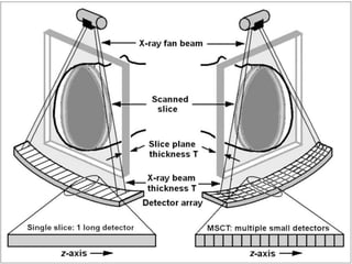
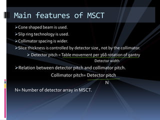
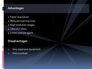
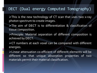
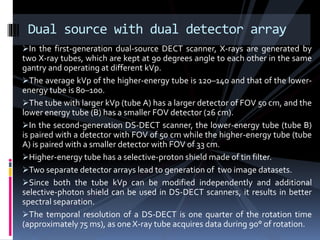
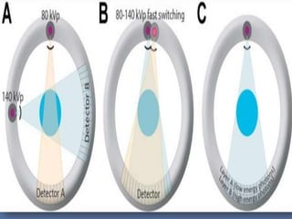
MSCT & DECT
MR. ROHIT BANSAL
ASISTANT PROFESSOR
MAMC (AGROHA)
MSCT (Multi slice computed tomography) / MDCT (Multi detector
computed tomography) are sixth generation CT Scan.
It uses helical scanning and low voltage slip rings.
In MSCT, multiple row of detectors are used instead of using single row
of detector.
The multiple row of detectors allows for registration of more than one
channel per gantry rotation.
Multiple detector array:
1. the detector element is a solid state scintillation detector.
2.They are designed in row and column.
Multi-detector array is available in two design:
1. Linear array (Equal width)
2. Adaptive array (Unequal array)
MSCT/MDCT
Cone shaped beam is used.
Slip ring technology is used.
Collimator spacing is wider.
Slice thickness is controlled by detector size , not by the collimator.
 Detector pitch =Table movement per 360̊ rotation of gantry
Detector width
Relation between detector pitch and collimator pitch.
Collimator pitch= Detector pitch
N
N= Number of detector array in MSCT.
Main features of MSCT
Advantages
1. Faster acquisition.
2. Reduced scanning time.
3. High resolution images.
4. Ultra thin slices.
5. Lesser contrast agent.
Disadvantages
1. Very expensive equipment.
2. Data overload.
This is the new technology of CT scan that uses two x-ray
photon spectrum to create images.
The aim of DECT is to differentiation & classification of
tissue composition.
Principle: Material separation of different composition is
achieved by DECT.
CT numbers at each voxel can be compared with different
energies.
Linear attenuation co-efficient of different elements will be
different so that unique absorption properties of two
materials permit their material classification.
DECT (Dual energy Computed Tomography)
The principle of dual-energy CT imaging is based on the differential
absorption of energy at variable kVp settings. For example, let us consider a
substance (A) with K-edge at 60 kVp and another (B) at 130 kVp. If we image
multiple combinations of A and/or B at 80 and 140 kVp, there will be
differential attenuation at both these energy settings depending on the
relative percentage of these substances. The object containing large amount
of substance A will show higher attenuation at 80 kVp and lower attenuation
at 140 kVp; whereas object containing larger amount of substance B will show
higher attenuation at 140 kVp. In clinical applications, the constituents of soft
tissues have a different K-edge (variable from 0.01 to 0.53), away from that of
iodine (33.2) or calcium (4); hence iodine or calcium can be distinguished from
soft tissues at dual energy imaging. In diagnostic imaging, once the datasets
at 80 kVp and 140 kVp are generated, the attenuation of the enhanced
structures containing iodine (vessels, highly perfused organs) are more on a
80 kVp image than on a 140 kVp image. This difference in attenuation varies
between different organs; for example, highly vascular organs and vessels
have higher difference than muscles Post-processing software's use this
information to generate a virtual non-contrast image, or to calculate the
material composition within a specific region of interest.
In the first-generation dual-source DECT scanner, X-rays are generated by
two X-ray tubes, which are kept at 90 degrees angle to each other in the same
gantry and operating at different kVp.
The average kVp of the higher-energy tube is 120–140 and that of the lower-
energy tube is 80–100.
The tube with larger kVp (tube A) has a larger detector of FOV 50 cm, and the
lower energy tube (B) has a smaller FOV detector (26 cm).
In the second-generation DS-DECT scanner, the lower-energy tube (tube B)
is paired with a detector with FOV of 50 cm while the higher-energy tube (tube
A) is paired with a smaller detector with FOV of 33 cm.
Higher-energy tube has a selective-proton shield made of tin filter.
Two separate detector arrays lead to generation of two image datasets.
Since both the tube kVp can be modified independently and additional
selective-photon shield can be used in DS-DECT scanners, it results in better
spectral separation.
The temporal resolution of a DS-DECT is one quarter of the rotation time
(approximately 75 ms), as one X-ray tube acquires data during 90° of rotation.
Dual source with dual detector array
Advantages
1. Better acquisition of images.
2. Effective in differentiating structures.
3. Better spatial resolution.
Disadvantages
1. However, the drawback of DS-DECT lies in term of temporal
misregistration, since the scans are not acquired simultaneously, but
at a small time gap (although in ms).
2. Another drawback is the limited dual-energy FOV (33 cm), which
makes it difficult to image patients with larger body habitus.
3. Complex technology.
4. More expensive.
Single-source DECT (SS-DECT) scanners use a single X-ray
tube, which generates high- and low-energy X-ray spectra by
rapid changing of the kVp settings (at an interval of 0.5 msec)
in the same rotation.
Since the tube current cannot be changed so rapidly, in order
to maximize the contrast to noise ratio, the exposure time ratio
is changed between two acquisitions. For example, 65%
exposure time is given for the 80 kVp beam and 35% for the
140 kVp beam.
The SS-DECT requires a very- fast detector and data-
acquisition system with fast-sampling capability.
The detector arrays used in SS-DECT are made of cesium-
activated garnet (Gemstone Spectral Imaging; GE Healthcare,
Piscataway, NJ).
Single source with fast KV switching
Better temporal registration between two datasets, as the images from
high and low-energy acquisition are acquired almost simultaneously.
Larger FOV of imaging (50 cm) and easier quantification of material
density.
Disadvantages
The disadvantage of SS-DECT includes a poorer spectral separation (hence
less accurate material characterization compared to DS-DECT with selective
photon shielding) as a selective-photon shield is not commercially available
in such scanners.
Advantage
This scanner contains a single X-ray source with hybrid
detector for high and low energy imaging.
The top layer captures low energy data and the bottom layer
captures high-energy data; which are then used to reconstruct
two separate image datasets.
Such scanners are under development with Phillips, not yet
commercially available.
Single source with dual detector array
Renal calculi characterization
Renal cysts differentiation.
Renal mass characterization and follow-up after ablation.
Adrenal Adenoma.
Hepatic Mass Characterization.
Follow-up after hepatic tumor ablation orTACE.
Hepatic steatosis or iron deposition.
Pulmonary thromboembolism.
Pulmonary nodule evaluation.
Diagnostic Applications
MSCT & DECT.pptx

MSCT & DECT.pptx

  • 1.
    MSCT & DECT MR.ROHIT BANSAL ASISTANT PROFESSOR MAMC (AGROHA)
  • 2.
    MSCT (Multi slicecomputed tomography) / MDCT (Multi detector computed tomography) are sixth generation CT Scan. It uses helical scanning and low voltage slip rings. In MSCT, multiple row of detectors are used instead of using single row of detector. The multiple row of detectors allows for registration of more than one channel per gantry rotation. Multiple detector array: 1. the detector element is a solid state scintillation detector. 2.They are designed in row and column. Multi-detector array is available in two design: 1. Linear array (Equal width) 2. Adaptive array (Unequal array) MSCT/MDCT
  • 6.
    Cone shaped beamis used. Slip ring technology is used. Collimator spacing is wider. Slice thickness is controlled by detector size , not by the collimator.  Detector pitch =Table movement per 360̊ rotation of gantry Detector width Relation between detector pitch and collimator pitch. Collimator pitch= Detector pitch N N= Number of detector array in MSCT. Main features of MSCT
  • 7.
    Advantages 1. Faster acquisition. 2.Reduced scanning time. 3. High resolution images. 4. Ultra thin slices. 5. Lesser contrast agent. Disadvantages 1. Very expensive equipment. 2. Data overload.
  • 8.
    This is thenew technology of CT scan that uses two x-ray photon spectrum to create images. The aim of DECT is to differentiation & classification of tissue composition. Principle: Material separation of different composition is achieved by DECT. CT numbers at each voxel can be compared with different energies. Linear attenuation co-efficient of different elements will be different so that unique absorption properties of two materials permit their material classification. DECT (Dual energy Computed Tomography)
  • 9.
    The principle ofdual-energy CT imaging is based on the differential absorption of energy at variable kVp settings. For example, let us consider a substance (A) with K-edge at 60 kVp and another (B) at 130 kVp. If we image multiple combinations of A and/or B at 80 and 140 kVp, there will be differential attenuation at both these energy settings depending on the relative percentage of these substances. The object containing large amount of substance A will show higher attenuation at 80 kVp and lower attenuation at 140 kVp; whereas object containing larger amount of substance B will show higher attenuation at 140 kVp. In clinical applications, the constituents of soft tissues have a different K-edge (variable from 0.01 to 0.53), away from that of iodine (33.2) or calcium (4); hence iodine or calcium can be distinguished from soft tissues at dual energy imaging. In diagnostic imaging, once the datasets at 80 kVp and 140 kVp are generated, the attenuation of the enhanced structures containing iodine (vessels, highly perfused organs) are more on a 80 kVp image than on a 140 kVp image. This difference in attenuation varies between different organs; for example, highly vascular organs and vessels have higher difference than muscles Post-processing software's use this information to generate a virtual non-contrast image, or to calculate the material composition within a specific region of interest.
  • 10.
    In the first-generationdual-source DECT scanner, X-rays are generated by two X-ray tubes, which are kept at 90 degrees angle to each other in the same gantry and operating at different kVp. The average kVp of the higher-energy tube is 120–140 and that of the lower- energy tube is 80–100. The tube with larger kVp (tube A) has a larger detector of FOV 50 cm, and the lower energy tube (B) has a smaller FOV detector (26 cm). In the second-generation DS-DECT scanner, the lower-energy tube (tube B) is paired with a detector with FOV of 50 cm while the higher-energy tube (tube A) is paired with a smaller detector with FOV of 33 cm. Higher-energy tube has a selective-proton shield made of tin filter. Two separate detector arrays lead to generation of two image datasets. Since both the tube kVp can be modified independently and additional selective-photon shield can be used in DS-DECT scanners, it results in better spectral separation. The temporal resolution of a DS-DECT is one quarter of the rotation time (approximately 75 ms), as one X-ray tube acquires data during 90° of rotation. Dual source with dual detector array
  • 11.
    Advantages 1. Better acquisitionof images. 2. Effective in differentiating structures. 3. Better spatial resolution. Disadvantages 1. However, the drawback of DS-DECT lies in term of temporal misregistration, since the scans are not acquired simultaneously, but at a small time gap (although in ms). 2. Another drawback is the limited dual-energy FOV (33 cm), which makes it difficult to image patients with larger body habitus. 3. Complex technology. 4. More expensive.
  • 13.
    Single-source DECT (SS-DECT)scanners use a single X-ray tube, which generates high- and low-energy X-ray spectra by rapid changing of the kVp settings (at an interval of 0.5 msec) in the same rotation. Since the tube current cannot be changed so rapidly, in order to maximize the contrast to noise ratio, the exposure time ratio is changed between two acquisitions. For example, 65% exposure time is given for the 80 kVp beam and 35% for the 140 kVp beam. The SS-DECT requires a very- fast detector and data- acquisition system with fast-sampling capability. The detector arrays used in SS-DECT are made of cesium- activated garnet (Gemstone Spectral Imaging; GE Healthcare, Piscataway, NJ). Single source with fast KV switching
  • 14.
    Better temporal registrationbetween two datasets, as the images from high and low-energy acquisition are acquired almost simultaneously. Larger FOV of imaging (50 cm) and easier quantification of material density. Disadvantages The disadvantage of SS-DECT includes a poorer spectral separation (hence less accurate material characterization compared to DS-DECT with selective photon shielding) as a selective-photon shield is not commercially available in such scanners. Advantage
  • 15.
    This scanner containsa single X-ray source with hybrid detector for high and low energy imaging. The top layer captures low energy data and the bottom layer captures high-energy data; which are then used to reconstruct two separate image datasets. Such scanners are under development with Phillips, not yet commercially available. Single source with dual detector array
  • 16.
    Renal calculi characterization Renalcysts differentiation. Renal mass characterization and follow-up after ablation. Adrenal Adenoma. Hepatic Mass Characterization. Follow-up after hepatic tumor ablation orTACE. Hepatic steatosis or iron deposition. Pulmonary thromboembolism. Pulmonary nodule evaluation. Diagnostic Applications