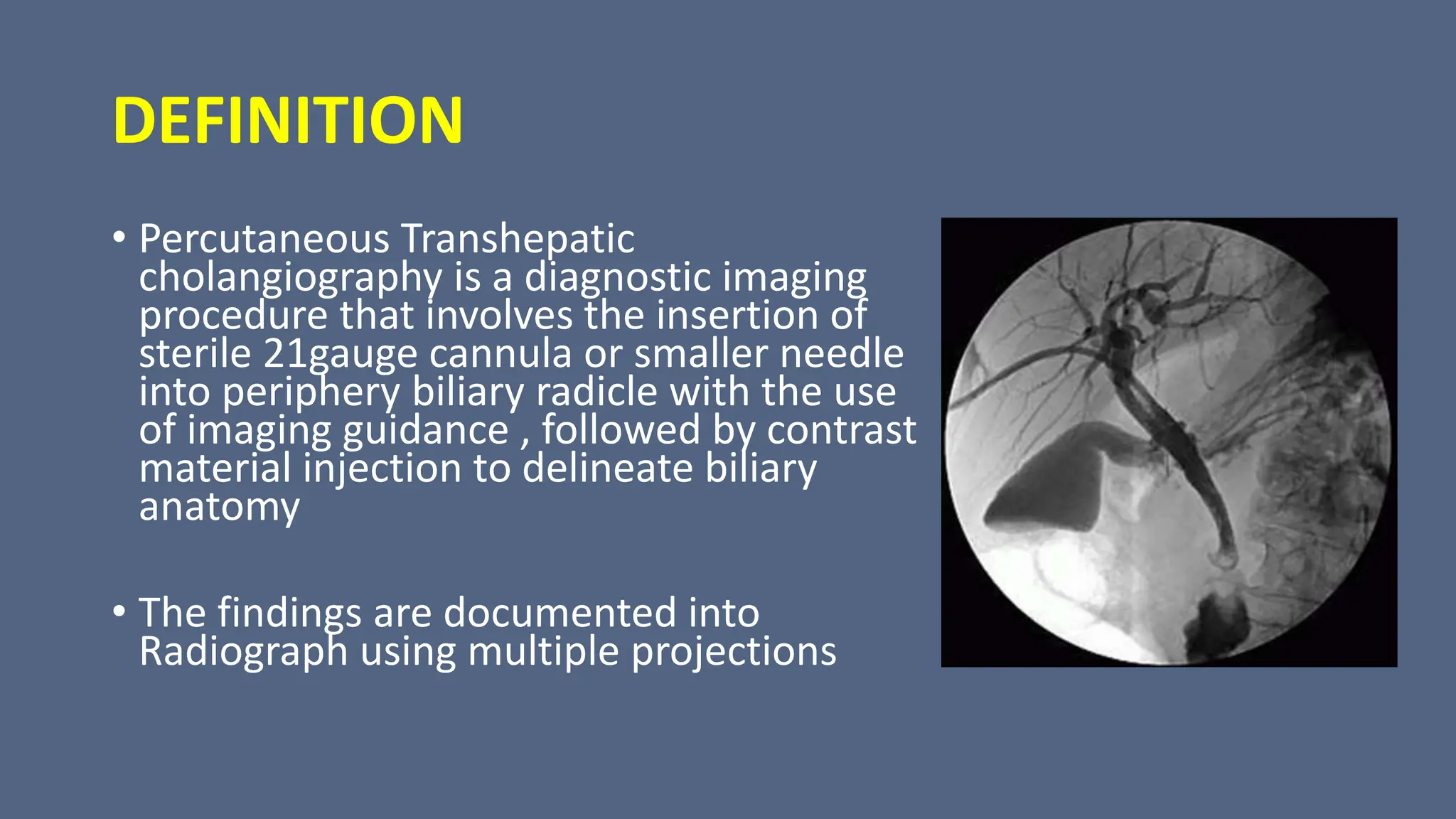
2. DEFINITION
• Percutaneous Transhepatic
cholangiography is a diagnostic imaging
procedure that involves the insertion of
sterile 21gauge cannula or smaller needle
into periphery biliary radicle with the use
of imaging guidance , followed by contrast
material injection to delineate biliary
anatomy
• The findings are documented into
Radiograph using multiple projections
5. METHODS OF IMAGING BILIARY TRACT
Indirect Imaging
• Involves Non Invasive Technique
• Ultrasound
• Plain Film
• CT
• MRCP
• Oral, IV Cholecystography
• Cholescintigraphy
Direct Imaging
• Involves Invasive Radiologic
Procedure
• ERCP
• Operative and T tube
Cholangiogram
• PTC
6. INDICATION
• To distinguish intrahepatic cholestasis from extrahepatic obstruction
• To determine the site , if possible the nature of extrahepatic obstruction
• Gold standard for bile duct stricture
• Prior to therapeutic intervention e.g. biliary drainage procedure.
• Determine cause of cholangitis
• For Whom ERCP is not Technically feasible
• Evaluate Suspected bile duct Inflammatory disease
• Chronic pancreatitis
• Post operative fistula
• Undiagnosed jaundice
7. CHOLESTASIS
• Cholestasis is a condition in which the flow of bile is slowed
or blocked
• Various Causes
• Extrahepatic Cholestasis occurs outside the liver and can be
caused through:
• Bile Duct Tumor, Cysts, Narrowing of bile duct ( Strictures),
Stones in common bile duct, pancreatitis, Pancreatic tumor
or pseudocysts, Pressure in bile duct due to nearby mass
tumor
9. CONTRAINDICATION
• Coagulation Problem( Platelet count< 100,000 mm3)
• Biliary Infection
• Hypersensitivity to contrast medium
• Severe heart disease and respiratory disease
• Poor General condition of patient
• Ascites
• Anemic
• Hydatid disease
10. Patient Preperation
• Haemogobin, prothrombin time, platelets are checked and
corrected if necessary
• Prophylactic Antibiotics: Ampicillin 500mg to commence 24
hours before and continue for 3 days after the examination.
• Nil by mouth for 5 hours prior to the procedure
• Preoxygenation , pulse optimetry should be used and
adequate nursing and radiographic support should be
available.
11.
12. Contrast Medium
• LOCM 150mg i/ml, 20-60 ml.
• Sedative: Midazolam and Analgesic: Lidnocane 3%
13. Equipment
• Ultrasound Machine, Fluroscopic Unit
with spot film device and tilting table
• Chiba needle ( 21F , 15cm long with
width outer and inner diameter 0.7 and
0.5 mm respectively)
• Betadine, cotton swabs, sterile gloves,
forceps
15. Technique
• Patient lie supine on the Fluoroscopy table
• Best to puncture liver in cranial position as possible. However it is
best to avoid traversing the pleura and it is essential not to puncture
the lungs as the pleural reflection are much deeper
• The Skin, intercoastal muscle and liver capsule are infiltrated
withlidocaine, after which 3mm skin incision is made
• Under US observation ,during suspended respiration the chiba needle
is inserted into liver but once it is within the liver parenchyma, patient
is allowed shallow respiration
• Once the needle is inserted the stellate is with drawn . If the bile
drips from the hub of needle , it indicates that a duct has been
entered
16. • If intrahepatic duct seems to be dilated , bile should be aspirated
and sent for microbiological examination
• Contrast media is injected to fill the duct system
• The needle is withdrawn , care should be taken not to over fill an
obstructed dust system , because septic shock may be precipitated
• Patient should remain in bed and be carefully monitored for
pulse,blood pressure and temperature and the wound inspected
for atleast 2 hrs
17.
18. Epigarstirc approach
It is preferred when:
• Left lobe cholangiogram is required or if right sided PTC has
failed to produce left lobe cholangiogram
• If there is right lobe atropy or previous right hepatectomy
which results in gall bladder or bowel lying deep to right
lateral wall where they are at risk of puncture with a right
flank approach
19. IMAGES
• With the Undercouch tube and
patient supine:
• PA
• LAO
• RAO
• Rolling the patients on left side will
fill the left ducts and common duct
20. After Care
• Dressing on the puncture site properly
• Bed Rest
• Pulse and Blood pressure measurement half hourly for 6 hrs
• Antibiotic Prophylaxis
21. COMPLICATION
• Leakage of bile into the peritoneal cavity
• Intraperitoneal haemorrhage
• Septicemia
• Hypotension
• Formation of biliary fistula
22. Succesful PTC
• Successful Percutaneous Transhepatic Cholangiography
involves as sufficient needle localization and contrast
material opacification to allow image based diagnosis and
planning of treatment
25. PTBD
• Percutaneous Transhepatic biliary drainage is a therapeutic
procedure that involves sterile cannulation of periphery biliary
radicle after percutaneous puncture followed by imaging guide
wires and catheter manipulation.
• Placement of External or Internal Stent or
tube completes the procedure
26. INDICATION
• Decompress Obstructed Biliary tree
• Dilate Biliary Strictures
• Remove the Bile duct stones when ERCP is contradicted or fails
• Divert Bile from Bile duct leak and stent bile duct defect
• Treatment of Acute Biliary Sepsis
• As preoperative procedure for resectable malignancies, prior to
neoadjuvant chemotherapy
• High biliary obstruction, failed endoscopic drainage, post-
operative cases with biliary obstruction, recurrent malignancies
and multiple segment stricture
• Undiagnosed jaundice
28. EQUIPMENT
• Ultrasound Machine
• Fluroscopic Unit
• Pigtail catheter because it have self retention capacity
• Urobag and Connecting tube for Urobag
31. Drain in PTBD
• External drain- biliary drain is in
the bile duct above a stenosis and
drains the bile externally into a bag
outside the patient
32.
33. • Internal drain-
• Bile flow through the drain to the digestive tract
• Achieved following transhepatic or endoscopic cannulation
of biliary tree
• Endoprosthesis with proximal and distal side holes or a
transhepatic catheter is sited across a stricture (making
internal-external drain)
34.
35.
36. • The purpose of biliary interventions is to decompress the
obstructed biliary system and if possible to develop
communication between biliary tree and bowel allowing
physiological bile flow.
• Biliary drainage help in improving liver function prior to
operation and neo-adjuvant chemotherapy.
37. Preprocedure Imaging
• A high quality imaging is preferred to determine the level of
obstruction like thin section Computed Tomography(CT) or
Magnetic Resonance Cholangiopancreatography(MRCP).
• Three dimentional constructions makes it easier to appreciate
the level of bile duct obstruction and normal variants of biliary
anatomy.
• CT and MR also provide an assessment of the functional hepatic
parenchyma. Drainage of the portion of the liver without an
intact portal venous blood supply will not result in improvement
of liver function.
• Ultrasound is useful to determine portal vein patency, the
presence of biliary dilation, and intraductal tumor but is itself not
usually adequate for procedure planning.
38. Preprocedure determination
• Preprocedure determination of the level of bile duct obstruction
has important therapeutic and prognostic implications.
• Biliary obstruction is divided into “low” and “high” bile duct
obstruction.
• Low bile duct obstruction occurs below the usual insertion of the
cystic duct
• In low bile duct obstruction complete drainage of the entire
biliary tree can be accomplished by a single, well-placed catheter
or stent because the obstruction is below the confluence of right
and left bile ducts. Low bile duct obstruction is best treated
endoscopically to avoid the risks associated with percutaneous
drainage and nuisance of an external catheter.
• When an endoscopic approach is not technically possible, a
percutaneous approach to low bile duct obstruction is indicated.
39. Preprocedure determination
• High bile duct obstruction occurs above the cystic duct
insertion
• High bile duct obstruction is best approached
percutaneously, because a specific duct can be targeted to
maximize drainage of functional parenchyma based on
preprocedure imaging. Percutaneous access allows for easier
and more reliable targeting than an endoscopic approach.
40. Sedation
• Biliary drainage is performed with conscious sedation, often
with short-acting benzodiazepines and narcotics.
41. Procedure
• Procedure is done with patient in supine position after
screening with USG
• Skin is cleaned and local anesthesia is injected in skin,
deeper tissue and liver capsule. (depending on right or left or
both ductal system needs drainage) then draped.
42. Approaches
• Two approaches are used
1. Right lateral approach
2. Anterior subcostal or left sided subxiphoid approach
43. Right lateral approach
Entry point:
• 2 cm below right costo-phrenic recess (on inspiration) at a
level ~2cm anterior to mid-axillary line (9th to 11th intercostal
space)
• Suspended respiration - chiba needle inserted into the liver.
• Advanced parallel to table top in direction of xiphisternum -
just short of right lateral margin of spine.
44. Anterior subcostal or subxiphoid approach
• Segment 3 bile duct lies close to anterior surface of left lobe
of liver and can be punctured by anterior subcostal approach
under USG guidance
• Skin entry point:
• Left of midline and needle directed posteromedially towards
the duct
45.
46. • The sectoral duct (usually segment 3 or 6 for left and right
sided PTBD respectively) is punctured by chiba needle (25
cm), using USG as guidance at approx. 1-3 cm away from the
secondary biliary confluence. (during suspended respiration
liver is punctured but once it is within the liver parenchyma
the patient is allowed shallow respirations.)
47. • Once there is backflow of bile a soft J tip guide wire (0.38
inch) is passed through the needle which is then exchanged
for a 5F or 6F dilator followed by removal of the guidewire.
• Cholangiogram is performed slowly to define the biliary
anatomy and type of obstruction (tapered, abrupt, irregular)
48. • Subsequently, the dilator is exchanged for a biliary
manipulation catheter or an angled tip angiographic catheter
over the wire.
• When the catheter tip is at the site of obstruction, attempt is
made to manipulate the soft hydrophilic guidewire (either
straight or J tip) to cross the stricture.
49. • Once the wire is across the stricture and in the duodenum,
the catheter is pushed over the wire into the duodenum.
• Soft wire is then replaced by Lunderquist guide-wire over
which the tract and stricture are dilated with 7F and 8F
dilators
50. • Then an 8.3F internal-external drainage catheter (ring biliary
catheter) is positioned across the stricture and the position is
confirmed with contrast injection.
• In patient where initial attempt to cross the stricture fails an
external drainage catheter is left with tip proximal to the
obstruction and internalization is attempted after a gap of 2-
4 days.
51. • For stricture distal to hilum i.e patent primary confluence,
technique is simple and single drainage is sufficient. Drainage
of single or both system can be done when primary biliary
confluence is completely occluded.
• Multiple segmental drainage may also be performed when
secondary confluence is involved.
• Chronically obstructed biliary segments with parenchymal
atrophy need not be drained as improvement in liver
function is unlikely.
52. Complication
Early complication (within 30 days)
• Pain at site of puncture
• Bile leak (risk of biliary peritonitis, biloma formation)
• Hemobilia
• Bilovenous fistula
• Arterial injury
• Acute pancreatitis
• Catheter-kinking, dislocation
• Pneumothorax, hemothorax
54. Difficulties
• If biliary dilation is mild or minimal, a micropuncture access
set with 21G needle and 0.018 wire can be used for initial
puncture.
• In grossly dilated biliary system, it is difficult to identify the
point of obstruction and to obtain stability for passing the
guide wire. (so external drainage is done initially to
decompress the system, reduces edema and increases
stability, straightens the wire course).
55. Aftercare
• Pulse and blood pressure half-hourly for 6 h.
• Antibiotics for at least 3 days.
• An externally draining catheter should be regularly flushed
through with normal saline and exchanged at 3-monthly
intervals.
A branching ductal system that collects bile from the hepatic parenchyma and transports it to the duodenum constitutes the biliary tree
By convention the biliary tree is divided into intra- and extra-hepatic bile ducts
Intrahepatic bile duct-network of small tubes that carry bile inside the liver-The right and left lobes of the liver are drained by these ducts.
Extrahepatic-from liver and gallbladder to small intestine-the common hepatic duct and the common bile duct.
magnetic resonance cholangio pancreatography
resoEndoscopic retrograde cholangiopancreatography
Cholangitis-inflammation of bile duct
Endoscopic retrograde cholangiopancretography
(Ercp-Endoscopic retrograde cholangiopancreatography)
Amyloid build up
Parasitic infection ..cyst in liver
Pt-prothrombin time
Aptt-activated partial prothrombin time
Iohexol(omnipaque)
22/23G
(Right flank approach)
Peripheral duct
Chiba neddle is inserted into peripheral duct
PTC showing the obstruction in the middle CBD and the presence of bilioenteric fistula (arrow)..
Liver-gall bladder
External biliary drainage catheter placed with common bile duct (CBD) obstruction secondary to pancreatic carcinoma.
Internal- external drain can be placed=drain is placed in the bile duct and the tip is localised in the duodenum allowing both bile flow through the drain to the digestive tract (internal) or into the bag (external). Capping the drain will stop the external drainage and results in internal drainage only.
Internal drainage through Transhepatic approach
Obstruction of CBD due to pancreatic head carcinoma….. Internal and external biliary drainage in patient with obstruction at CBD
A duct in the right lobe of the liver is chosen that has a horizontal or caudal course to the porta hepatis
Both procedure can be done in usg or fluoroscopic guidance
Use of stiff end or hard metallic guidewire to go across lesion is strictly contraindicated as this can perforate the bile duct causing hemorrhage and lead to false tract formation.
probing should be gentle using the floppy end
Communication betn hepatic duct and portal vein branches