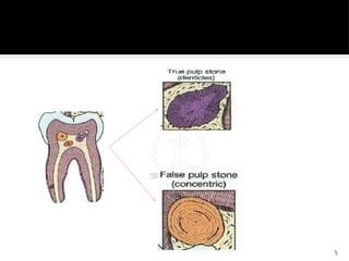
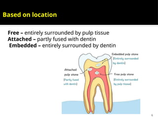
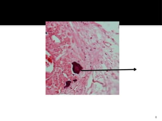
1
Regressive alterations –
Pulp calcification ,
Hypercementosis
2
Pulp calcifications
Masses of calcified tissue present on pulp
chamber and roots of teeth
Common phenomenon occurs with increasing
age
Classified
1. Based on morphologic forms
- Discrete pulp stones
- Diffuse calcifications
2. Based on location
- Free
- Attached
- Embedded
3
Pulp stones (Denticles)
 Nodular, calcified masses appearing in
either or both the coronal and root
portion of the pulp organ in teeth
 Seen in functional as well as
unerupteed embedded teeth
 Noted in with systemic or genetic
diseases – dentine dysplasia,
dentinogenesis imperfecta and Van der
Woude syndrome
 Exact cause unknown
4
Types of denticles
 True denticles
- made up of dentine that is lined by
odontoblast.
- more common in pulp chamber than in
root
 False denticles
- composed of localized masses of calcified
material
- arranged in concentric layers or lamellae
deposited around a central nidus
5
6
Free – entirely surrounded by pulp tissue
Attached – partly fused with dentin
Embedded – entirely surrounded by dentin
Based on location
7
Diffuse Calcifications
 Most commonly seen in root canals of
teeth
 Resemble calcification seen in other
tissues of the body following
degeneration.
 Also termed as calcific degeneration
 Usually seen as amorphous, unorganized
linear strands or columns paralleling the
blood vessels and nerves of pulp .
8
9
Etiology of Pulp
Calcification
 Pulp degeneration
 Increasing age
 Circulatory disturbances within the pulp ,
long standing local irritants such as
dental caries, pulp-capping procedures,
healed tooth fractures, tooth injury
restorations and periodontal diseases
 Orthodontic tooth movements
 Trauma
10
Mechanism
Local metabolic
dysfunction
Trauma
Hyalinization of injured
cells
Vascular damage
(Thrombosis,Vessel wall
damage)
Fibrosis
Mineralization (Nidus
formation)
Pulp stone
Growth with
time
11
Hypercementosis
12
13
14
Etiology
 Variety of circumstances favour
deposition of excessive amounts of
cementum. Include :
 Accelerated elongation of a tooth
 Inflammation about tooth
 Tooth repair
 Osteitis deformans or paget’s disease
of bone
 Spike formation of cementum
Local
factors
15
Clinical featurs
 No clinical signs and symptoms
 No visible signs
 No remarked tooth sensitivity on
percussion
 Tooth with hypercementosis extracted –
roots appear larger in diameter than
normal with rounded apices.
16
Radiographic features
 Thickening and apparent blunting of
root with rounding of apex
 Apex appears bulbous
 Lamina dura will follow the outline of
teeth in normal periodontal ligament
space
 Irregular accumulation of cementum
that is accommodated by related area
of bone resorption
17
18
Histologic features
 Microscopic appearance – excessive
amount of secondary or cellular
cementum deposited directly over the
thin layer of primary cementum
 Involved area – entire root or a portion
mainly apical region
 Secondary cementum –
osteocementum because of its cellular
nature and its resemblance to bone
19
 Cementum – arranged
in concentric layers
around root and
frequently shows
numerous resting lines,
indicated by deeply
staining hematoxyphilic
lines parallel to root
surface
20
Treatment
 No treatment
21
22
Types of cementicles
 Free Cementicles – lamellated cemental
bodies that lie freely in PDL
 Attached Cementicles – cemental
bodies which are attached to root
surface
23
Resorption of teeth
 Tooth resorption – process where all or
part of tooth structure lost due to
activation of body’s innate capacity to
remove mineralization tissue as
mediated by cells such as osteoclasts
 Physiological or pathological
24
 Physiological tooth resorption – affects
deciduous teeth results into loosening
and shedding off due to pressure
arising from the underlying successors
 Pathological tooth resorption –
resorption of permanent teeth
25
External resorption
 Resorption occurring in PDL (external
surface)
 Extremely common
 Many local factors are involved in
external resorption
26
Causes of external
resorption
27
Periapical inflammation
Caries/ trauma
Increases inflammatory response
Protective layer is damaged ,osteoclasts
acts on the damaged area.
resorption.
28
Reimplantation of teeth
 Extensive resorption
 Tooth outside socket – PDL cells
undergo necrosis
 No PDL cells – bone view tooth as
foreign body thus resorption occurs
29
Tumours and Cysts
Pressure
phenomenon
30
Excessive mechanical or occlusal
forces
 Resorption from orthodontic
treatment
Maxillary incisor region
31
Impaction of teeth
32
Idiopathic resorption
 Root resorption without any etiological
factor
 When multiple teeth involved – multiple
idiopathic root resorption
33
Radiographic feature
 Moth – eaten appearance
 Borders irregular & ill defined
 Outline of root canal is normal
 Root canal is seen running through the defect
 Almost always accompanied by resorption of
bone
 Radiolucency appear in root and adjacent
bone
 Lesion moves away from canal as angulation
changes
34
Treatment
 Removal of tooth
 Identification and elimination of
accelerating factor
35
Internal root resorption
 Other synonyms
Chronic perforating hyperplasia of pulp
Internal granuloma
Odontoclastoma
Pink tooth of Mummery
 The progressive destruction of intraradicular
dentin
and dentinal tubules along the middle and apical
thirds of the canal walls as a result of clastic
activities.
 Resorption begins centrally within tooth (dental pulp)
 Cause unknown – but may be associated with carious
exposure and pulpal infection
36
Clinical features
 No early clinical symptoms
 First evidence of lesion – appearance of
pink – hued area on crown of tissue
filling resorbed area
 Incisors, cuspids, bicuspids and molars
– resorption reported one time or
another
37
Types of internal resorption
38
Radiographic features
 Margins are smooth & clearly defined
 Root canal walls appear to balloon out
 Outline of root canal distorted
 Root canal & resorptive defect appear
continuous
 Radiolucency confined to root (does not
involve bone)
Bone lesion seen only if resorption
perforate tooth
Lesion appear close to root canal in
different angulations
39
40
Histopathology
 An internal resorption lesion mainly
consists of granulation tissue.
 The pulpal connective tissue is highly
vascularized with varying degrees of
inflammation, infiltrated by lymphocytes,
macrophages, neutrophilic leukocytes, and
plasma cells.
 Neutrophils and macrophages are
attached to the mineralized dentin surface.
 “Resorptive bays” with numerous
odontoclasts are also seen
41
42
Treatment
 Early identification – endodontic
treatment
 Later removal of teeth

Regressive alterations pulp stones ppt..pptx

  • 1.
    1 Regressive alterations – Pulpcalcification , Hypercementosis
  • 2.
    2 Pulp calcifications Masses ofcalcified tissue present on pulp chamber and roots of teeth Common phenomenon occurs with increasing age Classified 1. Based on morphologic forms - Discrete pulp stones - Diffuse calcifications 2. Based on location - Free - Attached - Embedded
  • 3.
    3 Pulp stones (Denticles) Nodular, calcified masses appearing in either or both the coronal and root portion of the pulp organ in teeth  Seen in functional as well as unerupteed embedded teeth  Noted in with systemic or genetic diseases – dentine dysplasia, dentinogenesis imperfecta and Van der Woude syndrome  Exact cause unknown
  • 4.
    4 Types of denticles True denticles - made up of dentine that is lined by odontoblast. - more common in pulp chamber than in root  False denticles - composed of localized masses of calcified material - arranged in concentric layers or lamellae deposited around a central nidus
  • 5.
  • 6.
    6 Free – entirelysurrounded by pulp tissue Attached – partly fused with dentin Embedded – entirely surrounded by dentin Based on location
  • 7.
    7 Diffuse Calcifications  Mostcommonly seen in root canals of teeth  Resemble calcification seen in other tissues of the body following degeneration.  Also termed as calcific degeneration  Usually seen as amorphous, unorganized linear strands or columns paralleling the blood vessels and nerves of pulp .
  • 8.
  • 9.
    9 Etiology of Pulp Calcification Pulp degeneration  Increasing age  Circulatory disturbances within the pulp , long standing local irritants such as dental caries, pulp-capping procedures, healed tooth fractures, tooth injury restorations and periodontal diseases  Orthodontic tooth movements  Trauma
  • 10.
    10 Mechanism Local metabolic dysfunction Trauma Hyalinization ofinjured cells Vascular damage (Thrombosis,Vessel wall damage) Fibrosis Mineralization (Nidus formation) Pulp stone Growth with time
  • 11.
  • 12.
  • 13.
  • 14.
    14 Etiology  Variety ofcircumstances favour deposition of excessive amounts of cementum. Include :  Accelerated elongation of a tooth  Inflammation about tooth  Tooth repair  Osteitis deformans or paget’s disease of bone  Spike formation of cementum Local factors
  • 15.
    15 Clinical featurs  Noclinical signs and symptoms  No visible signs  No remarked tooth sensitivity on percussion  Tooth with hypercementosis extracted – roots appear larger in diameter than normal with rounded apices.
  • 16.
    16 Radiographic features  Thickeningand apparent blunting of root with rounding of apex  Apex appears bulbous  Lamina dura will follow the outline of teeth in normal periodontal ligament space  Irregular accumulation of cementum that is accommodated by related area of bone resorption
  • 17.
  • 18.
    18 Histologic features  Microscopicappearance – excessive amount of secondary or cellular cementum deposited directly over the thin layer of primary cementum  Involved area – entire root or a portion mainly apical region  Secondary cementum – osteocementum because of its cellular nature and its resemblance to bone
  • 19.
    19  Cementum –arranged in concentric layers around root and frequently shows numerous resting lines, indicated by deeply staining hematoxyphilic lines parallel to root surface
  • 20.
  • 21.
  • 22.
    22 Types of cementicles Free Cementicles – lamellated cemental bodies that lie freely in PDL  Attached Cementicles – cemental bodies which are attached to root surface
  • 23.
    23 Resorption of teeth Tooth resorption – process where all or part of tooth structure lost due to activation of body’s innate capacity to remove mineralization tissue as mediated by cells such as osteoclasts  Physiological or pathological
  • 24.
    24  Physiological toothresorption – affects deciduous teeth results into loosening and shedding off due to pressure arising from the underlying successors  Pathological tooth resorption – resorption of permanent teeth
  • 25.
    25 External resorption  Resorptionoccurring in PDL (external surface)  Extremely common  Many local factors are involved in external resorption
  • 26.
  • 27.
    27 Periapical inflammation Caries/ trauma Increasesinflammatory response Protective layer is damaged ,osteoclasts acts on the damaged area. resorption.
  • 28.
    28 Reimplantation of teeth Extensive resorption  Tooth outside socket – PDL cells undergo necrosis  No PDL cells – bone view tooth as foreign body thus resorption occurs
  • 29.
  • 30.
    30 Excessive mechanical orocclusal forces  Resorption from orthodontic treatment Maxillary incisor region
  • 31.
  • 32.
    32 Idiopathic resorption  Rootresorption without any etiological factor  When multiple teeth involved – multiple idiopathic root resorption
  • 33.
    33 Radiographic feature  Moth– eaten appearance  Borders irregular & ill defined  Outline of root canal is normal  Root canal is seen running through the defect  Almost always accompanied by resorption of bone  Radiolucency appear in root and adjacent bone  Lesion moves away from canal as angulation changes
  • 34.
    34 Treatment  Removal oftooth  Identification and elimination of accelerating factor
  • 35.
    35 Internal root resorption Other synonyms Chronic perforating hyperplasia of pulp Internal granuloma Odontoclastoma Pink tooth of Mummery  The progressive destruction of intraradicular dentin and dentinal tubules along the middle and apical thirds of the canal walls as a result of clastic activities.  Resorption begins centrally within tooth (dental pulp)  Cause unknown – but may be associated with carious exposure and pulpal infection
  • 36.
    36 Clinical features  Noearly clinical symptoms  First evidence of lesion – appearance of pink – hued area on crown of tissue filling resorbed area  Incisors, cuspids, bicuspids and molars – resorption reported one time or another
  • 37.
  • 38.
    38 Radiographic features  Marginsare smooth & clearly defined  Root canal walls appear to balloon out  Outline of root canal distorted  Root canal & resorptive defect appear continuous  Radiolucency confined to root (does not involve bone) Bone lesion seen only if resorption perforate tooth Lesion appear close to root canal in different angulations
  • 39.
  • 40.
    40 Histopathology  An internalresorption lesion mainly consists of granulation tissue.  The pulpal connective tissue is highly vascularized with varying degrees of inflammation, infiltrated by lymphocytes, macrophages, neutrophilic leukocytes, and plasma cells.  Neutrophils and macrophages are attached to the mineralized dentin surface.  “Resorptive bays” with numerous odontoclasts are also seen
  • 41.
  • 42.
    42 Treatment  Early identification– endodontic treatment  Later removal of teeth