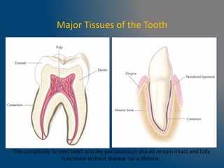
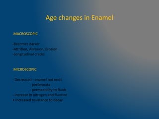
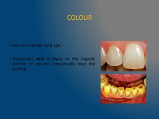
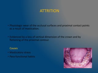
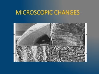
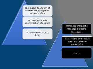
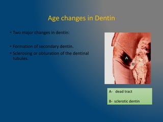
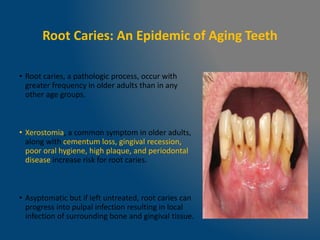
AGE
CHANGES IN
TOOTH
• Ageing is defined as a process of morphological and physiological
disintegration as distinguished from infant, childhood and adolescence
which are typified by processes of integration and coordination.
Carranza.
Major Tissues of the Tooth
The completely formed teeth and the periodontium should remain intact and fully
functional without disease for a lifetime.
Age changes in Enamel
MACROSCOPIC
-Becomes darker
-Attrition, Abrasion, Erosion
-Longitudinal cracks
MICROSCOPIC
- Decreased - enamel rod ends
- perikymata
- permeability to fluids
- Increase in nitrogen and fluorine
• Increased resistance to decay
MACROSCOPIC CHANGES
COLOUR
• Becomes darker with age.
• Associated with changes in the organic
portion of enamel, presumably near the
surface.
ATTRITION ,ABRASION AND EROSION
Attrition Abrasion
Erosion
ATTRITION
• Physiologic wear of the occlusal surfaces and proximal contact points
as a result of mastication.
• Evidenced by a loss of vertical dimension of the crown and by
flattening of the proximal contour.
Causes
• Masticatory stress
• Para-functional habits
Stages of Attrition
Stage I- Wear of enamel at cusps and incisal edges without exposure of
dentin.
Stage II Wear of enamel and exposure of dentin on incisal edges and
isolated area over individual cusps.
Stage III Wear of enamel forming a broad strip on incisal edges and the
confluence of two are more areas of wear over adjacent cusps.
Stage IV Wear of enamel and dentin on incisors to form a plateau on the
teeth to form a central area of dentin surrounded by a peripheral rim of
enamel.
ABRASION
• Pathological wearing away of tooth through abnormal mechanical
processes.
e.g.- abrasive dentifrice
- occupational
- improper flossing
EROSION
• Loss of tooth substance by a chemical process that does not involve
known bacterial action.
• Lingual erosion
e.g. -chronic vomiting
-acidic carbonated
beverages
• Labial erosion
LONGITUDINAL CRACKS
• May be developmental in origin.
• Although their numbers do not increase with age , they become
more obvious.
•Thinning of enamel at the
extent of cementoenamel
junction.
•Thickening at the incisal
edge (maximum facial-palatal
width) due to wear with
advancing age.
•This results in an overall
reduction in the height of the
tooth crown.
Site-specific thickness of enamel
MICROSCOPIC CHANGES
PERIKYMATA
• Transverse wave like grooves which lie parallel to each other and also to
cemento -enamel junction.
• The surfaces of unerupted and recently erupted teeth are covered
completely with pronounced rod ends and perikymata.
• Advancing age shows generalized loss of rod ends and slower flattening
of perikymata which are eventually lost.
• More rapid loss of structure
Facial and lingual surfaces
Anterior teeth
PERMEABILITY TO FLUIDS AND
WATER CONTENT
Due to acquisition of ions from oral fluids.
Crystal size increases
Reduction in pore size within the substance of enamel.
Decrease in permeability and water content
Continuous deposition of
fluoride and nitrogen on
enamel surface
Increase in fluoride
concentration of enamel
Increased resistance to
decay
Hardness and Elastic
modulus of enamel
increases .
Increases the brittleness of
teeth and decreases
permeability.
Cracks
Age changes in Dentin
• Two major changes in dentin:
• Formation of secondary dentin.
• Sclerosing or obturation of the dentinal
tubules.
A- dead tract
B- sclerotic dentin
Secondary dentin
• Secondary dentin forms after the complete formation of the tooth.
Types:
• Physiologic secondary dentin, which forms with normal stimulus,
• Reparative secondary dentin, which forms with traumatic or
abnormal stimulus.
Types of secondary dentin
Regular
Cause: mild stimuli (slow attrition,
slowly progressing caries)
Site of formation: entire pulpal
surface (thicker on pulp roof
and floor)
Tubules: wavy course, decrease in
number
Clinically:
The increase of the dentin
thickness and the closure of the
pulp horns make it much less
possible to expose the pulp
chamber during preparation.
Irregular
Cause: severe stimuli, severe
attrition, erosion, deep caries.
Site of formation: localized (eg pulp
horn)
Tubules: wavy and twisted course,
decrease in number or
atubular
Clinically:
Functions as a barrier for
against caries.
Physiologic regular secondary dentin
Secondary D
Primary D
REPARATIVE /REACTIONARY DENTIN
Odontoblast
s are cut
Degenerate
Replaced by the
migration of
undifferentiated
cells from cell rich
zone
Odontoblasts
are cut
Live odontoblasts
Reparative
Dentin
Reactionary/Re
generated
Dentin
Extensive
abrasion ,
Erosion
,caries and
Operative
process
DEAD TRACTS
• Odontoblastic cell processes in the dentinal
tubules are degenerated, leaving behind
empty, air-filled tubules referred to as ā€œdead
tractsā€.
• Appear black in transmitted light and white
in reflected light.
• Probably the initial step to form sclerotic
dentin.
• Demonstrate decreased sensitivity.
TRANSLUCENT OR SCLEROTIC DENTIN
• Physiological change or
pathological change (caries,
attrition, deep fillings, ) in
primary or secondary dentin.
• More highly mineralized,
harder and denser than normal
dentin
• Seen first near the root apex
in a middle aged person.
• Spreads upwards from the
apex with advancing age.
• This is one of the criteria used
in forensic odontology to
assess the age of an individual
Young dentin Adult dentin Sclerotic dentin
Sclerotic dentin
Appears light in transmitted light and dark in
reflected light
Decrease in sensation to hot and cold stimuli.
Tubular lumen becomes obturated by peritubular dentin.
By age 80, almost all dentinal tubules are fully occluded.
Secondary dentin grows inwardly into the pulp chamber
decreasing the chamber’s size .
Age related Sensitivity changes
Age changes in the pulp
• Reduction in the size and volume of the pulp as a result of a continuous
deposition of dentin.
• Decrease in the number of cells and apparent increased fibrosis with
time, may not be from continued formation of collagen but may be due
to the persistence of connective tissue sheath in an increasingly narrow
pulp space.
• Cellular composition of the pulp is modified.
• Fibroblasts and Odontoblasts show degeneration with decrease in
size and decrease in the number of cell organelles.
• Ultrastructural studies reveal an increase in vacuole numbers and
gradual degenerative changes leading to the absence of cells.
Changes in vascular distribution:
• There is a narrowing of the circumference of the blood vessels.
• Atherosclerotic changes are seen in small arteries in the root pulp of
aging teeth.
• Intimal layer of the vessel is thickened resulting in a small lumen.
Changes in nerve distribution:
• Degeneration and loss of pulpal nerve fibres may affect transmission
from pulpal structures, resulting in increased thresholds to pain
stimuli.
• Myelin sheath changes and terminal axon remolding due to age
related axon injury could be sources of abnormal pain in the oral
region.
Pulp stones
• These are nodular, calcified masses appearing in either or both
the coronal or root portions of the pulp organ.
• True -Made of dentin and lined by odontoblasts ,found close to
root apex
• False -Formed from degenerating cells which mineralize, usually
found in the pulp chamber
Free -Stone not related to pulp
space wall, surrounded by soft
tissue.
Adherent -Stone attached to wall
of pulp space, not fully enclosed
by dentin.
Embedded -Stone enclosed within
canal wall.
• Fibrodentine -Material produced by fibroblast-like cells against dentin
prior to differentiation of a new generation of odontoblast-like cells.
• Dystrophic calcification -Inappropriate biomineralization of the pulp
in the absence of mineral imbalance.
Root Caries: An Epidemic of Aging Teeth
• Root caries, a pathologic process, occur with
greater frequency in older adults than in any
other age groups.
• Xerostomia, a common symptom in older adults,
along with cementum loss, gingival recession,
poor oral hygiene, high plaque, and periodontal
disease increase risk for root caries.
• Asyptomatic but if left untreated, root caries can
progress into pulpal infection resulting in local
infection of surrounding bone and gingival tissue.
ENDODONTIC CONSIDERATIONS
• Formation of a permanent tooth
generally completes in three years
after its eruption into the oral cavity
but this doesnot apply to maturation
of apex.
• Remodeling/deposition of the
cementum occuring at the apex is an
aging process.
• This probably occurs to compensate
for the attrited enamel, or due to
physiological mesial migration of
teeth, or as a response to occlusal
forces.
• As a sequelae to depostion, there is an increase in the overall
distance from the root apex to the apical constriction of the root
canal.
• Working length of a tooth is relatively shorter from the radiographic
apex for an aged tooth than it is for a young adult.
• Diameter of the apical foramen does not change with age.
• D. Arola, R.K. Reprogel carried out a study to evaluate effects of
aging on the mechanical behavior of human dentin and concluded
:
• The maximum flexure strength and energy to fracture dentin
decreases with age.
• The mean flexural strength of dentin beams from the youngest
patients (17) exceeded 140MPa, whereas dentin beams from the
oldest patients exhibited a mean strength of less than 80MPa.
• There is a reduction in the fatigue strength of dentin and
becomes more brittle with age.
• The old dentin was less tolerant to damage than young dentin.
• Microcracks were more prevalent in young dentin and provided
evidence of an increased ability to withstand fatigue damage.
• Based on differences in the stiffness history and microcrack density,
aging appears to result in an increase in both the rate of damage
initiation and propagation in dentin.
Summary
• Although the dentinal thickness may aid in pulpal protection, the
pulp itself decreases in its reparative capabilities with age.
• The pulpal blood flow declines due to a decrease in the number of
blood vessels, and an increase in calcified tissues in pulp.
• MRI findings suggest a decline in pulp signal intensity.
• Pulp stones, benign masses of mineralization within the pulp chamber,
occur in approximately 6–7% of normal pulp in older adults.
• The results of these physiologic changes along with dentinal thickness
decrease pulpal resiliency and its ability to sense insult.
REFERENCES
• Orbans textbook of oral histology and embryology 12
th edition.
• Pathways of pulp by Stephen Cohen : 9th edition.
• Normal Aging of Teeth Gregory An, Biology of Aging.
• Effects of aging on the mechanical behavior of human
dentin D. Arola, R.K. Reprogel. Biomaterials 26 (2005)
4051–4061
Age changes in enamel, dentin and pulp1.pptx

Age changes in enamel, dentin and pulp1.pptx

  • 1.
  • 2.
    • Ageing isdefined as a process of morphological and physiological disintegration as distinguished from infant, childhood and adolescence which are typified by processes of integration and coordination. Carranza.
  • 3.
    Major Tissues ofthe Tooth The completely formed teeth and the periodontium should remain intact and fully functional without disease for a lifetime.
  • 4.
    Age changes inEnamel MACROSCOPIC -Becomes darker -Attrition, Abrasion, Erosion -Longitudinal cracks MICROSCOPIC - Decreased - enamel rod ends - perikymata - permeability to fluids - Increase in nitrogen and fluorine • Increased resistance to decay
  • 5.
  • 6.
    COLOUR • Becomes darkerwith age. • Associated with changes in the organic portion of enamel, presumably near the surface.
  • 7.
    ATTRITION ,ABRASION ANDEROSION Attrition Abrasion Erosion
  • 8.
    ATTRITION • Physiologic wearof the occlusal surfaces and proximal contact points as a result of mastication. • Evidenced by a loss of vertical dimension of the crown and by flattening of the proximal contour. Causes • Masticatory stress • Para-functional habits
  • 9.
    Stages of Attrition StageI- Wear of enamel at cusps and incisal edges without exposure of dentin. Stage II Wear of enamel and exposure of dentin on incisal edges and isolated area over individual cusps. Stage III Wear of enamel forming a broad strip on incisal edges and the confluence of two are more areas of wear over adjacent cusps. Stage IV Wear of enamel and dentin on incisors to form a plateau on the teeth to form a central area of dentin surrounded by a peripheral rim of enamel.
  • 10.
    ABRASION • Pathological wearingaway of tooth through abnormal mechanical processes. e.g.- abrasive dentifrice - occupational - improper flossing
  • 11.
    EROSION • Loss oftooth substance by a chemical process that does not involve known bacterial action. • Lingual erosion e.g. -chronic vomiting -acidic carbonated beverages • Labial erosion
  • 12.
    LONGITUDINAL CRACKS • Maybe developmental in origin. • Although their numbers do not increase with age , they become more obvious.
  • 13.
    •Thinning of enamelat the extent of cementoenamel junction. •Thickening at the incisal edge (maximum facial-palatal width) due to wear with advancing age. •This results in an overall reduction in the height of the tooth crown. Site-specific thickness of enamel
  • 14.
  • 15.
    PERIKYMATA • Transverse wavelike grooves which lie parallel to each other and also to cemento -enamel junction. • The surfaces of unerupted and recently erupted teeth are covered completely with pronounced rod ends and perikymata. • Advancing age shows generalized loss of rod ends and slower flattening of perikymata which are eventually lost. • More rapid loss of structure Facial and lingual surfaces Anterior teeth
  • 17.
    PERMEABILITY TO FLUIDSAND WATER CONTENT Due to acquisition of ions from oral fluids. Crystal size increases Reduction in pore size within the substance of enamel. Decrease in permeability and water content
  • 18.
    Continuous deposition of fluorideand nitrogen on enamel surface Increase in fluoride concentration of enamel Increased resistance to decay Hardness and Elastic modulus of enamel increases . Increases the brittleness of teeth and decreases permeability. Cracks
  • 19.
    Age changes inDentin • Two major changes in dentin: • Formation of secondary dentin. • Sclerosing or obturation of the dentinal tubules. A- dead tract B- sclerotic dentin
  • 20.
    Secondary dentin • Secondarydentin forms after the complete formation of the tooth. Types: • Physiologic secondary dentin, which forms with normal stimulus, • Reparative secondary dentin, which forms with traumatic or abnormal stimulus.
  • 21.
    Types of secondarydentin Regular Cause: mild stimuli (slow attrition, slowly progressing caries) Site of formation: entire pulpal surface (thicker on pulp roof and floor) Tubules: wavy course, decrease in number Clinically: The increase of the dentin thickness and the closure of the pulp horns make it much less possible to expose the pulp chamber during preparation. Irregular Cause: severe stimuli, severe attrition, erosion, deep caries. Site of formation: localized (eg pulp horn) Tubules: wavy and twisted course, decrease in number or atubular Clinically: Functions as a barrier for against caries.
  • 22.
    Physiologic regular secondarydentin Secondary D Primary D
  • 23.
    REPARATIVE /REACTIONARY DENTIN Odontoblast sare cut Degenerate Replaced by the migration of undifferentiated cells from cell rich zone Odontoblasts are cut Live odontoblasts Reparative Dentin Reactionary/Re generated Dentin Extensive abrasion , Erosion ,caries and Operative process
  • 24.
    DEAD TRACTS • Odontoblasticcell processes in the dentinal tubules are degenerated, leaving behind empty, air-filled tubules referred to as ā€œdead tractsā€. • Appear black in transmitted light and white in reflected light. • Probably the initial step to form sclerotic dentin. • Demonstrate decreased sensitivity.
  • 25.
    TRANSLUCENT OR SCLEROTICDENTIN • Physiological change or pathological change (caries, attrition, deep fillings, ) in primary or secondary dentin. • More highly mineralized, harder and denser than normal dentin • Seen first near the root apex in a middle aged person. • Spreads upwards from the apex with advancing age. • This is one of the criteria used in forensic odontology to assess the age of an individual Young dentin Adult dentin Sclerotic dentin
  • 26.
    Sclerotic dentin Appears lightin transmitted light and dark in reflected light
  • 28.
    Decrease in sensationto hot and cold stimuli. Tubular lumen becomes obturated by peritubular dentin. By age 80, almost all dentinal tubules are fully occluded. Secondary dentin grows inwardly into the pulp chamber decreasing the chamber’s size . Age related Sensitivity changes
  • 29.
    Age changes inthe pulp • Reduction in the size and volume of the pulp as a result of a continuous deposition of dentin. • Decrease in the number of cells and apparent increased fibrosis with time, may not be from continued formation of collagen but may be due to the persistence of connective tissue sheath in an increasingly narrow pulp space.
  • 30.
    • Cellular compositionof the pulp is modified. • Fibroblasts and Odontoblasts show degeneration with decrease in size and decrease in the number of cell organelles. • Ultrastructural studies reveal an increase in vacuole numbers and gradual degenerative changes leading to the absence of cells.
  • 31.
    Changes in vasculardistribution: • There is a narrowing of the circumference of the blood vessels. • Atherosclerotic changes are seen in small arteries in the root pulp of aging teeth. • Intimal layer of the vessel is thickened resulting in a small lumen.
  • 32.
    Changes in nervedistribution: • Degeneration and loss of pulpal nerve fibres may affect transmission from pulpal structures, resulting in increased thresholds to pain stimuli. • Myelin sheath changes and terminal axon remolding due to age related axon injury could be sources of abnormal pain in the oral region.
  • 33.
    Pulp stones • Theseare nodular, calcified masses appearing in either or both the coronal or root portions of the pulp organ. • True -Made of dentin and lined by odontoblasts ,found close to root apex • False -Formed from degenerating cells which mineralize, usually found in the pulp chamber
  • 34.
    Free -Stone notrelated to pulp space wall, surrounded by soft tissue. Adherent -Stone attached to wall of pulp space, not fully enclosed by dentin. Embedded -Stone enclosed within canal wall.
  • 36.
    • Fibrodentine -Materialproduced by fibroblast-like cells against dentin prior to differentiation of a new generation of odontoblast-like cells. • Dystrophic calcification -Inappropriate biomineralization of the pulp in the absence of mineral imbalance.
  • 37.
    Root Caries: AnEpidemic of Aging Teeth • Root caries, a pathologic process, occur with greater frequency in older adults than in any other age groups. • Xerostomia, a common symptom in older adults, along with cementum loss, gingival recession, poor oral hygiene, high plaque, and periodontal disease increase risk for root caries. • Asyptomatic but if left untreated, root caries can progress into pulpal infection resulting in local infection of surrounding bone and gingival tissue.
  • 38.
    ENDODONTIC CONSIDERATIONS • Formationof a permanent tooth generally completes in three years after its eruption into the oral cavity but this doesnot apply to maturation of apex. • Remodeling/deposition of the cementum occuring at the apex is an aging process. • This probably occurs to compensate for the attrited enamel, or due to physiological mesial migration of teeth, or as a response to occlusal forces.
  • 39.
    • As asequelae to depostion, there is an increase in the overall distance from the root apex to the apical constriction of the root canal. • Working length of a tooth is relatively shorter from the radiographic apex for an aged tooth than it is for a young adult. • Diameter of the apical foramen does not change with age.
  • 40.
    • D. Arola,R.K. Reprogel carried out a study to evaluate effects of aging on the mechanical behavior of human dentin and concluded : • The maximum flexure strength and energy to fracture dentin decreases with age. • The mean flexural strength of dentin beams from the youngest patients (17) exceeded 140MPa, whereas dentin beams from the oldest patients exhibited a mean strength of less than 80MPa. • There is a reduction in the fatigue strength of dentin and becomes more brittle with age.
  • 41.
    • The olddentin was less tolerant to damage than young dentin. • Microcracks were more prevalent in young dentin and provided evidence of an increased ability to withstand fatigue damage. • Based on differences in the stiffness history and microcrack density, aging appears to result in an increase in both the rate of damage initiation and propagation in dentin.
  • 42.
    Summary • Although thedentinal thickness may aid in pulpal protection, the pulp itself decreases in its reparative capabilities with age. • The pulpal blood flow declines due to a decrease in the number of blood vessels, and an increase in calcified tissues in pulp. • MRI findings suggest a decline in pulp signal intensity.
  • 43.
    • Pulp stones,benign masses of mineralization within the pulp chamber, occur in approximately 6–7% of normal pulp in older adults. • The results of these physiologic changes along with dentinal thickness decrease pulpal resiliency and its ability to sense insult.
  • 44.
    REFERENCES • Orbans textbookof oral histology and embryology 12 th edition. • Pathways of pulp by Stephen Cohen : 9th edition. • Normal Aging of Teeth Gregory An, Biology of Aging. • Effects of aging on the mechanical behavior of human dentin D. Arola, R.K. Reprogel. Biomaterials 26 (2005) 4051–4061

Editor's Notes

  • #8Ā Physiological wear of the occlusal surfaces Abrasion ; pathological wearing of tooth through mechanical processes Erosion; loss of tooth substance by chemical process that does not involve known bacterial action