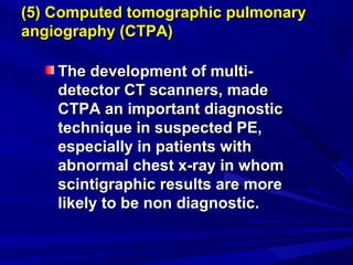
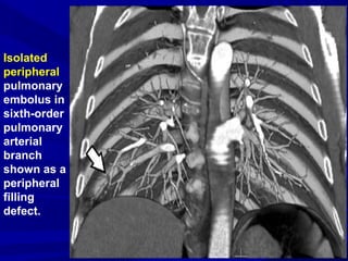
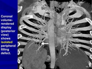
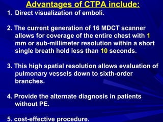
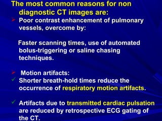
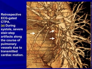
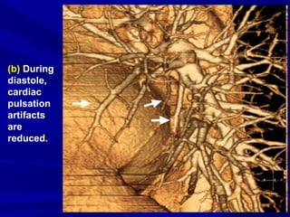
SlideShare a Scribd company logo
CT Angiography ofCT Angiography of
PulmonaryPulmonary EmbolismEmbolism
By
DR. Naglaa Mahmoud
KCCC
IntroductionIntroduction
PE is the third most common
acute CVS disease that results in
thousands of deaths each year
because it often goes undetected.
The majority of pulmonary emboli
begin in the pelvic or lower
extremity veins.
Importance of early diagnosis:
10% of symptomatic PE are fatal in the
first hour.
Most of the deaths occur when the
diagnosis is delayed or never made.
While if diagnosedif diagnosed, they are rarely fatal, they are rarely fatal
and rarely recurand rarely recur.
Clinical picture:
 Dyspnea 73%
 Pleuritic Pain 66%
 Cough 43%
 Leg Swelling 33%
 Leg Pain 30%
 Hemoptysis 15%
 Palpitations 12%
 Wheezing 10%
 Angina-Like pain 5%
The signs and symptoms are non specific and
serve only to raise the suspicion of PE.
Predisposing Factors:Predisposing Factors:
• Immobility: post surgery, major
trauma, obesity.
• Blood coagulopathy: malignancy.
• Slow circulation: pregnancy,
congestive heart failure.
DIFFERENTIAL DIAGNOSISDIFFERENTIAL DIAGNOSIS
CHFCHF
Asthma / COPD exacerbationAsthma / COPD exacerbation
PneumothoraxPneumothorax
PneumoniaPneumonia
Pleural diseasePleural disease
Chest wall PainChest wall Pain
Myocardial infarctionMyocardial infarction
PericarditisPericarditis
Infradiaphragmatic processInfradiaphragmatic process
e.g. cholecystitis, splenic infarction,e.g. cholecystitis, splenic infarction,
pancreatitis.pancreatitis.
Imaging of pulmonaryImaging of pulmonary
embolismembolism
(1) Chest x-ray findings, non specific, may
include:
• Normal
• Diaphragm elevation
A diaphragm mayA diaphragm may
be elevated,be elevated,
reflecting volumereflecting volume
loss in the affectedloss in the affected
lung.lung.
Atelectasis is
quite common.
Pleural based opacity (Hampton’s hump).
Pleural effusions are common and most often
unilateral occupying less than 15% of a
hemithorax.
Westermark’s Sign is a sign that represents a
focus of oligemia seen distal to a pulmonary
embolus.
Prominent central pulmonary artery (with
extensive PE).
(2) Ventilation-perfusion scintigraphy
• It has to be correlated with chest x-ray.
•There are 3 V/Q lung scan patterns:
1. A normal perfusion scan rule out PE:
2. High probability scan:
3. All other lung scan patterns:
• (~ 60% of all the scans) are non
diagnostic, include:
“low probability”,
“intermediate probability”, and
“indeterminate probability”.
• Further testing is required in patients
with this V/Q scan.
Advantages of V/Q Lung Scans include:Advantages of V/Q Lung Scans include:
1. A normal V/Q scan rules out PE.1. A normal V/Q scan rules out PE.
2. Low radiation dose.2. Low radiation dose.
3. Iodine-based contrast is not used.3. Iodine-based contrast is not used.
Limitations of V/Q Lung Scans include:Limitations of V/Q Lung Scans include:
1. The majority of V/Q scans are non1. The majority of V/Q scans are non
diagnostic.diagnostic.
2. Do not provide an alternate diagnosis in the2. Do not provide an alternate diagnosis in the
patients without PE.patients without PE.
3. High cost.3. High cost.
(3) Pulmonary angiography
• Some still consider it as the standard
technique for diagnosis of PE, but in reality
it is infrequently performed as it is:
• Expensive.
• Technically more difficult.
• Risky.
• Moreover there are limitations for
unequivocal diagnosis of isolated peripheral
pulmonary emboli.
(4) Lower limb ultrasonography
Since the treatment for DVT and
PE is the same,
the demonstration of clot in the
leg using Duplex US in a patient
who is clinically suspected to
have PE, usually no further
diagnostic work-up is pursued.
(5) Computed tomographic pulmonary(5) Computed tomographic pulmonary
angiography (CTPA)angiography (CTPA)
The development of multi-The development of multi-
detector CT scanners, madedetector CT scanners, made
CTPA an important diagnosticCTPA an important diagnostic
technique in suspected PE,technique in suspected PE,
especially in patients withespecially in patients with
abnormal chest x-ray in whomabnormal chest x-ray in whom
scintigraphic results are morescintigraphic results are more
likely to be non diagnostic.likely to be non diagnostic.
Technique of CTPATechnique of CTPA
 Pre-scanning RFT and history of allergy to CM
must be checked.
 Light-speed 16-section CT scanning of the
thorax in a caudo-cranial direction.
 Introduction of an 18- or 20-gauge catheter
into an antecubital vein.
 The chest field of view is the widest rib-to rib
distance acquired during breath hold after
inspiration.
 Rate of CM injection is 4 ml / sec for a totalCM injection is 4 ml / sec for a total
dose of 135 ml.dose of 135 ml.
 Scanning delay is determined
by:
Dividing the acquisition time
for lung imaging by 2 and
subtracting the result from
the total injection time (34
seconds).
 Images are displayed with three
different gray scales for
interpretation:
 Lung window (1500/600).
 Mediastinal window (400/40).
 Pulmonary embolism–specific
(700/100) window.
 Multiplanar Reformatted images
through the longitudinal axis of
a vessel are used to:
Overcome difficulties
encountered with axial sections
of obliquely or axially oriented
arteries.
 Reformatted images can help
differentiation between:
 True pulmonary embolism
and
 Patient-related, technical,
anatomic, and pathologic factors
that can mimic pulmonary
embolism.
 Contrast-enhanced CT of LL veins
can be performed using the same
contrast bolus used for CTPA.
 Images of the iliac, femoral, and
popliteal veins are obtained 4
minutes after the onset of contrast
injection.
CTV in a case with PE shows acute LCF DVT. Expansion of the vein is
evident with marked wall thickening and enhancement, and the
adjacent fat is edematous.
CTV shows DVT in right external iliac vein.
CT Diagnostic Criteria for
Acute Pulmonary
Embolism:
1. Arterial
occlusion
with failure
to enhance
the entire
lumen due
to a large
filling
defect; the
artery may
be
enlarged
compared
with
adjacent
patent
vessels.
2. A partial
filling defect
surrounded
by contrast
material,
producing
the “polo
mint” sign on
images
acquired
perpendicular
to the long
axis of a
vessel
3. The
“railway
track” sign
on
longitudinal
images of
the vessel.
4. A
peripheral
intralumin
al filling
defect
that forms
acute
angles
with the
arterial
wall.
Peripheral
wedge-shaped
areas of
hyperattenuat-
ion (infarcts),
with linear
bands, are
ancillary
findings with
acute
pulmonary
embolism.
However,
these
radiologic
features are
not specific.
A diagnosis of PE is established
on the basis of individual
transverse sections, although,
extensive or
isolated PE,
as well as normal pulmonary
vasculature can be visualized by
means of 3D reconstructions.
Extensive
acute
central PE
with "saddle
embolus"
extending
into both
central
pulmonary
arteries.
Isolated
peripheral
pulmonary
embolus in
sixth-order
pulmonary
arterial
branch
shown as a
peripheral
filling
defect.
Normal pulmonary vessels.
What about smallWhat about small
peripheral clots?peripheral clots?
Recent studies proved that,
small peripheral clots that might
have gone unnoticed in the past are
now frequently detected, often in
patients with minor symptoms.
Consecutive transverse sections show isolated peripheral pulmonary
embolus in a subsegmental pulmonary artery in the left lung.
Oblique
sagittal
multiplanar
reformation
also shows
embolus.
Coronal
volume-
rendered
display
(posterior
view)
shows
isolated
peripheral
filling
defect.
Advantages of CTPA include:
1. Direct visualization of emboli.
2. The current generation of 16 MDCT scanner
allows for coverage of the entire chest with 1
mm or sub-millimeter resolution within a short
single breath hold less than 10 seconds.
3. This high spatial resolution allows evaluation of
pulmonary vessels down to sixth-order
branches.
4. Provide the alternate diagnosis in patients
without PE.
5. cost-effective procedure.
Limitations of CTPA include:Limitations of CTPA include:
1. Allergy to CM.
2. Patients with renal insufficiency.
3. Pregnancy.
4. Severe obesity.
CTPA Trouble Shooting
The most common reasons for nonThe most common reasons for non
diagnostic CT images are:diagnostic CT images are:
 Poor contrast enhancement of pulmonaryPoor contrast enhancement of pulmonary
vessels, overcome by:vessels, overcome by:
Faster scanning timesFaster scanning times, use of automated
bolus-triggering or saline chasing
techniques.
 Motion artifacts:
 Shorter breath-hold times reduce the
occurrence of respiratory motion artifacts.
 Artifacts due to transmitted cardiac pulsation
are reduced by retrospective ECG gating of
the CT.
Retrospective
ECG-gated
CTPA.
(a) During
systole, severe
stair-step
artifacts along
the course of
pulmonary
vessels due to
transmitted
cardiac motion.
(b) During
diastole,
cardiac
pulsation
artifacts
are
reduced.
How to proceed ?How to proceed ?
Before proceeding to imaging,Before proceeding to imaging,
the most important first-linethe most important first-line forfor
diagnosis of PE isdiagnosis of PE is D-dimerD-dimer testing, thetesting, the
results of which, if negative, rules outresults of which, if negative, rules out
PE.PE.
A patient with a normal D-dimer and aA patient with a normal D-dimer and a
low pre-test probability does notlow pre-test probability does not
require further diagnostic imaging.require further diagnostic imaging.
PE symptoms +/- DVT SYMPTOMS
Lower Extremity Evaluation (Duplex u/s)
(-) (+)
TreatCTPA
(+) (-)
Treat
Poor quality
CTPA
Repeat CTPA/ (Angio?)
Good quality
CTPA
STOP
High clinical suspicion
(Angio?)
conclusionconclusion
• The development of MDCT
technique has made CTPA the
first-line imaging test in daily
clinical routine for patients
suspected of having PE as it is:
• Fast
• Non invasive
• Has a high sensitivity and
specificity
• Can detect small emboli in
peripheral pulmonary arteries.
THANK YOUTHANK YOU

More Related Content

What's hot

Signs in Chest Xray
Signs in Chest Xray Signs in Chest Xray
Signs in Chest Xray
Archana Koshy
 
HRCT TECHNIQUE AND INTERPRETATION
HRCT TECHNIQUE AND INTERPRETATIONHRCT TECHNIQUE AND INTERPRETATION
HRCT TECHNIQUE AND INTERPRETATION
Sahil Chaudhry
 
Chest radiology in intensive care
Chest radiology in intensive careChest radiology in intensive care
Chest radiology in intensive careAndrew Ferguson
 
Pneumomediastinum
PneumomediastinumPneumomediastinum
Pneumomediastinum
Gamal Agmy
 
chest radiology in ICU
   chest radiology in ICU   chest radiology in ICU
chest radiology in ICU
Eman Mahmoud
 
Imaging of pulmonary embolism
Imaging of pulmonary embolismImaging of pulmonary embolism
Imaging of pulmonary embolism
Thorsang Chayovan
 
USG chest
USG chestUSG chest
USG chest
Prashant Gupta
 
Doppler ultrasound of A-V access for hemodialysis
Doppler ultrasound of A-V access for hemodialysisDoppler ultrasound of A-V access for hemodialysis
Doppler ultrasound of A-V access for hemodialysisSamir Haffar
 
Pulmonary interstitium
Pulmonary interstitiumPulmonary interstitium
Pulmonary interstitium
Thorsang Chayovan
 
Pulmonary embolism imaging
Pulmonary embolism imagingPulmonary embolism imaging
Pulmonary embolism imaging
Dr. Soe Moe Htoo
 
Collapse- RADIOLOGY
Collapse- RADIOLOGYCollapse- RADIOLOGY
Collapse- RADIOLOGYNavdeep Shah
 
The Basics of Lung Ultrasound
The Basics of Lung UltrasoundThe Basics of Lung Ultrasound
The Basics of Lung Ultrasound
ICNUploads
 
Figure 3 sign fleischner sign-fissure sign-headcheese sign-hilar overlay sign...
Figure 3 sign fleischner sign-fissure sign-headcheese sign-hilar overlay sign...Figure 3 sign fleischner sign-fissure sign-headcheese sign-hilar overlay sign...
Figure 3 sign fleischner sign-fissure sign-headcheese sign-hilar overlay sign...
Minstry of health ,Ibn alnafis hoapital, Damascus
 
HRCT Chest - By Dr. Tinku Joseph
HRCT Chest - By Dr. Tinku JosephHRCT Chest - By Dr. Tinku Joseph
HRCT Chest - By Dr. Tinku Joseph
Dr.Tinku Joseph
 
Basics of CT Chest
Basics of CT Chest Basics of CT Chest
Interstitial lung diseases- HRCT
Interstitial lung diseases- HRCTInterstitial lung diseases- HRCT
Interstitial lung diseases- HRCTNavdeep Shah
 
Lung ultrasound
Lung ultrasoundLung ultrasound
Lung ultrasound
SCGH ED CME
 
Ct pulmonary angiogram
Ct pulmonary angiogramCt pulmonary angiogram
Ct pulmonary angiogrammuhammed Yasar
 

What's hot (20)

Signs in Chest Xray
Signs in Chest Xray Signs in Chest Xray
Signs in Chest Xray
 
HRCT TECHNIQUE AND INTERPRETATION
HRCT TECHNIQUE AND INTERPRETATIONHRCT TECHNIQUE AND INTERPRETATION
HRCT TECHNIQUE AND INTERPRETATION
 
Normal chest ct
Normal chest ctNormal chest ct
Normal chest ct
 
Chest radiology in intensive care
Chest radiology in intensive careChest radiology in intensive care
Chest radiology in intensive care
 
Pneumomediastinum
PneumomediastinumPneumomediastinum
Pneumomediastinum
 
Chest ultrasound
Chest ultrasoundChest ultrasound
Chest ultrasound
 
chest radiology in ICU
   chest radiology in ICU   chest radiology in ICU
chest radiology in ICU
 
Imaging of pulmonary embolism
Imaging of pulmonary embolismImaging of pulmonary embolism
Imaging of pulmonary embolism
 
USG chest
USG chestUSG chest
USG chest
 
Doppler ultrasound of A-V access for hemodialysis
Doppler ultrasound of A-V access for hemodialysisDoppler ultrasound of A-V access for hemodialysis
Doppler ultrasound of A-V access for hemodialysis
 
Pulmonary interstitium
Pulmonary interstitiumPulmonary interstitium
Pulmonary interstitium
 
Pulmonary embolism imaging
Pulmonary embolism imagingPulmonary embolism imaging
Pulmonary embolism imaging
 
Collapse- RADIOLOGY
Collapse- RADIOLOGYCollapse- RADIOLOGY
Collapse- RADIOLOGY
 
The Basics of Lung Ultrasound
The Basics of Lung UltrasoundThe Basics of Lung Ultrasound
The Basics of Lung Ultrasound
 
Figure 3 sign fleischner sign-fissure sign-headcheese sign-hilar overlay sign...
Figure 3 sign fleischner sign-fissure sign-headcheese sign-hilar overlay sign...Figure 3 sign fleischner sign-fissure sign-headcheese sign-hilar overlay sign...
Figure 3 sign fleischner sign-fissure sign-headcheese sign-hilar overlay sign...
 
HRCT Chest - By Dr. Tinku Joseph
HRCT Chest - By Dr. Tinku JosephHRCT Chest - By Dr. Tinku Joseph
HRCT Chest - By Dr. Tinku Joseph
 
Basics of CT Chest
Basics of CT Chest Basics of CT Chest
Basics of CT Chest
 
Interstitial lung diseases- HRCT
Interstitial lung diseases- HRCTInterstitial lung diseases- HRCT
Interstitial lung diseases- HRCT
 
Lung ultrasound
Lung ultrasoundLung ultrasound
Lung ultrasound
 
Ct pulmonary angiogram
Ct pulmonary angiogramCt pulmonary angiogram
Ct pulmonary angiogram
 

Similar to Pulmonary embolism

Pulmonary Embolism- Diagnosis by Dr.Tinku Joseph
Pulmonary Embolism- Diagnosis  by Dr.Tinku JosephPulmonary Embolism- Diagnosis  by Dr.Tinku Joseph
Pulmonary Embolism- Diagnosis by Dr.Tinku Joseph
Dr.Tinku Joseph
 
Update on Pulmonary Embolism
Update on Pulmonary EmbolismUpdate on Pulmonary Embolism
Update on Pulmonary Embolism
Muhanad Majeed
 
Pulmonary Embolism lecture 2022
Pulmonary Embolism lecture 2022Pulmonary Embolism lecture 2022
Pulmonary Embolism lecture 2022
Dr.Marwan Sneymeh
 
6. radiologic imaging in ecmo #beach2019 (nieboer)
6. radiologic imaging in ecmo #beach2019 (nieboer)6. radiologic imaging in ecmo #beach2019 (nieboer)
6. radiologic imaging in ecmo #beach2019 (nieboer)
International Fluid Academy
 
Pulmonary embolism@ghanem@.2013
Pulmonary embolism@ghanem@.2013Pulmonary embolism@ghanem@.2013
Pulmonary embolism@ghanem@.2013
Islam Ghanem
 
CLINICAL MEET IVC thrombosis.pptx
CLINICAL MEET IVC thrombosis.pptxCLINICAL MEET IVC thrombosis.pptx
CLINICAL MEET IVC thrombosis.pptx
Mehak783822
 
Pulmonary embolism radiology imaging
Pulmonary embolism radiology imagingPulmonary embolism radiology imaging
Pulmonary embolism radiology imaging
harshvardhan
 
deep vein thrombosis and pe
deep vein thrombosis and pedeep vein thrombosis and pe
deep vein thrombosis and pe
Praveen Shukla
 
PULMONARY EMBOLISM (Masteral reporting for finals)
PULMONARY EMBOLISM (Masteral reporting for finals)PULMONARY EMBOLISM (Masteral reporting for finals)
PULMONARY EMBOLISM (Masteral reporting for finals)
Mary Ann Adiong
 
Pulmonary angiography
Pulmonary angiographyPulmonary angiography
Pulmonary angiography
Pankaj Kaira
 
Vte 2014
Vte 2014Vte 2014
Vte 2014
Ihsaan Peer
 
Venous thromboembolism, THROMBOPROPHYLAXIS and management
Venous thromboembolism, THROMBOPROPHYLAXIS and managementVenous thromboembolism, THROMBOPROPHYLAXIS and management
Venous thromboembolism, THROMBOPROPHYLAXIS and management
mauryaramgopal
 
Updates in venous thromboembolism
Updates in venous thromboembolismUpdates in venous thromboembolism
Updates in venous thromboembolism
Gamal Agmy
 
Pulmonary embolism
Pulmonary embolism Pulmonary embolism
Pulmonary embolism
RahulGupta1687
 
Pulmonaryembolism ....tanmay new
Pulmonaryembolism ....tanmay newPulmonaryembolism ....tanmay new
Pulmonaryembolism ....tanmay new
Tanmay Jain
 
Diagnosis of Pulmonary Embolism - by Dr KD DELE
Diagnosis of Pulmonary Embolism - by Dr KD DELEDiagnosis of Pulmonary Embolism - by Dr KD DELE
Diagnosis of Pulmonary Embolism - by Dr KD DELE
Kemi Dele-Ijagbulu
 
Locally advanced lung ca
Locally advanced lung caLocally advanced lung ca
Locally advanced lung ca
amanuelasefa1
 
Ultrasound in ricu
Ultrasound in ricuUltrasound in ricu
Ultrasound in ricu
Manal Refaat Hafez
 
Pulmonary embolism
Pulmonary embolismPulmonary embolism
Pulmonary embolism
Ramin Sadeghi
 
BALKAN MCO 2011 - D. Jovanovic - Diagnostic procedures, staging and surgery o...
BALKAN MCO 2011 - D. Jovanovic - Diagnostic procedures, staging and surgery o...BALKAN MCO 2011 - D. Jovanovic - Diagnostic procedures, staging and surgery o...
BALKAN MCO 2011 - D. Jovanovic - Diagnostic procedures, staging and surgery o...European School of Oncology
 

Similar to Pulmonary embolism (20)

Pulmonary Embolism- Diagnosis by Dr.Tinku Joseph
Pulmonary Embolism- Diagnosis  by Dr.Tinku JosephPulmonary Embolism- Diagnosis  by Dr.Tinku Joseph
Pulmonary Embolism- Diagnosis by Dr.Tinku Joseph
 
Update on Pulmonary Embolism
Update on Pulmonary EmbolismUpdate on Pulmonary Embolism
Update on Pulmonary Embolism
 
Pulmonary Embolism lecture 2022
Pulmonary Embolism lecture 2022Pulmonary Embolism lecture 2022
Pulmonary Embolism lecture 2022
 
6. radiologic imaging in ecmo #beach2019 (nieboer)
6. radiologic imaging in ecmo #beach2019 (nieboer)6. radiologic imaging in ecmo #beach2019 (nieboer)
6. radiologic imaging in ecmo #beach2019 (nieboer)
 
Pulmonary embolism@ghanem@.2013
Pulmonary embolism@ghanem@.2013Pulmonary embolism@ghanem@.2013
Pulmonary embolism@ghanem@.2013
 
CLINICAL MEET IVC thrombosis.pptx
CLINICAL MEET IVC thrombosis.pptxCLINICAL MEET IVC thrombosis.pptx
CLINICAL MEET IVC thrombosis.pptx
 
Pulmonary embolism radiology imaging
Pulmonary embolism radiology imagingPulmonary embolism radiology imaging
Pulmonary embolism radiology imaging
 
deep vein thrombosis and pe
deep vein thrombosis and pedeep vein thrombosis and pe
deep vein thrombosis and pe
 
PULMONARY EMBOLISM (Masteral reporting for finals)
PULMONARY EMBOLISM (Masteral reporting for finals)PULMONARY EMBOLISM (Masteral reporting for finals)
PULMONARY EMBOLISM (Masteral reporting for finals)
 
Pulmonary angiography
Pulmonary angiographyPulmonary angiography
Pulmonary angiography
 
Vte 2014
Vte 2014Vte 2014
Vte 2014
 
Venous thromboembolism, THROMBOPROPHYLAXIS and management
Venous thromboembolism, THROMBOPROPHYLAXIS and managementVenous thromboembolism, THROMBOPROPHYLAXIS and management
Venous thromboembolism, THROMBOPROPHYLAXIS and management
 
Updates in venous thromboembolism
Updates in venous thromboembolismUpdates in venous thromboembolism
Updates in venous thromboembolism
 
Pulmonary embolism
Pulmonary embolism Pulmonary embolism
Pulmonary embolism
 
Pulmonaryembolism ....tanmay new
Pulmonaryembolism ....tanmay newPulmonaryembolism ....tanmay new
Pulmonaryembolism ....tanmay new
 
Diagnosis of Pulmonary Embolism - by Dr KD DELE
Diagnosis of Pulmonary Embolism - by Dr KD DELEDiagnosis of Pulmonary Embolism - by Dr KD DELE
Diagnosis of Pulmonary Embolism - by Dr KD DELE
 
Locally advanced lung ca
Locally advanced lung caLocally advanced lung ca
Locally advanced lung ca
 
Ultrasound in ricu
Ultrasound in ricuUltrasound in ricu
Ultrasound in ricu
 
Pulmonary embolism
Pulmonary embolismPulmonary embolism
Pulmonary embolism
 
BALKAN MCO 2011 - D. Jovanovic - Diagnostic procedures, staging and surgery o...
BALKAN MCO 2011 - D. Jovanovic - Diagnostic procedures, staging and surgery o...BALKAN MCO 2011 - D. Jovanovic - Diagnostic procedures, staging and surgery o...
BALKAN MCO 2011 - D. Jovanovic - Diagnostic procedures, staging and surgery o...
 

More from Naglaa Mahmoud

Breast bpe
Breast bpe Breast bpe
Breast bpe
Naglaa Mahmoud
 
Radiology night 10/2015
Radiology night 10/2015Radiology night 10/2015
Radiology night 10/2015
Naglaa Mahmoud
 
Gestational trophoblastic disease
Gestational trophoblastic diseaseGestational trophoblastic disease
Gestational trophoblastic disease
Naglaa Mahmoud
 
Treatment related complications from Chemothearpy
Treatment related complications from ChemothearpyTreatment related complications from Chemothearpy
Treatment related complications from Chemothearpy
Naglaa Mahmoud
 
Positive Oral Contrast for Oncology Patients
Positive Oral Contrast for Oncology Patients Positive Oral Contrast for Oncology Patients
Positive Oral Contrast for Oncology Patients Naglaa Mahmoud
 
Secondary Peritoneal Disease
Secondary Peritoneal DiseaseSecondary Peritoneal Disease
Secondary Peritoneal DiseaseNaglaa Mahmoud
 
DWI borderline / malignant epithelial ovarian tumors
DWI borderline / malignant epithelial ovarian tumorsDWI borderline / malignant epithelial ovarian tumors
DWI borderline / malignant epithelial ovarian tumorsNaglaa Mahmoud
 
Digital Breast Tomosynthesis, Microcalcifications
Digital Breast Tomosynthesis, MicrocalcificationsDigital Breast Tomosynthesis, Microcalcifications
Digital Breast Tomosynthesis, MicrocalcificationsNaglaa Mahmoud
 
Breast Cancer not Detected on MRI
Breast Cancer not Detected on MRIBreast Cancer not Detected on MRI
Breast Cancer not Detected on MRINaglaa Mahmoud
 
Incidental Adnexal Masses at CT
Incidental Adnexal Masses at CTIncidental Adnexal Masses at CT
Incidental Adnexal Masses at CTNaglaa Mahmoud
 
Contrast Induced Nephropathy
Contrast Induced NephropathyContrast Induced Nephropathy
Contrast Induced NephropathyNaglaa Mahmoud
 

More from Naglaa Mahmoud (12)

Breast bpe
Breast bpe Breast bpe
Breast bpe
 
Radiology night 10/2015
Radiology night 10/2015Radiology night 10/2015
Radiology night 10/2015
 
Gestational trophoblastic disease
Gestational trophoblastic diseaseGestational trophoblastic disease
Gestational trophoblastic disease
 
Treatment related complications from Chemothearpy
Treatment related complications from ChemothearpyTreatment related complications from Chemothearpy
Treatment related complications from Chemothearpy
 
Positive Oral Contrast for Oncology Patients
Positive Oral Contrast for Oncology Patients Positive Oral Contrast for Oncology Patients
Positive Oral Contrast for Oncology Patients
 
Secondary Peritoneal Disease
Secondary Peritoneal DiseaseSecondary Peritoneal Disease
Secondary Peritoneal Disease
 
DWI borderline / malignant epithelial ovarian tumors
DWI borderline / malignant epithelial ovarian tumorsDWI borderline / malignant epithelial ovarian tumors
DWI borderline / malignant epithelial ovarian tumors
 
Digital Breast Tomosynthesis, Microcalcifications
Digital Breast Tomosynthesis, MicrocalcificationsDigital Breast Tomosynthesis, Microcalcifications
Digital Breast Tomosynthesis, Microcalcifications
 
Breast Cancer not Detected on MRI
Breast Cancer not Detected on MRIBreast Cancer not Detected on MRI
Breast Cancer not Detected on MRI
 
Incidental Adnexal Masses at CT
Incidental Adnexal Masses at CTIncidental Adnexal Masses at CT
Incidental Adnexal Masses at CT
 
H&N post ttt
H&N post tttH&N post ttt
H&N post ttt
 
Contrast Induced Nephropathy
Contrast Induced NephropathyContrast Induced Nephropathy
Contrast Induced Nephropathy
 

Recently uploaded

Evaluation of antidepressant activity of clitoris ternatea in animals
Evaluation of antidepressant activity of clitoris ternatea in animalsEvaluation of antidepressant activity of clitoris ternatea in animals
Evaluation of antidepressant activity of clitoris ternatea in animals
Shweta
 
Lung Cancer: Artificial Intelligence, Synergetics, Complex System Analysis, S...
Lung Cancer: Artificial Intelligence, Synergetics, Complex System Analysis, S...Lung Cancer: Artificial Intelligence, Synergetics, Complex System Analysis, S...
Lung Cancer: Artificial Intelligence, Synergetics, Complex System Analysis, S...
Oleg Kshivets
 
Ocular injury ppt Upendra pal optometrist upums saifai etawah
Ocular injury  ppt  Upendra pal  optometrist upums saifai etawahOcular injury  ppt  Upendra pal  optometrist upums saifai etawah
Ocular injury ppt Upendra pal optometrist upums saifai etawah
pal078100
 
Non-respiratory Functions of the Lungs.pdf
Non-respiratory Functions of the Lungs.pdfNon-respiratory Functions of the Lungs.pdf
Non-respiratory Functions of the Lungs.pdf
MedicoseAcademics
 
BRACHYTHERAPY OVERVIEW AND APPLICATORS
BRACHYTHERAPY OVERVIEW  AND  APPLICATORSBRACHYTHERAPY OVERVIEW  AND  APPLICATORS
BRACHYTHERAPY OVERVIEW AND APPLICATORS
Krishan Murari
 
For Better Surat #ℂall #Girl Service ❤85270-49040❤ Surat #ℂall #Girls
For Better Surat #ℂall #Girl Service ❤85270-49040❤ Surat #ℂall #GirlsFor Better Surat #ℂall #Girl Service ❤85270-49040❤ Surat #ℂall #Girls
For Better Surat #ℂall #Girl Service ❤85270-49040❤ Surat #ℂall #Girls
Savita Shen $i11
 
ARTIFICIAL INTELLIGENCE IN HEALTHCARE.pdf
ARTIFICIAL INTELLIGENCE IN  HEALTHCARE.pdfARTIFICIAL INTELLIGENCE IN  HEALTHCARE.pdf
ARTIFICIAL INTELLIGENCE IN HEALTHCARE.pdf
Anujkumaranit
 
263778731218 Abortion Clinic /Pills In Harare ,
263778731218 Abortion Clinic /Pills In Harare ,263778731218 Abortion Clinic /Pills In Harare ,
263778731218 Abortion Clinic /Pills In Harare ,
sisternakatoto
 
Are There Any Natural Remedies To Treat Syphilis.pdf
Are There Any Natural Remedies To Treat Syphilis.pdfAre There Any Natural Remedies To Treat Syphilis.pdf
Are There Any Natural Remedies To Treat Syphilis.pdf
Little Cross Family Clinic
 
Pulmonary Thromboembolism - etilogy, types, medical- Surgical and nursing man...
Pulmonary Thromboembolism - etilogy, types, medical- Surgical and nursing man...Pulmonary Thromboembolism - etilogy, types, medical- Surgical and nursing man...
Pulmonary Thromboembolism - etilogy, types, medical- Surgical and nursing man...
VarunMahajani
 
The Normal Electrocardiogram - Part I of II
The Normal Electrocardiogram - Part I of IIThe Normal Electrocardiogram - Part I of II
The Normal Electrocardiogram - Part I of II
MedicoseAcademics
 
Hemodialysis: Chapter 3, Dialysis Water Unit - Dr.Gawad
Hemodialysis: Chapter 3, Dialysis Water Unit - Dr.GawadHemodialysis: Chapter 3, Dialysis Water Unit - Dr.Gawad
Hemodialysis: Chapter 3, Dialysis Water Unit - Dr.Gawad
NephroTube - Dr.Gawad
 
Couples presenting to the infertility clinic- Do they really have infertility...
Couples presenting to the infertility clinic- Do they really have infertility...Couples presenting to the infertility clinic- Do they really have infertility...
Couples presenting to the infertility clinic- Do they really have infertility...
Sujoy Dasgupta
 
Alcohol_Dr. Jeenal Mistry MD Pharmacology.pdf
Alcohol_Dr. Jeenal Mistry MD Pharmacology.pdfAlcohol_Dr. Jeenal Mistry MD Pharmacology.pdf
Alcohol_Dr. Jeenal Mistry MD Pharmacology.pdf
Dr Jeenal Mistry
 
How to Give Better Lectures: Some Tips for Doctors
How to Give Better Lectures: Some Tips for DoctorsHow to Give Better Lectures: Some Tips for Doctors
How to Give Better Lectures: Some Tips for Doctors
LanceCatedral
 
Surat @ℂall @Girls ꧁❤8527049040❤꧂@ℂall @Girls Service Vip Top Model Safe
Surat @ℂall @Girls ꧁❤8527049040❤꧂@ℂall @Girls Service Vip Top Model SafeSurat @ℂall @Girls ꧁❤8527049040❤꧂@ℂall @Girls Service Vip Top Model Safe
Surat @ℂall @Girls ꧁❤8527049040❤꧂@ℂall @Girls Service Vip Top Model Safe
Savita Shen $i11
 
Triangles of Neck and Clinical Correlation by Dr. RIG.pptx
Triangles of Neck and Clinical Correlation by Dr. RIG.pptxTriangles of Neck and Clinical Correlation by Dr. RIG.pptx
Triangles of Neck and Clinical Correlation by Dr. RIG.pptx
Dr. Rabia Inam Gandapore
 
Prix Galien International 2024 Forum Program
Prix Galien International 2024 Forum ProgramPrix Galien International 2024 Forum Program
Prix Galien International 2024 Forum Program
Levi Shapiro
 
Pharynx and Clinical Correlations BY Dr.Rabia Inam Gandapore.pptx
Pharynx and Clinical Correlations BY Dr.Rabia Inam Gandapore.pptxPharynx and Clinical Correlations BY Dr.Rabia Inam Gandapore.pptx
Pharynx and Clinical Correlations BY Dr.Rabia Inam Gandapore.pptx
Dr. Rabia Inam Gandapore
 
ARTHROLOGY PPT NCISM SYLLABUS AYURVEDA STUDENTS
ARTHROLOGY PPT NCISM SYLLABUS AYURVEDA STUDENTSARTHROLOGY PPT NCISM SYLLABUS AYURVEDA STUDENTS
ARTHROLOGY PPT NCISM SYLLABUS AYURVEDA STUDENTS
Dr. Vinay Pareek
 

Recently uploaded (20)

Evaluation of antidepressant activity of clitoris ternatea in animals
Evaluation of antidepressant activity of clitoris ternatea in animalsEvaluation of antidepressant activity of clitoris ternatea in animals
Evaluation of antidepressant activity of clitoris ternatea in animals
 
Lung Cancer: Artificial Intelligence, Synergetics, Complex System Analysis, S...
Lung Cancer: Artificial Intelligence, Synergetics, Complex System Analysis, S...Lung Cancer: Artificial Intelligence, Synergetics, Complex System Analysis, S...
Lung Cancer: Artificial Intelligence, Synergetics, Complex System Analysis, S...
 
Ocular injury ppt Upendra pal optometrist upums saifai etawah
Ocular injury  ppt  Upendra pal  optometrist upums saifai etawahOcular injury  ppt  Upendra pal  optometrist upums saifai etawah
Ocular injury ppt Upendra pal optometrist upums saifai etawah
 
Non-respiratory Functions of the Lungs.pdf
Non-respiratory Functions of the Lungs.pdfNon-respiratory Functions of the Lungs.pdf
Non-respiratory Functions of the Lungs.pdf
 
BRACHYTHERAPY OVERVIEW AND APPLICATORS
BRACHYTHERAPY OVERVIEW  AND  APPLICATORSBRACHYTHERAPY OVERVIEW  AND  APPLICATORS
BRACHYTHERAPY OVERVIEW AND APPLICATORS
 
For Better Surat #ℂall #Girl Service ❤85270-49040❤ Surat #ℂall #Girls
For Better Surat #ℂall #Girl Service ❤85270-49040❤ Surat #ℂall #GirlsFor Better Surat #ℂall #Girl Service ❤85270-49040❤ Surat #ℂall #Girls
For Better Surat #ℂall #Girl Service ❤85270-49040❤ Surat #ℂall #Girls
 
ARTIFICIAL INTELLIGENCE IN HEALTHCARE.pdf
ARTIFICIAL INTELLIGENCE IN  HEALTHCARE.pdfARTIFICIAL INTELLIGENCE IN  HEALTHCARE.pdf
ARTIFICIAL INTELLIGENCE IN HEALTHCARE.pdf
 
263778731218 Abortion Clinic /Pills In Harare ,
263778731218 Abortion Clinic /Pills In Harare ,263778731218 Abortion Clinic /Pills In Harare ,
263778731218 Abortion Clinic /Pills In Harare ,
 
Are There Any Natural Remedies To Treat Syphilis.pdf
Are There Any Natural Remedies To Treat Syphilis.pdfAre There Any Natural Remedies To Treat Syphilis.pdf
Are There Any Natural Remedies To Treat Syphilis.pdf
 
Pulmonary Thromboembolism - etilogy, types, medical- Surgical and nursing man...
Pulmonary Thromboembolism - etilogy, types, medical- Surgical and nursing man...Pulmonary Thromboembolism - etilogy, types, medical- Surgical and nursing man...
Pulmonary Thromboembolism - etilogy, types, medical- Surgical and nursing man...
 
The Normal Electrocardiogram - Part I of II
The Normal Electrocardiogram - Part I of IIThe Normal Electrocardiogram - Part I of II
The Normal Electrocardiogram - Part I of II
 
Hemodialysis: Chapter 3, Dialysis Water Unit - Dr.Gawad
Hemodialysis: Chapter 3, Dialysis Water Unit - Dr.GawadHemodialysis: Chapter 3, Dialysis Water Unit - Dr.Gawad
Hemodialysis: Chapter 3, Dialysis Water Unit - Dr.Gawad
 
Couples presenting to the infertility clinic- Do they really have infertility...
Couples presenting to the infertility clinic- Do they really have infertility...Couples presenting to the infertility clinic- Do they really have infertility...
Couples presenting to the infertility clinic- Do they really have infertility...
 
Alcohol_Dr. Jeenal Mistry MD Pharmacology.pdf
Alcohol_Dr. Jeenal Mistry MD Pharmacology.pdfAlcohol_Dr. Jeenal Mistry MD Pharmacology.pdf
Alcohol_Dr. Jeenal Mistry MD Pharmacology.pdf
 
How to Give Better Lectures: Some Tips for Doctors
How to Give Better Lectures: Some Tips for DoctorsHow to Give Better Lectures: Some Tips for Doctors
How to Give Better Lectures: Some Tips for Doctors
 
Surat @ℂall @Girls ꧁❤8527049040❤꧂@ℂall @Girls Service Vip Top Model Safe
Surat @ℂall @Girls ꧁❤8527049040❤꧂@ℂall @Girls Service Vip Top Model SafeSurat @ℂall @Girls ꧁❤8527049040❤꧂@ℂall @Girls Service Vip Top Model Safe
Surat @ℂall @Girls ꧁❤8527049040❤꧂@ℂall @Girls Service Vip Top Model Safe
 
Triangles of Neck and Clinical Correlation by Dr. RIG.pptx
Triangles of Neck and Clinical Correlation by Dr. RIG.pptxTriangles of Neck and Clinical Correlation by Dr. RIG.pptx
Triangles of Neck and Clinical Correlation by Dr. RIG.pptx
 
Prix Galien International 2024 Forum Program
Prix Galien International 2024 Forum ProgramPrix Galien International 2024 Forum Program
Prix Galien International 2024 Forum Program
 
Pharynx and Clinical Correlations BY Dr.Rabia Inam Gandapore.pptx
Pharynx and Clinical Correlations BY Dr.Rabia Inam Gandapore.pptxPharynx and Clinical Correlations BY Dr.Rabia Inam Gandapore.pptx
Pharynx and Clinical Correlations BY Dr.Rabia Inam Gandapore.pptx
 
ARTHROLOGY PPT NCISM SYLLABUS AYURVEDA STUDENTS
ARTHROLOGY PPT NCISM SYLLABUS AYURVEDA STUDENTSARTHROLOGY PPT NCISM SYLLABUS AYURVEDA STUDENTS
ARTHROLOGY PPT NCISM SYLLABUS AYURVEDA STUDENTS
 

Pulmonary embolism

  • 1. CT Angiography ofCT Angiography of PulmonaryPulmonary EmbolismEmbolism By DR. Naglaa Mahmoud KCCC
  • 3. PE is the third most common acute CVS disease that results in thousands of deaths each year because it often goes undetected. The majority of pulmonary emboli begin in the pelvic or lower extremity veins.
  • 4. Importance of early diagnosis: 10% of symptomatic PE are fatal in the first hour. Most of the deaths occur when the diagnosis is delayed or never made. While if diagnosedif diagnosed, they are rarely fatal, they are rarely fatal and rarely recurand rarely recur.
  • 5. Clinical picture:  Dyspnea 73%  Pleuritic Pain 66%  Cough 43%  Leg Swelling 33%  Leg Pain 30%  Hemoptysis 15%  Palpitations 12%  Wheezing 10%  Angina-Like pain 5% The signs and symptoms are non specific and serve only to raise the suspicion of PE.
  • 6. Predisposing Factors:Predisposing Factors: • Immobility: post surgery, major trauma, obesity. • Blood coagulopathy: malignancy. • Slow circulation: pregnancy, congestive heart failure.
  • 7. DIFFERENTIAL DIAGNOSISDIFFERENTIAL DIAGNOSIS CHFCHF Asthma / COPD exacerbationAsthma / COPD exacerbation PneumothoraxPneumothorax PneumoniaPneumonia Pleural diseasePleural disease Chest wall PainChest wall Pain Myocardial infarctionMyocardial infarction PericarditisPericarditis Infradiaphragmatic processInfradiaphragmatic process e.g. cholecystitis, splenic infarction,e.g. cholecystitis, splenic infarction, pancreatitis.pancreatitis.
  • 8. Imaging of pulmonaryImaging of pulmonary embolismembolism
  • 9. (1) Chest x-ray findings, non specific, may include: • Normal • Diaphragm elevation A diaphragm mayA diaphragm may be elevated,be elevated, reflecting volumereflecting volume loss in the affectedloss in the affected lung.lung.
  • 11. Pleural based opacity (Hampton’s hump).
  • 12. Pleural effusions are common and most often unilateral occupying less than 15% of a hemithorax.
  • 13. Westermark’s Sign is a sign that represents a focus of oligemia seen distal to a pulmonary embolus.
  • 14. Prominent central pulmonary artery (with extensive PE).
  • 15. (2) Ventilation-perfusion scintigraphy • It has to be correlated with chest x-ray. •There are 3 V/Q lung scan patterns: 1. A normal perfusion scan rule out PE:
  • 17. 3. All other lung scan patterns: • (~ 60% of all the scans) are non diagnostic, include: “low probability”, “intermediate probability”, and “indeterminate probability”. • Further testing is required in patients with this V/Q scan.
  • 18.
  • 19. Advantages of V/Q Lung Scans include:Advantages of V/Q Lung Scans include: 1. A normal V/Q scan rules out PE.1. A normal V/Q scan rules out PE. 2. Low radiation dose.2. Low radiation dose. 3. Iodine-based contrast is not used.3. Iodine-based contrast is not used. Limitations of V/Q Lung Scans include:Limitations of V/Q Lung Scans include: 1. The majority of V/Q scans are non1. The majority of V/Q scans are non diagnostic.diagnostic. 2. Do not provide an alternate diagnosis in the2. Do not provide an alternate diagnosis in the patients without PE.patients without PE. 3. High cost.3. High cost.
  • 20. (3) Pulmonary angiography • Some still consider it as the standard technique for diagnosis of PE, but in reality it is infrequently performed as it is: • Expensive. • Technically more difficult. • Risky. • Moreover there are limitations for unequivocal diagnosis of isolated peripheral pulmonary emboli.
  • 21.
  • 22. (4) Lower limb ultrasonography Since the treatment for DVT and PE is the same, the demonstration of clot in the leg using Duplex US in a patient who is clinically suspected to have PE, usually no further diagnostic work-up is pursued.
  • 23. (5) Computed tomographic pulmonary(5) Computed tomographic pulmonary angiography (CTPA)angiography (CTPA) The development of multi-The development of multi- detector CT scanners, madedetector CT scanners, made CTPA an important diagnosticCTPA an important diagnostic technique in suspected PE,technique in suspected PE, especially in patients withespecially in patients with abnormal chest x-ray in whomabnormal chest x-ray in whom scintigraphic results are morescintigraphic results are more likely to be non diagnostic.likely to be non diagnostic.
  • 25.  Pre-scanning RFT and history of allergy to CM must be checked.  Light-speed 16-section CT scanning of the thorax in a caudo-cranial direction.  Introduction of an 18- or 20-gauge catheter into an antecubital vein.  The chest field of view is the widest rib-to rib distance acquired during breath hold after inspiration.  Rate of CM injection is 4 ml / sec for a totalCM injection is 4 ml / sec for a total dose of 135 ml.dose of 135 ml.
  • 26.  Scanning delay is determined by: Dividing the acquisition time for lung imaging by 2 and subtracting the result from the total injection time (34 seconds).
  • 27.  Images are displayed with three different gray scales for interpretation:  Lung window (1500/600).  Mediastinal window (400/40).  Pulmonary embolism–specific (700/100) window.
  • 28.  Multiplanar Reformatted images through the longitudinal axis of a vessel are used to: Overcome difficulties encountered with axial sections of obliquely or axially oriented arteries.
  • 29.  Reformatted images can help differentiation between:  True pulmonary embolism and  Patient-related, technical, anatomic, and pathologic factors that can mimic pulmonary embolism.
  • 30.  Contrast-enhanced CT of LL veins can be performed using the same contrast bolus used for CTPA.  Images of the iliac, femoral, and popliteal veins are obtained 4 minutes after the onset of contrast injection.
  • 31. CTV in a case with PE shows acute LCF DVT. Expansion of the vein is evident with marked wall thickening and enhancement, and the adjacent fat is edematous.
  • 32. CTV shows DVT in right external iliac vein.
  • 33. CT Diagnostic Criteria for Acute Pulmonary Embolism:
  • 34. 1. Arterial occlusion with failure to enhance the entire lumen due to a large filling defect; the artery may be enlarged compared with adjacent patent vessels.
  • 35. 2. A partial filling defect surrounded by contrast material, producing the “polo mint” sign on images acquired perpendicular to the long axis of a vessel
  • 37. 4. A peripheral intralumin al filling defect that forms acute angles with the arterial wall.
  • 38. Peripheral wedge-shaped areas of hyperattenuat- ion (infarcts), with linear bands, are ancillary findings with acute pulmonary embolism. However, these radiologic features are not specific.
  • 39. A diagnosis of PE is established on the basis of individual transverse sections, although, extensive or isolated PE, as well as normal pulmonary vasculature can be visualized by means of 3D reconstructions.
  • 43. What about smallWhat about small peripheral clots?peripheral clots?
  • 44. Recent studies proved that, small peripheral clots that might have gone unnoticed in the past are now frequently detected, often in patients with minor symptoms.
  • 45. Consecutive transverse sections show isolated peripheral pulmonary embolus in a subsegmental pulmonary artery in the left lung.
  • 48. Advantages of CTPA include: 1. Direct visualization of emboli. 2. The current generation of 16 MDCT scanner allows for coverage of the entire chest with 1 mm or sub-millimeter resolution within a short single breath hold less than 10 seconds. 3. This high spatial resolution allows evaluation of pulmonary vessels down to sixth-order branches. 4. Provide the alternate diagnosis in patients without PE. 5. cost-effective procedure.
  • 49.
  • 50. Limitations of CTPA include:Limitations of CTPA include: 1. Allergy to CM. 2. Patients with renal insufficiency. 3. Pregnancy. 4. Severe obesity.
  • 52. The most common reasons for nonThe most common reasons for non diagnostic CT images are:diagnostic CT images are:  Poor contrast enhancement of pulmonaryPoor contrast enhancement of pulmonary vessels, overcome by:vessels, overcome by: Faster scanning timesFaster scanning times, use of automated bolus-triggering or saline chasing techniques.  Motion artifacts:  Shorter breath-hold times reduce the occurrence of respiratory motion artifacts.  Artifacts due to transmitted cardiac pulsation are reduced by retrospective ECG gating of the CT.
  • 53. Retrospective ECG-gated CTPA. (a) During systole, severe stair-step artifacts along the course of pulmonary vessels due to transmitted cardiac motion.
  • 55. How to proceed ?How to proceed ?
  • 56. Before proceeding to imaging,Before proceeding to imaging, the most important first-linethe most important first-line forfor diagnosis of PE isdiagnosis of PE is D-dimerD-dimer testing, thetesting, the results of which, if negative, rules outresults of which, if negative, rules out PE.PE. A patient with a normal D-dimer and aA patient with a normal D-dimer and a low pre-test probability does notlow pre-test probability does not require further diagnostic imaging.require further diagnostic imaging.
  • 57. PE symptoms +/- DVT SYMPTOMS Lower Extremity Evaluation (Duplex u/s) (-) (+) TreatCTPA (+) (-) Treat Poor quality CTPA Repeat CTPA/ (Angio?) Good quality CTPA STOP High clinical suspicion (Angio?)
  • 59. • The development of MDCT technique has made CTPA the first-line imaging test in daily clinical routine for patients suspected of having PE as it is: • Fast • Non invasive • Has a high sensitivity and specificity • Can detect small emboli in peripheral pulmonary arteries.