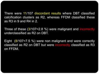
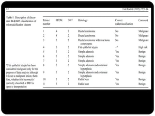
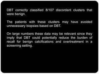
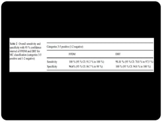
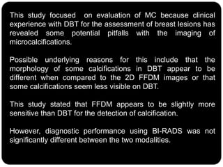
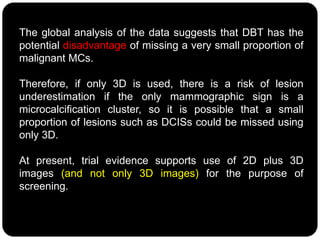
This study compared the characterization of microcalcification clusters using 2D digital mammography (FFDM) and digital breast tomosynthesis (DBT) in 107 cases. There were 11 discordant results where DBT classified clusters lower than FFDM. DBT incorrectly underclassified 3 clusters as benign that were malignant. However, DBT correctly classified 8 clusters as benign that FFDM misclassified as suspicious. While diagnostic performance between the two modalities was similar, the authors conclude DBT has the potential to underestimate a small portion of malignant lesions, so 2D plus 3D imaging is recommended for breast screening to avoid missing microcalcification clusters.
Digital Breast Tomosynthesis with Minimal CompressionDavid Scaduto
Breast compression is utilized in mammography to improve image quality and reduce radiation dose. Lesion conspicuity is improved by reducing scatter effects on contrast and by reducing the superposition of tissue structures. However, patient discomfort due to breast compression has been cited as a potential cause of noncompliance with recommended screening practices. Further, compression may also occlude blood flow in the breast, complicating imaging with intravenous contrast agents and preventing accurate quantification of contrast enhancement and kinetics. Previous studies have investigated reducing breast compression in planar mammography and digital breast tomosynthesis (DBT), though this typically comes at the expense of degradation in image quality or increase in mean glandular dose (MGD). We propose to optimize the image acquisition technique for reduced compression in DBT without compromising image quality or increasing MGD. A zero-frequency signal-difference-to-noise ratio model is employed to investigate the relationship between tube potential, SDNR and MGD. Phantom and patient images are acquired on a prototype DBT system using the optimized imaging parameters and are assessed for image quality and lesion conspicuity. A preliminary assessment of patient motion during DBT with minimal compression is presented.
A comprehensive study about new and upcoming modalities in imaging and screening of breast lesions with description about every new modalities with their advantages and pitfalls.
Quality Assurance Programme in Computed TomographyRamzee Small
Introduction to Computed Tomography
Basic description of the components of a CT System
Introduction to Quality Assurance
Quality Assurance and Quality Control Tests in Computed Tomography base on frequency
Objective of QA/QC Test
Quality Assurance Programme in Computed TomographyRamzee Small
Introduction to Computed Tomography
Basic description of the components of a CT System
Introduction to Quality Assurance
Quality Assurance and Quality Control Tests in Computed Tomography base on frequency
Objective of QA/QC Test
Role of Breast Tomosynthesis in the Morphological Analysis of Breast LesionsApollo Hospitals
1. To assess the role of Breast Tomosynthesis (by 3D Combined View) versus 2D Full
Field Digital Mammogram alone in the morphological analysis of breast lesions.
2. To evaluate the potential role of Tomosynthesis in BIRADS Categorisation and Final
Histopathology.
In May 2011 we migrated from an Analogue Mammogram with a dedicated Mammogram
CR system to a Full Field Digital System with 3D Tomosynthesis.
In India there is no official screening programme. All screening is opportunistic, self-
initiated and self-funded. Most Mammograms done at our hospital, a Corporate Tertiary
care Oncology facility, are performed as Diagnostic Mammograms followed by mandatory Breast Ultrasound and additional views, if necessary, on the same day obviating the need for recall.
Reducing the number of cases for additional views and breast ultrasound will help
in decreasing the patient's waiting time, making reporting more efficient, without
compromising on the accuracy. We used BIRADS categorisation as an evaluating tool
and compared the BIRADS categorisation with the final HPE.
Signs of Benign Breast Disease in 3D TomosynthesisApollo Hospitals
The role of three dimensional tomosynthesis in margin analysis especially for malignant lesions is well known (reference 2) .Three dimensional tomosynthesis has specific signs in lesions for categorization of Benign & Malignant pathology resulting in efficient & effective diagnosis. Three dimensional Tomosynthesis has been found to reduce recall rates(reference 3 , 4 ) because of better assessment and categorisation of breast lesions.
Role of Tomosynthesis in Assessing the Size of the Breast LesionApollo Hospitals
To assess the role of 3D tomosynthesis in the evaluation of the size of malignant breast lesions and to compare it with the size in 2D, Ultrasound and final Histopatholgy.
International Journal of Computational Engineering Research (IJCER) is dedicated to protecting personal information and will make every reasonable effort to handle collected information appropriately. All information collected, as well as related requests, will be handled as carefully and efficiently as possible in accordance with IJCER standards for integrity and objectivity.
Reduced Radiation Exposure in Dual-Energy Computed Tomography of the Chest: ...MehranMouzam
ABSTRACT:
Objective: This study purports to answer the question: Does a dual-energy CT scan of the chest using reduced radiation result in images of equal or better quality compared to those produced by the gold standard of care?
Methods: With the agreement of the Ethical Review Committee and written informed consent from 32 patients, who received dual-energy CT (DECT) scan of the chest in a dual-source scanner, a second set of images was taken at a reduced radiation dose. On virtual monochromatic images at 40 and 60 keV, three thoracic radiologists evaluated image quality, normal thoracic structures, and pulmonary and mediastinal aberrations. Students analyzed the data using analysis of variance, Kappa statistics, and Wilcoxon signed-rank tests.
Results: No irregularities in the scans were missed in the virtual monochrome photographs of all patients at a lower radiation dose, and the images were found to be of sufficient quality. At 40 and 60 keV, standard-of-care pictures produced equal contrast enhancement and lesion detection. Observers were entirely consistent with one another. Among other characteristics, reduced-dose DECT had a CTDIvol of 3.0 ±0.6 mGy, and a size specified dose estimate (SSDE) of 4.0 ±0.6 mGy, a dose-length product (DLP) of 107 ±30 mgy.cm, and an effective dose (ED) of 1.15 ±0.4 mSv.
Conclusion: Dual-energy computed tomography of the chest allows for the administration of lower radiation doses (CTDIvol <3 mGy).
MEDICAL IMAGING MUTIFRACTAL ANALYSIS IN PREDICTION OF EFFICIENCY OF CANCER TH...cscpconf
Based on pressing need for predictive performance improvement, we explored the value of pretherapy
tumour histology image analysis to predict chemotherapy response. It was shown that
multifractal analysis of breast tumour tissue prior to chemotherapy indeed has the capacity to
distinguish between histological images of the different chemotherapy responder groups with
accuracies of 91.4% for pPR, 82.9% for pCR and 82.1% for PD/SD.
MEDICAL IMAGING MUTIFRACTAL ANALYSIS IN PREDICTION OF EFFICIENCY OF CANCER TH...csandit
Based on pressing need for predictive performance improvement, we explored the value of pretherapy
tumour histology image analysis to predict chemotherapy response. It was shown that
multifractal analysis of breast tumour tissue prior to chemotherapy indeed has the capacity to
distinguish between histological images of the different chemotherapy responder groups with
accuracies of 91.4% for pPR, 82.9% for pCR and 82.1% for PD/SD.
PERSONALIZED MEDICINE 66515 2013Molecular Imaging .docxherbertwilson5999
PERSONALIZED MEDICINE
665
15 2013
Molecular Imaging in the Framework of Personalized
Cancer Medicine
Dževad Belkić PhD1 and Karen Belkić MD PhD1,2,3
1Department of Oncology/Pathology, Karolinska Institute, Stockholm, Sweden
2Claremont Graduate University, School of Community and Global Health, and 3Institute for Prevention Research, University of Southern California School of Medicine, Los
Angeles, CA, USA
M olecular imaging is an emerging medical discipline that integrates cellular and molecular biology with diagnos-
tic imaging. It encompasses several imaging modalities that
provide critical information for early detection and progres-
sion of disease, for predicting response to therapy and for
evaluating treatment efficacy through its cellular and molecu-
lar pathways. Given the rapidly growing sophistication in
our understanding of the cell biology of cancer, molecular
imaging offers a strategic bridge to clinical oncology. It is of
critical importance to non-invasively assess the behavior of
tumors, for which in vivo molecular imaging is a key meth-
odology. Molecular imaging detects not only the presence
of the disease process, but can potentially also quantify its
extent and severity, as well as follow the course of disease over
time. Besides its diagnostic possibilities, molecular imaging
is vital for target definition within dose-planning systems
for radiotherapy. Molecular imaging is also an invaluable
tool during and after therapy for assessing dose delivery
and the overall success of treatment, as well as for deciding
post-radiotherapy whether the patient needs further treat-
ment and, if so, with which modalities. Recently, molecular
imaging has proven to be pivotal, as well, for image-guided
biopsy and surgery. This is particularly crucial since there
may be multiple sites of cancer and image guiding can help
detect them. Imaging is likely to become an important tool
for cancer staging. Molecular targeting can be especially help-
ful in visualizing the extent of certain malignancies such as
androgen-sensitive prostate cancers, neuroendocrine tumors,
among others.
The strategic importance of molecular imaging in provid-
ing the best possible care for patients with or at risk for cancer
has been emphasized [1-3]. As recently stated: “Molecular
imaging is rapidly gaining recognition as a tool that has the
capacity to improve every facet of cancer care. The growing
demands among physicians, patients and society for per-
sonalized care are increasing the importance of molecular
imaging and shaping the development of biomedical imaging
as a whole.” [3] (p. 182)
LIMITATIONS OF PURELY ANATOMIC IMAGING
Anatomic imaging is vital for cancer detection, as well as for
staging and evaluation of response to therapy. Magnetic reso-
nance imaging, ultrasound and computerized tomography
are anatomic imaging modalities used routinely in clinical
With our increased understanding of cancer cell biology, .
1. Characterisation of microcalcification clusters
on 2D digital mammography (FFDM) and
digital breast tomosynthesis (DBT): does DBT
underestimate microcalcification clusters?
Results of a multicentre study.
4. Digital breast tomosynthesis (DBT) is a novel mammography
technique capable of studying the breast using 3D
reconstructions of the tissue from multiple low-dose digital
mammographic images, acquired along a 15 to 50 degree
arc.
This process increases lesion visibility because of the
reduction of overlapping breast tissue.
Recently, digital breast tomosynthesis alone or combined with
digital mammography has been shown to detect cancers
occult to 2D mammography.
5. Its potential role as a screening test has also been
recently demonstrated in large scale prospective
screening trials.
In addition, a recent meta-analysis showed that DBT may
have a higher sensitivity and specificity in breast
diagnosis than digital mammography.
Regarding radiation dose, an increase in radiation dose
due to the 3D acquisition has been detected with an
increase of entrance skin air kerma and average
glandular dose.
6. Very few reports in the literature have compared FFDM and
DBT for classification of microcalcification clusters.
Some authors have outlined that DBT may underestimate BI-
RADS cluster classification compared to FFDM.
In theory, using DBT, the radiologist may be more accurate in
characterising some breast lesions to reduce the rate of false
positive breast biopsies but it is unknown whether this applies
to microcalcifications.
The purpose of this study was to assess differences between
DBT and FFDM in the characterisation of microcalcification
clusters using the standard BI-RADS classification.
8. Institutional Review Board approval and written informed
consent were received.
The study was undertaken in three different centres: IRCCS
San Martino Hospital-University of Genoa, IRCCS Molinette
Hospital Torino and Bolzano Central Hospital.
10. A total of 120 mammographic examinations showing
microcalcification clusters (MCs) were identified based on
searching the electronic databases of the radiological reports
between January 2010 and June 2012.
All women had digital mammography using a Selenia
Dimensions unit with integrated 2D and 3D mammography
done in the COMBO mode (acquisition of 2D plus 3D images
in the same session).
The X-ray tube moves over a 15° arc.
The 2D and 3D images are acquired at the same examination
with a single breast position and compression.
Each 2D and 3D image consisted of a bilateral two-view
(mediolateral oblique and craniocaudal) mammogram.
11. Only MCs with both DBT and FFDM images available and with
a follow-up of at least 12 months were included in this study.
Clearly benign calcified lesions such as calcified
fibroadenomas were not included in the analysis.
Final histology was used to confirm malignant cases.
Bilateral mediolateral oblique and craniocaudal views were
considered an image set for FFDM, whereas the 3D
mediolateral oblique and craniocaudal view reconstructed
images and the video clip were considered the DBT image set.
12. Six breast radiologists, each with at least 7 years of
experience in digital mammography and 2 years of
experience in the clinical use of DBT, read the FFDM and
DBT image sets prospectively and assigned a BI-RADS
score to each cluster.
The two image sets (FFDM and DBT) were read
independently in sessions separated by at least 4 weeks to
minimise recall bias.
13. Data analysis
The overall sensitivity and specificity with a 95 %
confidence interval were calculated for FFDM and DBT
using final histology and clinical follow-up as the reference
standard.
Comparative sensitivity of FFDM and DBT was calculated
classifying results as positive or negative as follows:
benign (R1-R2) vs. malignant (R3-R4-R5).
15. The calcification clusters with complete data comprised 107
of 120, with 13 clusters excluded because of incomplete
follow-up (n=10 patients lost to follow-up) or to missing
DBT data in the hospital PACS (n=3).
All clusters were visible on both DBT and FFDM.
The mean age was 51.7 years.
The benign/malignant ratio of MC was 66/41.
16. There were 11/107 discordant results where DBT classified
calcification clusters as R2, whereas FFDM classified these
as R3 in 9 and R4 in 2.
Three of these (3/107=2.8 %) were malignant and incorrectly
underclassified as R2 on DBT.
Eight (8/107=7.5 %) were non malignant and were correctly
classified as R2 on DBT but were incorrectly classified as R3
on FFDM.
17.
18. Left craniocaudal (CC) view in FFDM and DBT shows a microcalcification
cluster that was classified with the BI-RADS classification as R4 in FFDM
and R2 in DBT (ductal carcinoma at histology)
19. DBT correctly classified 8/107 discordant clusters that
were benign.
The patients with these clusters may have avoided
unnecessary biopsies based on DBT.
On large numbers these data may be relevant since they
imply that DBT could potentially reduce the burden of
recall for benign calcifications and overtreatment in a
screening setting.
22. This study focused on evaluation of MC because clinical
experience with DBT for the assessment of breast lesions has
revealed some potential pitfalls with the imaging of
microcalcifications.
Possible underlying reasons for this include that the
morphology of some calcifications in DBT appear to be
different when compared to the 2D FFDM images or that
some calcifications seem less visible on DBT.
This study stated that FFDM appears to be slightly more
sensitive than DBT for the detection of calcification.
However, diagnostic performance using BI-RADS was not
significantly different between the two modalities.
23. The global analysis of the data suggests that DBT has the
potential disadvantage of missing a very small proportion of
malignant MCs.
Therefore, if only 3D is used, there is a risk of lesion
underestimation if the only mammographic sign is a
microcalcification cluster, so it is possible that a small
proportion of lesions such as DCISs could be missed using
only 3D.
At present, trial evidence supports use of 2D plus 3D
images (and not only 3D images) for the purpose of
screening.
25. Conclusion
This study suggests that microcalcification clusters may be
classified differently on FFDM and DBT using the BI-RADS
classification, although this only occurred in a minority of
cases.
In discordant cases, DBT-assigned lower BI-RADS classes
compared to FFDM, and DBT may have missed some
malignant and premalignant lesions.
On the other hand, DBT may have the advantage of avoiding
unnecessary biopsies in patients with benign conditions
manifest as microcalcifications.
26. Conclusion
Most MCs are similarly scored on BI-RADS on FFDM and
DBT, but DBT has the potential disadvantage of
underclassifying a very small proportion of malignant
lesions that manifest as MC.
Authors suggest careful evaluation and further research
on the use of DBT for assessment of microcalcifications.
27. Take home message
2D plus 3D images (and not only 3D images)
should be used for the purpose of screening
to avoid underestimation of MC clusters if 3D
images are used alone.