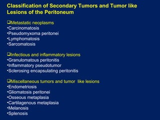
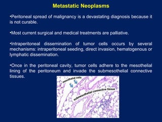
Imaging of Secondary Tumors and Tumor like
Lesions of the Peritoneal Cavity
By
Dr. Naglaa Mahmoud
Registrar of Clinical Radiology
KCCC
introduction
 Tumors and tumor like lesions that involve the peritoneum are a
diverse group of disorders that range from benign to highly
malignant.
 The anatomy of peritoneal ligaments and mesenteries and the
normal circulation of peritoneal fluid dictate location and
distribution of these diseases within the peritoneal cavity.
 The pathologic processes that may secondarily affect the
peritoneum and peritoneal cavity can be loosely categorized into
three broad groups: metastatic neoplasms, infectious and
inflammatory lesions and miscellaneous tumors and tumor like
lesions.
Anatomic Spaces and Fluid Flow in the Peritoneal Cavity
 The peritoneal cavity is the potential space between the layers of visceral and
parietal peritoneum. The visceral peritoneum lines the intraperitoneal organs,
mesenteries, and omenta, and the parietal peritoneum lines the undersurface
of the hemidiaphragm, abdominal wall, the retroperitoneum and pelvis.
 The peritoneal ligaments (coronary, gastrohepatic, hepatoduodenal, falciform,
gastrocolic, duodenocolic, gastrosplenic, splenorenal, and phrenicocolic) and
mesenteries (transverse mesocolon, small bowel mesentery, and sigmoid
mesentery) are double folds of peritoneum.
 They suspend and support the intraperitoneal organs and subdivide the
peritoneal cavity into interconnected compartments that dictate the flow of
fluid and location of disease.
 The peritoneal cavity normally contains a very small volume of sterile fluid,
which is similar to plasma.
 The clearance of fluid from the peritoneal cavity is augmented by continual
circulation of the fluid upward to the subdiaphragmatic spaces where the
subphrenic submesothelial lymphatic provide most of the lymphatic clearance
from the peritoneal cavity.
Anatomic Spaces and Fluid Flow in the Peritoneal Cavity
 Initially, peritoneal fluid preferentially seeks gravity-dependent spaces, such as
the deep recesses of the pelvis (the pouch of Douglas in women and the
retrovesical space in men), and the lateral paravesical spaces, and then
ascends cephalad in the paracolic gutters to reach the subdiaphragmatic
spaces.
 Most of the fluid ascends via the right paracolic gutter into the right
subdiaphragmatic space, because the left paracolic gutter is shallow and
discontinuous with the left subdiaphragmatic space at the phrenicocolic ligament
and because direct passage from the right to left subdiaphragmatic space is
prevented by the falciform ligament.
 In pathologic conditions that result in ascites, fluid collects in well-defined areas
of stasis or arrested flow: the peritoneal recesses of the pelvis (pouch of
Douglas in women and retrovesical space in men), the right lower quadrant
(near the termination of the small bowel mesentery at the ileocecal junction), the
superior aspect of the sigmoid mesocolon and the right paracolic gutter.
 Relative stasis of ascites at these sites promotes seeding of malignant cells or
accumulation of infection at these locations.
(a) Illustration of the normal posterior peritoneal reflections and intraabdominal spaces shows the cut
surface of the transverse mesocolon (arrow), which divides the peritoneum into supramesocolic
and inframesocolic compartments. The root of the small bowel mesentery (arrowhead) divides the
inframesocolic compartment into the right and left infracolic spaces. The cut surface of the sigmoid
mesentery is also shown. The sigmoid mesentery serves as a watershed to channel peritoneal fluid
into the pelvis.
(b) Illustration shows the pathways of flow of intraperitoneal fluid and the four predominant sites (*) of
stasis of ascitic fluid in the lower abdomen.
Classification of Secondary Tumors and Tumor like
Lesions of the Peritoneum
Metastatic neoplasms
•Carcinomatosis
•Pseudomyxoma peritonei
•Lymphomatosis
•Sarcomatosis
Infectious and inflammatory lesions
•Granulomatous peritonitis
•Inflammatory pseudotumor
•Sclerosing encapsulating peritonitis
Miscellaneous tumors and tumor like lesions
•Endometriosis
•Gliomatosis peritonei
•Osseous metaplasia
•Cartilagenous metaplasia
•Melanosis
•Splenosis
Metastatic Neoplasms
•Peritoneal spread of malignancy is a devastating diagnosis because it
is not curable.
•Most current surgical and medical treatments are palliative.
•Intraperitoneal dissemination of tumor cells occurs by several
mechanisms: intraperitoneal seeding, direct invasion, hematogenous or
lymphatic dissemination.
•Once in the peritoneal cavity, tumor cells adhere to the mesothelial
lining of the peritoneum and invade the submesothelial connective
tissues.
Primary intraperitoneal seeding
• Occurs most commonly from gastrointestinal and ovarian
malignancies as a consequence of intramural tumor growth and
full thickness invasion of the tumor through the bowel wall or
extension of the tumor through the peritoneal lining of the ovary,
pancreas or liver.
Secondary seeding of the peritoneum
• Occurs iatrogenically during surgery or biopsy.
• Once in the peritoneal cavity, freely floating tumor cells migrate
through the peritoneal cavity with the normal circulation of fluid.
• Tumor cells initially tend to coalesce in gravity dependent
recesses of the peritoneum and in regions of stasis as well as
the subdiaphragmatic spaces.
• Eventually, tumor deposits may diffusely stud both visceral and
parietal peritoneum.
1- Carcinomatosis
•Carcinomas from the gastrointestinal tract (stomach, colon, appendix,
gallbladder and pancreas), ovary, breast, lung and uterus.
Clinical picture:
•Asymptomatic at the onset of the lesions.
•Progressive involvement of the peritoneum will cause abdominal
enlargement from ascites, nausea, vomiting and abdominal pain from bowel
obstruction which frequently occurs with peritoneal carcinomatosis from CRC.
Imaging features:
US:
• Ascites is a common finding in peritoneal carcinomatosis.
• It may be anechoic or may contain hypoechoic particles that
reflects proteinaceous exudate.
• Tumor nodules on the visceral or parietal peritoneal surfaces are
generally hypoechoic nodules or sheet like masses.
• Tumor infiltrating the omentum produces an echogenic, plaque
like structure, which can be observed floating in ascitic fluid or
adherent to the anterior abdominal wall.
Peritoneal carcinomatosis from mucinous adenocarcinoma of the colon.
US of the pelvis shows a large amount of ascites that contains fixed echoes and nodular, sheet
like thickening of the posterior peritoneum (arrows).
CT scan:
• CT is the imaging modality of choice for evaluating patients with diffuse
abdominal disorders and for staging malignancies that may involve the
peritoneal cavity.
• In absence of ascites, careful inspection of areas of stasis of peritoneal
fluid is important during staging CT because peritoneal tumors can be
subtle and difficult to identify.
• The pouch of Douglas or retrovesical space, ileocecal region, paracolic
gutters, subhepatic space, right subdiaphragmatic space and root of the
small bowel mesentery are important sites of occult tumor.
Colonic adenocarcinoma with peritoneal carcinomatosis.
CT shows the primary lesion in the transverse colon (* in b) with pericolonic lymphadenopathy; nodular
peritoneal thickening in the left upper quadrant (arrow in a); and tumor nodules in the gastrosplenic ligament
(arrow in b), left paracolic gutter (arrow in c) and retrovesical space (arrow in d).
Peritoneal carcinomatosis from adenocarcinoma of the colon.
CT shows a small amount of ascites, nodular soft-tissue thickening of the right subdiaphragmatic
peritoneum (arrows in a), and soft tissue infiltrating the greater omentum (arrows in b).
• The imaging findings of peritoneal carcinomatosis vary from multifocal
discrete nodules to infiltrative masses.
Photograph of the resected greater omentum shows tumor
nodules that stud the omental surface and replace normal fat.
• Small bowel obstruction is the most common complication of peritoneal
carcinomatosis and may be secondary to diffusely infiltrating tumor or
focal tumor masses.
Peritoneal mucinous carcinomatosis that caused small bowel obstruction .
CT shows low-attenuation mucinous ascites that infiltrates between the folds of the small bowel mesentery.
There are low-attenuation mucinous metastatic deposits in the greater omentum (arrows in a) and along the
peritoneal surfaces and in the paracolic gutters (arrows in b).
The small bowel is partially obstructed from metastatic tumor that encases a segment of distal small bowel
(arrowhead in b).
MRI:
• Gadolinium-enhanced MRI is the examination of choice for depicting peritoneal
carcinomatosis due to its superior contrast resolution especially in tumor nodules
less than 1 cm.
• Peritoneal carcinomatosis enhances slowly and therefore is best shown 5–10
minutes after contrast injection.
• Abnormal enhancement should be suspected when the peritoneum is enhancing
greater than the liver or has associated thickening, nodularity or masses.
Patient with abdominal distension.
Multidetector helical CT scan shows
perihepatic ascites without evidence
of tumor.
Arterial Phase
Non enhancing ascites is present around the
liver. No abnormal peritoneal enhancement is
present on these images obtained immediately
following injection of 0.2mmol/kg gadolinium.
Delayed Gad
Fat suppressed MR image obtained 5
min after injection of IV gadolinium
depicts abnormal enhancement and
thickening of the right subphrenic
peritoneum. MR findings indicate
peritoneal carcinomatosis.
This patient underwent laparotomy
confirming stage III ovarian cancer.
FDG PET/CT:
•The reported sensitivities for the detection of peritoneal carcinomatosis are 78%–
100%, with the highest sensitivities in patients with clinically suspected recurrent
tumor because of elevation in tumor markers.
•Metastatic foci appear as round, oval or globular areas of increased activity.
•Subcentimeter lesions may not demonstrate sufficient uptake to be visible.
Peritoneal carcinomatosis from ovarian carcinoma .
CT scan shows loculated ascites. (b) Fused PET/CT image shows focal, intense uptake in the left mid
abdomen (arrow) from a peritoneal metastatic deposit that is nearly isoattenuating relative to the ascites and
difficult to visualize with CT alone.
2- Pseudomyxoma Peritonei (jelly belly)
•A rare condition with a reported incidence of one case per million
population per year.
•Copious, thick mucinous or gelatinous material on the surfaces of the
peritoneal cavity.
•The majority of cases of classic pseudomyxoma peritonei develop from
low-grade mucinous carcinomas that arise in the appendix and that
penetrate or rupture into the peritoneal cavity.
•Common in women with mean age of 49 years.
Intraoperative photograph shows globules of
mucin adherent to the greater omentum.
Classification:
1- Pseudomyxoma peritonei (PMP)
•Contains benign or borderline epithelial cells or cells from well-
differentiated (low-grade) mucinous carcinomas.
•This form does not invade the stroma and is amenable to surgical
debulking.
2- Peritoneal mucinous carcinomatosis
•Invasive, high grade, moderately or poorly differentiated mucinous
carcinoma.
•Originates from a mucinous carcinoma of the gastrointestinal tract,
gallbladder, pancreas or ovary and its clinical course is fatal.
Clinical picture:
•Progressive abdominal pain, increasing abdominal girth and weight loss.
•In some cases, patients complain of predominantly right-sided pain or they
may have signs and symptoms similar to those of appendicitis.
US:
•Echogenic ascitic fluid.
•In contrast to echoes that may be secondary to proteinaceous, bloody or fibrinous
exudates or infection, the echoes within pseudomyxoma peritonei are not mobile.
•Echogenic septations within the gelatinous ascites are frequently observed.
•The intestine is usually displaced centrally and posteriorly and may have a
starburst appearance in which the central highly echogenic bowel is in star like
configuration surrounded by hypoechoic gelatinous fluid.
Pseudomyxoma peritonei in a 70-year-old woman who complained of increasing abdominal girth.
US shows complex, hypoechoic ascites that contains non mobile echoes and centrally displaced
small bowel that has a starburst appearance.
CT scan:
• Scalloping of the hepatic and splenic margins is an important diagnostic
finding that helps differentiate pseudomyxoma from simple ascites.
• Mucinous deposits of pseudomyxoma peritonei accumulate in areas of
stasis in the peritoneal cavity: the pouch of Douglas in women or the
retrovesical space in men, the right lower quadrant near the ileocecal
junction, the right subhepatic space (Morison pouch) and the right
subphrenic space.
• Mucin within the peritoneum is usually low in CT attenuation, but areas
of soft-tissue attenuation may be present that represent solid tumor
elements, fibrosis or compression of the mesentery.
• Curvilinear or amorphous calcifications may be present.
Pseudomyxoma peritonei .
CT scans show low-attenuation mucinous ascites that scallops the margins of the liver, spleen, and
gastric fundus; displaces bowel and mesentery; and contains coarse calcifications.
The greater omentum (arrow in b) has increased attenuation from omental infiltration or
fibrosis.
• Differentiation between true pseudomyxoma peritonei from mucinous
carcinomatosis is important as patients with PMP from an
appendiceal low-grade mucinous neoplasm have a 5-year survival
rate of 50% and those who have mucinous carcinomatosis have a 5-
year survival rate of less than 10%.
• Difficult differentiation by CT alone due to overlap in findings.
• Mucinous carcinomatosis tends to involve the chest more
frequently with effusions or pleural masses and may also be
accompanied by mesenteric or retroperitoneal lymphadenopathy,
omental caking and invasion into parenchymal organs.
• In contrast, pseudomyxoma peritonei typically does not invade
visceral organs or spread by lymphatic or hematogenous routes.
3- Lymphomatosis
•Lymphomas may involve the peritoneal surfaces as a primary or secondary
process.
•Secondary peritoneal involvement may occur in the general population of
patients with lymphoma as well as in immunocompromised patients.
•Primary lymphomas of the peritoneum are uncommon and nearly
exclusively found in immunocompromised patients, most of whom are
infected with HIV.
•Primary lymphoma is mainly confined to the cavity of origin, but end stage
primary lymphoma is associated with generalized lymph node involvement.
•High grade and aggressive and have a poor prognosis.
Imaging Features:
•Peritoneal lymphomatosis secondary to a preexisting lymphoma is
characterized by diffusely thickened peritoneal surfaces with
multifocal nodules and masses that mimic peritoneal carcinomatosis.
•Other CT features include ascites, peritoneal enhancement and
thickening, omental caking, infiltration of the small bowel mesentery
and fine nodularity in the mesenteric and omental fat.
•The presence of extensive adenopathy in lymph node chains
typically involved with lymphoma, such as those in the retrocrural
region and small bowel mesentery, may suggest lymphomatosis
over carcinomatosis.
•In contrast, the most common imaging manifestation of primary
peritoneal lymphoma is malignant ascites without associated
lymphadenopathy, organomegaly or masses.
Intravenous and oral contrast-enhanced CT scan shows soft tissue
diffusely infiltrating through the peritoneum, encasing the small
bowel, and lining the folds of the small bowel mesentery. Ascites
and diffuse peritoneal thickening are present.
4- Sarcomatosis
•Sarcomas are malignant neoplasms that arise from tissue derived from
primitive mesoderm.
•Metastasis to the peritoneal cavity from soft-tissue and extremity
sarcomas is unusual and is presumed to occur through a hematogenous
route.
•In contrast, sarcomas arising from the GIT, namely malignant GIST,
commonly spread to the peritoneum by extension through the serosal
surface of the bowel and direct seeding of the peritoneal cavity.
•Peritoneal metastases are focal soft-tissue masses, nodules, or confluent
areas of increased attenuation within the mesentery and omentum.
•Visceral implantation may occur, which results in soft-tissue masses on
serosal margins of the liver and spleen.
•Ascites is minimal and occurs late in the course of disease.
Gastrointestinal stromal tumor metastatic to the peritoneum.
CT scans show a metastatic deposit of tumor on the serosal surface of the liver (arrow in a) and a
large, partially cystic metastatic deposit in the left mid abdomen (* in b). No significant ascites is
present.
Infectious and Inflammatory Conditions
1- Granulomatous Peritonitis
•A wide range of unusual forms of peritoneal inflammation and
infection that have overlapping clinical, pathologic and imaging
features.
•Infectious agents such as tuberculous, Histoplasma, or
Pneumocystis organisms; foreign material such as talc and
barium; meconium; bowel contents; contents of ruptured ovarian
cysts; bile or gallstones may cause granulomatous peritonitis.
•Peritoneal histoplasmosis and Pneumocystis infections are rare
and usually occur in immunocompromised patients.
• Tuberculosis may reach the peritoneal cavity as part of a systemic infection (i.e.
miliary tuberculosis), direct extension from the bowel to the peritoneum, or
lymphatic dissemination.
• Tuberculous peritonitis has been described as having three imaging patterns
(wet, fibrotic fixed, and dry plastic) depending on the amount of ascites and soft-
tissue component.
• Ascites in the wet form may be diffuse or loculated and is characteristically has
high attenuation.
• peritoneal soft-tissue masses or nodules have soft-tissue attenuation and may
show enhancement post IV contrast.
• Concomitant lymph node enlargement with central low attenuation from caseous
necrosis in the peripancreatic and periportal regions, mesenteries, or
retroperitoneum may also be present.
• Other findings in the abdomen such as miliary microabscesses in the liver or
spleen, splenomegaly, splenic or lymph node calcification, inflammatory
thickening of the terminal ileum and cecum may help suggest tuberculosis as the
etiology of diffuse peritoneal disease.
Intravenous and oral contrast-enhanced CT scan of a 22-year-old man who complained of intermittent
fevers for 1 month and a new onset of abdominal pain shows loculated ascites.
There is diffuse enhancement of the peritoneum and nodularity of the greater omentum (arrow). The
visualized small bowel segments are fixed with mild dilatation.
2- Inflammatory Pseudotumor
•An unusual, benign, chronic inflammatory lesion can occur in mesentery
and peritoneum.
•Patients present with symptoms secondary to mass effect, fever, weight
loss, anemia, thrombocytosis and a polyclonal hypergammaglobulinemia
which resolve after complete surgical resection.
•Local recurrence has been reported with incomplete surgical resection.
•Non specific imaging features. It may have well-defined or infiltrating
margins that extend up to adjacent bowel segments and into the
surrounding mesentery.
US:
• Solid mixed echotexture mass within the mesentery.
Color Doppler:
• Prominent vascularity.
CT: Variable enhancement
• Non enhancing, heterogeneous enhancement or peripheral
enhancement.
• Larger lesions may have central areas of hypoattenuation
due to necrosis.
Inflammatory pseudotumor in a 36-year-old man who complained of left flank pain, diarrhea,
nausea, and vomiting. (a) Intravenous contrast-enhanced CT scan shows a 6-cm soft-tissue
attenuation mass in the small bowel mesentery that has central low attenuation (arrow).
(b) Photograph of the resected surgical specimen shows the tan mass with central necrosis in
the small bowel mesentery.
3- Sclerosing Encapsulating Peritonitis
•A rare chronic inflammatory disorder of the peritoneum that occurs
in patients who undergo continuous ambulatory peritoneal dialysis.
•It may also be idiopathic, associated with ventriculoperitoneal
shunts, liver transplantation, tuberculosis, foreign material, and use
of beta-blocker drugs (as a rare complication).
Clinical picture:
•Abdominal pain, nausea, vomiting and repeated episodes of bowel
obstruction with the diagnosis is clinically established by loss of ultrafiltration
capacity during peritoneal dialysis.
US:
•Thin echogenic strands can be seen within ascitic fluid with tethered small
bowel. Over time, the peritoneum becomes thick and echogenic.
CT:
•Diffusely thickened peritoneum, ascites; small bowel may be tethered or
matted within loculated fluid collections.
•Multiple foci of linear calcification may develop as the disease progresses.
Sclerosing encapsulating
peritonitis in a liver transplant
recipient who complained of
bloating and symptoms
of bowel obstruction.
(a, b) Intravenous contrast-
enhanced CT scans show
encapsulation of the small bowel
by a thickened and enhancing
peritoneum (arrows in b). The
small bowel is tethered in a radial
configuration and has mural
thickening. A small amount of
ascites is present. (c, d)
Intraoperative photographs show
the thick, opaque peritoneum
unopened (c) and opened (d).
Miscellaneous Conditions
1- Endometriosis
•A common condition that affects the peritoneal cavity of women.
•It is defined as the occurrence of functional endometrial glands and
stroma outside the uterus, typically in the peritoneal surfaces of the
pelvis and pelvic organs. However, all visceral and parietal peritoneal
surfaces may be affected.
•Affects 10% of women of childbearing age with mean age at diagnosis
is 25–29 years.
Clinical picture:
•Asymptomatic.
•Infertility, pelvic pain, dysmenorrhea, dyspareunia, and dysfunctional
uterine bleeding.
•Pelvic adhesions and implants on the peritoneal surfaces of the
bowel may cause bowel obstruction.
US and CT findings:
•Non specific, masses may appear solid, cystic or mixed solid and
cystic.
MRI:
•Relies on the detection of blood products within the lesions.
•The SI varies depending on the stage of hemorrhage degradation.
•The most common signal intensity pattern is hyperintensity on T1WIs and
hypointensity on T2WIs due to deoxyhemoglobin and methemoglobin. The
loss of signal on T2WIs reflects chronic and recurrent hemorrhage within the
lesion and the accumulation of iron and protein.
• Other MR imaging features include a low-signal-intensity rim on both T1-
and T2 caused by the presence of surrounding fibrosis and hemosiderin.
•Use of fat-suppressed and chemical-shift imaging techniques improves the
conspicuity and detection rate of peritoneal disease and small
endometriomas.
•Use of gadolinium-based contrast material does not add specificity to the
diagnosis of endometriosis because the enhancement patterns are
nonspecific and overlap with those of other benign and malignant processes.
Endometriosis in the right subhepatic space in a 43-year-old woman who complained of intermittent
right upper quadrant pain. (a) Axial T1-weighted image shows a small low-signal-intensity mass in the
right subhepatic space (arrow). (b) On an axial opposed phase T1-weighted fast multiplanar image, the
lesion has high signal intensity with a low-signal-intensity rim (arrow). (c) On an axial fast spin-echo
T2-weighted image with fat saturation, the lesion has low signal intensity (arrow). (d) Axial T1-weighted
image with fat saturation obtained during the portal venous phase of gadolinium enhancement shows
that the lesion enhances (arrow).
2- Gliomatosis Peritonei
•Presence of benign mature glial implants throughout the peritoneum.
•Occurs almost exclusively in association with solid or immature ovarian
teratomas but it has also been reported to occur in association with
ventriculoperitoneal shunts.
•Glial tissue may enter the peritoneal cavity from rupture of an ovarian
teratoma or it may occur independent of the material in the teratoma
and that the implants were thought to be metaplasia of the peritoneal
tissue.
•Malignant transformation is exceedingly rare.
CT:
• Soft-tissue peritoneal nodules and masses, with omental caking
and ascites in the setting of a pelvic mass with appearance
suggestive of a teratoma.
• The imaging findings of gliomatosis peritonei are identical to those
of peritoneal carcinomatosis.
• In the setting of an ovarian mass, imaging findings alone cannot aid
in differentiation of diffuse peritoneal malignant seeding from
benign glial deposits.
Gliomatosis peritonei in a 13-year-old girl who complained of abdominal pain, distention, and constipation.
(a, b) CT scans (a obtained at a more cephalic level than b) show a large, well-defined, complex cystic mass
in the lower abdomen that contains fat and calcifications and extends into the upper abdomen. Ascites is present.
(c) Intraoperative photograph shows a complex solid and cystic mass that mimics cerebral tissue. The
mass is admixed with fibrosis, fat, cartilaginous tissue, and bone, content similar to that of a teratoma.
(d) Intraoperative photograph of the omentum shows innumerable tiny nodules of gliomatosis throughout.
3- Osseous Metaplasia
•Osseous metaplasia of the mesentery and peritoneum.
•An unusual lesion that can develop after trauma or repeated
intraabdominal operative procedures.
•More frequently in men.
•It may represent metaplasia of the submesothelial mesenchyme or
implantation of osteoblasts or periosteum during trauma or procedures.
CT:
•Multiple high-attenuation, linear-branching structures within the
mesentery that extend to the peritoneal surfaces.
Heterotopic ossification and osseous metaplasia in a 53-year-old man who sustained extensive
abdominal injuries in a motor vehicle accident and underwent multiple laparotomies.
Intravenous and oral contrast enhanced CT scan shows multiple, high-attenuation, branching
and linear structures (arrows) in the small bowel mesentery and along the peritoneal surface.
These structures extend into the open abdominal wall.
4- Splenosis
•Intraperitoneal dissemination or implantation of splenic tissue after disruption
of the splenic capsule by trauma or surgery.
•It can be found anywhere in the peritoneal cavity, implanted in visceral
organs, within the thorax and in surgical scars.
•Most patients are asymptomatic.
•In rare cases, splenosis can result in bowel obstruction.
•Large lesions may undergo torsion and infarction or rupture.
•Splenosis may be the site of recurrent disease in patients who have
undergone splenectomy to control a hematologic disorder.
•US, CT and MR imaging features are identical to those of the normal spleen.
Splenosis in a 47-year-old man with hepatitis C and cirrhosis who had undergone traumatic splenectomy at
age 13 years.
(a) Intravenous and oral contrast enhanced CT scan shows multiple, lobular and round,
homogeneously enhancing masses (arrows) in the anterior abdomen. (b) Photograph of the resected
omentum shows multiple foci of splenic tissue.
Conclusions
Secondary peritoneal tumors and tumor like lesions are a diverse group of
pathologic disorders.
Secondary tumors are more common than primary lesions.
Metastatic disease should be the initial concern in a patient with ascites,
peritoneal nodularity and omental masses in patients with positive history of
primary neoplasm.
Mucinous carcinomatosis and pseudomyxoma peritonei should be
considered when peritoneal disease is of low attenuation on CT and when
evidence of a primary mucinous neoplasm is noted.
Infectious conditions such as disseminated tuberculosis should be
considered if the clinical history is appropriate or if supportive findings such as
miliary microabscesses in the liver or spleen or low-attenuation
lymphadenopathy are present.
Miscellaneous disorders such as endometriosis, splenosis, and osseous
metaplasia of the mesentery should be considered when the appropriate
findings and clinical history are present.
Secondary Peritoneal Disease

Secondary Peritoneal Disease

  • 1.
    Imaging of SecondaryTumors and Tumor like Lesions of the Peritoneal Cavity By Dr. Naglaa Mahmoud Registrar of Clinical Radiology KCCC
  • 2.
    introduction  Tumors andtumor like lesions that involve the peritoneum are a diverse group of disorders that range from benign to highly malignant.  The anatomy of peritoneal ligaments and mesenteries and the normal circulation of peritoneal fluid dictate location and distribution of these diseases within the peritoneal cavity.  The pathologic processes that may secondarily affect the peritoneum and peritoneal cavity can be loosely categorized into three broad groups: metastatic neoplasms, infectious and inflammatory lesions and miscellaneous tumors and tumor like lesions.
  • 3.
    Anatomic Spaces andFluid Flow in the Peritoneal Cavity  The peritoneal cavity is the potential space between the layers of visceral and parietal peritoneum. The visceral peritoneum lines the intraperitoneal organs, mesenteries, and omenta, and the parietal peritoneum lines the undersurface of the hemidiaphragm, abdominal wall, the retroperitoneum and pelvis.  The peritoneal ligaments (coronary, gastrohepatic, hepatoduodenal, falciform, gastrocolic, duodenocolic, gastrosplenic, splenorenal, and phrenicocolic) and mesenteries (transverse mesocolon, small bowel mesentery, and sigmoid mesentery) are double folds of peritoneum.  They suspend and support the intraperitoneal organs and subdivide the peritoneal cavity into interconnected compartments that dictate the flow of fluid and location of disease.  The peritoneal cavity normally contains a very small volume of sterile fluid, which is similar to plasma.  The clearance of fluid from the peritoneal cavity is augmented by continual circulation of the fluid upward to the subdiaphragmatic spaces where the subphrenic submesothelial lymphatic provide most of the lymphatic clearance from the peritoneal cavity.
  • 4.
    Anatomic Spaces andFluid Flow in the Peritoneal Cavity  Initially, peritoneal fluid preferentially seeks gravity-dependent spaces, such as the deep recesses of the pelvis (the pouch of Douglas in women and the retrovesical space in men), and the lateral paravesical spaces, and then ascends cephalad in the paracolic gutters to reach the subdiaphragmatic spaces.  Most of the fluid ascends via the right paracolic gutter into the right subdiaphragmatic space, because the left paracolic gutter is shallow and discontinuous with the left subdiaphragmatic space at the phrenicocolic ligament and because direct passage from the right to left subdiaphragmatic space is prevented by the falciform ligament.  In pathologic conditions that result in ascites, fluid collects in well-defined areas of stasis or arrested flow: the peritoneal recesses of the pelvis (pouch of Douglas in women and retrovesical space in men), the right lower quadrant (near the termination of the small bowel mesentery at the ileocecal junction), the superior aspect of the sigmoid mesocolon and the right paracolic gutter.  Relative stasis of ascites at these sites promotes seeding of malignant cells or accumulation of infection at these locations.
  • 6.
    (a) Illustration ofthe normal posterior peritoneal reflections and intraabdominal spaces shows the cut surface of the transverse mesocolon (arrow), which divides the peritoneum into supramesocolic and inframesocolic compartments. The root of the small bowel mesentery (arrowhead) divides the inframesocolic compartment into the right and left infracolic spaces. The cut surface of the sigmoid mesentery is also shown. The sigmoid mesentery serves as a watershed to channel peritoneal fluid into the pelvis. (b) Illustration shows the pathways of flow of intraperitoneal fluid and the four predominant sites (*) of stasis of ascitic fluid in the lower abdomen.
  • 7.
    Classification of SecondaryTumors and Tumor like Lesions of the Peritoneum Metastatic neoplasms •Carcinomatosis •Pseudomyxoma peritonei •Lymphomatosis •Sarcomatosis Infectious and inflammatory lesions •Granulomatous peritonitis •Inflammatory pseudotumor •Sclerosing encapsulating peritonitis Miscellaneous tumors and tumor like lesions •Endometriosis •Gliomatosis peritonei •Osseous metaplasia •Cartilagenous metaplasia •Melanosis •Splenosis
  • 8.
    Metastatic Neoplasms •Peritoneal spreadof malignancy is a devastating diagnosis because it is not curable. •Most current surgical and medical treatments are palliative. •Intraperitoneal dissemination of tumor cells occurs by several mechanisms: intraperitoneal seeding, direct invasion, hematogenous or lymphatic dissemination. •Once in the peritoneal cavity, tumor cells adhere to the mesothelial lining of the peritoneum and invade the submesothelial connective tissues.
  • 9.
    Primary intraperitoneal seeding •Occurs most commonly from gastrointestinal and ovarian malignancies as a consequence of intramural tumor growth and full thickness invasion of the tumor through the bowel wall or extension of the tumor through the peritoneal lining of the ovary, pancreas or liver. Secondary seeding of the peritoneum • Occurs iatrogenically during surgery or biopsy. • Once in the peritoneal cavity, freely floating tumor cells migrate through the peritoneal cavity with the normal circulation of fluid. • Tumor cells initially tend to coalesce in gravity dependent recesses of the peritoneum and in regions of stasis as well as the subdiaphragmatic spaces. • Eventually, tumor deposits may diffusely stud both visceral and parietal peritoneum.
  • 10.
    1- Carcinomatosis •Carcinomas fromthe gastrointestinal tract (stomach, colon, appendix, gallbladder and pancreas), ovary, breast, lung and uterus. Clinical picture: •Asymptomatic at the onset of the lesions. •Progressive involvement of the peritoneum will cause abdominal enlargement from ascites, nausea, vomiting and abdominal pain from bowel obstruction which frequently occurs with peritoneal carcinomatosis from CRC.
  • 11.
    Imaging features: US: • Ascitesis a common finding in peritoneal carcinomatosis. • It may be anechoic or may contain hypoechoic particles that reflects proteinaceous exudate. • Tumor nodules on the visceral or parietal peritoneal surfaces are generally hypoechoic nodules or sheet like masses. • Tumor infiltrating the omentum produces an echogenic, plaque like structure, which can be observed floating in ascitic fluid or adherent to the anterior abdominal wall.
  • 12.
    Peritoneal carcinomatosis frommucinous adenocarcinoma of the colon. US of the pelvis shows a large amount of ascites that contains fixed echoes and nodular, sheet like thickening of the posterior peritoneum (arrows).
  • 13.
    CT scan: • CTis the imaging modality of choice for evaluating patients with diffuse abdominal disorders and for staging malignancies that may involve the peritoneal cavity. • In absence of ascites, careful inspection of areas of stasis of peritoneal fluid is important during staging CT because peritoneal tumors can be subtle and difficult to identify. • The pouch of Douglas or retrovesical space, ileocecal region, paracolic gutters, subhepatic space, right subdiaphragmatic space and root of the small bowel mesentery are important sites of occult tumor.
  • 14.
    Colonic adenocarcinoma withperitoneal carcinomatosis. CT shows the primary lesion in the transverse colon (* in b) with pericolonic lymphadenopathy; nodular peritoneal thickening in the left upper quadrant (arrow in a); and tumor nodules in the gastrosplenic ligament (arrow in b), left paracolic gutter (arrow in c) and retrovesical space (arrow in d).
  • 15.
    Peritoneal carcinomatosis fromadenocarcinoma of the colon. CT shows a small amount of ascites, nodular soft-tissue thickening of the right subdiaphragmatic peritoneum (arrows in a), and soft tissue infiltrating the greater omentum (arrows in b). • The imaging findings of peritoneal carcinomatosis vary from multifocal discrete nodules to infiltrative masses.
  • 16.
    Photograph of theresected greater omentum shows tumor nodules that stud the omental surface and replace normal fat.
  • 17.
    • Small bowelobstruction is the most common complication of peritoneal carcinomatosis and may be secondary to diffusely infiltrating tumor or focal tumor masses. Peritoneal mucinous carcinomatosis that caused small bowel obstruction . CT shows low-attenuation mucinous ascites that infiltrates between the folds of the small bowel mesentery. There are low-attenuation mucinous metastatic deposits in the greater omentum (arrows in a) and along the peritoneal surfaces and in the paracolic gutters (arrows in b). The small bowel is partially obstructed from metastatic tumor that encases a segment of distal small bowel (arrowhead in b).
  • 18.
    MRI: • Gadolinium-enhanced MRIis the examination of choice for depicting peritoneal carcinomatosis due to its superior contrast resolution especially in tumor nodules less than 1 cm. • Peritoneal carcinomatosis enhances slowly and therefore is best shown 5–10 minutes after contrast injection. • Abnormal enhancement should be suspected when the peritoneum is enhancing greater than the liver or has associated thickening, nodularity or masses. Patient with abdominal distension. Multidetector helical CT scan shows perihepatic ascites without evidence of tumor.
  • 19.
    Arterial Phase Non enhancingascites is present around the liver. No abnormal peritoneal enhancement is present on these images obtained immediately following injection of 0.2mmol/kg gadolinium. Delayed Gad Fat suppressed MR image obtained 5 min after injection of IV gadolinium depicts abnormal enhancement and thickening of the right subphrenic peritoneum. MR findings indicate peritoneal carcinomatosis. This patient underwent laparotomy confirming stage III ovarian cancer.
  • 20.
    FDG PET/CT: •The reportedsensitivities for the detection of peritoneal carcinomatosis are 78%– 100%, with the highest sensitivities in patients with clinically suspected recurrent tumor because of elevation in tumor markers. •Metastatic foci appear as round, oval or globular areas of increased activity. •Subcentimeter lesions may not demonstrate sufficient uptake to be visible. Peritoneal carcinomatosis from ovarian carcinoma . CT scan shows loculated ascites. (b) Fused PET/CT image shows focal, intense uptake in the left mid abdomen (arrow) from a peritoneal metastatic deposit that is nearly isoattenuating relative to the ascites and difficult to visualize with CT alone.
  • 21.
    2- Pseudomyxoma Peritonei(jelly belly) •A rare condition with a reported incidence of one case per million population per year. •Copious, thick mucinous or gelatinous material on the surfaces of the peritoneal cavity. •The majority of cases of classic pseudomyxoma peritonei develop from low-grade mucinous carcinomas that arise in the appendix and that penetrate or rupture into the peritoneal cavity. •Common in women with mean age of 49 years. Intraoperative photograph shows globules of mucin adherent to the greater omentum.
  • 22.
    Classification: 1- Pseudomyxoma peritonei(PMP) •Contains benign or borderline epithelial cells or cells from well- differentiated (low-grade) mucinous carcinomas. •This form does not invade the stroma and is amenable to surgical debulking. 2- Peritoneal mucinous carcinomatosis •Invasive, high grade, moderately or poorly differentiated mucinous carcinoma. •Originates from a mucinous carcinoma of the gastrointestinal tract, gallbladder, pancreas or ovary and its clinical course is fatal. Clinical picture: •Progressive abdominal pain, increasing abdominal girth and weight loss. •In some cases, patients complain of predominantly right-sided pain or they may have signs and symptoms similar to those of appendicitis.
  • 23.
    US: •Echogenic ascitic fluid. •Incontrast to echoes that may be secondary to proteinaceous, bloody or fibrinous exudates or infection, the echoes within pseudomyxoma peritonei are not mobile. •Echogenic septations within the gelatinous ascites are frequently observed. •The intestine is usually displaced centrally and posteriorly and may have a starburst appearance in which the central highly echogenic bowel is in star like configuration surrounded by hypoechoic gelatinous fluid. Pseudomyxoma peritonei in a 70-year-old woman who complained of increasing abdominal girth. US shows complex, hypoechoic ascites that contains non mobile echoes and centrally displaced small bowel that has a starburst appearance.
  • 24.
    CT scan: • Scallopingof the hepatic and splenic margins is an important diagnostic finding that helps differentiate pseudomyxoma from simple ascites. • Mucinous deposits of pseudomyxoma peritonei accumulate in areas of stasis in the peritoneal cavity: the pouch of Douglas in women or the retrovesical space in men, the right lower quadrant near the ileocecal junction, the right subhepatic space (Morison pouch) and the right subphrenic space. • Mucin within the peritoneum is usually low in CT attenuation, but areas of soft-tissue attenuation may be present that represent solid tumor elements, fibrosis or compression of the mesentery. • Curvilinear or amorphous calcifications may be present.
  • 25.
    Pseudomyxoma peritonei . CTscans show low-attenuation mucinous ascites that scallops the margins of the liver, spleen, and gastric fundus; displaces bowel and mesentery; and contains coarse calcifications. The greater omentum (arrow in b) has increased attenuation from omental infiltration or fibrosis.
  • 26.
    • Differentiation betweentrue pseudomyxoma peritonei from mucinous carcinomatosis is important as patients with PMP from an appendiceal low-grade mucinous neoplasm have a 5-year survival rate of 50% and those who have mucinous carcinomatosis have a 5- year survival rate of less than 10%. • Difficult differentiation by CT alone due to overlap in findings. • Mucinous carcinomatosis tends to involve the chest more frequently with effusions or pleural masses and may also be accompanied by mesenteric or retroperitoneal lymphadenopathy, omental caking and invasion into parenchymal organs. • In contrast, pseudomyxoma peritonei typically does not invade visceral organs or spread by lymphatic or hematogenous routes.
  • 27.
    3- Lymphomatosis •Lymphomas mayinvolve the peritoneal surfaces as a primary or secondary process. •Secondary peritoneal involvement may occur in the general population of patients with lymphoma as well as in immunocompromised patients. •Primary lymphomas of the peritoneum are uncommon and nearly exclusively found in immunocompromised patients, most of whom are infected with HIV. •Primary lymphoma is mainly confined to the cavity of origin, but end stage primary lymphoma is associated with generalized lymph node involvement. •High grade and aggressive and have a poor prognosis.
  • 28.
    Imaging Features: •Peritoneal lymphomatosissecondary to a preexisting lymphoma is characterized by diffusely thickened peritoneal surfaces with multifocal nodules and masses that mimic peritoneal carcinomatosis. •Other CT features include ascites, peritoneal enhancement and thickening, omental caking, infiltration of the small bowel mesentery and fine nodularity in the mesenteric and omental fat. •The presence of extensive adenopathy in lymph node chains typically involved with lymphoma, such as those in the retrocrural region and small bowel mesentery, may suggest lymphomatosis over carcinomatosis. •In contrast, the most common imaging manifestation of primary peritoneal lymphoma is malignant ascites without associated lymphadenopathy, organomegaly or masses.
  • 29.
    Intravenous and oralcontrast-enhanced CT scan shows soft tissue diffusely infiltrating through the peritoneum, encasing the small bowel, and lining the folds of the small bowel mesentery. Ascites and diffuse peritoneal thickening are present.
  • 30.
    4- Sarcomatosis •Sarcomas aremalignant neoplasms that arise from tissue derived from primitive mesoderm. •Metastasis to the peritoneal cavity from soft-tissue and extremity sarcomas is unusual and is presumed to occur through a hematogenous route. •In contrast, sarcomas arising from the GIT, namely malignant GIST, commonly spread to the peritoneum by extension through the serosal surface of the bowel and direct seeding of the peritoneal cavity. •Peritoneal metastases are focal soft-tissue masses, nodules, or confluent areas of increased attenuation within the mesentery and omentum. •Visceral implantation may occur, which results in soft-tissue masses on serosal margins of the liver and spleen. •Ascites is minimal and occurs late in the course of disease.
  • 31.
    Gastrointestinal stromal tumormetastatic to the peritoneum. CT scans show a metastatic deposit of tumor on the serosal surface of the liver (arrow in a) and a large, partially cystic metastatic deposit in the left mid abdomen (* in b). No significant ascites is present.
  • 32.
    Infectious and InflammatoryConditions 1- Granulomatous Peritonitis •A wide range of unusual forms of peritoneal inflammation and infection that have overlapping clinical, pathologic and imaging features. •Infectious agents such as tuberculous, Histoplasma, or Pneumocystis organisms; foreign material such as talc and barium; meconium; bowel contents; contents of ruptured ovarian cysts; bile or gallstones may cause granulomatous peritonitis. •Peritoneal histoplasmosis and Pneumocystis infections are rare and usually occur in immunocompromised patients.
  • 33.
    • Tuberculosis mayreach the peritoneal cavity as part of a systemic infection (i.e. miliary tuberculosis), direct extension from the bowel to the peritoneum, or lymphatic dissemination. • Tuberculous peritonitis has been described as having three imaging patterns (wet, fibrotic fixed, and dry plastic) depending on the amount of ascites and soft- tissue component. • Ascites in the wet form may be diffuse or loculated and is characteristically has high attenuation. • peritoneal soft-tissue masses or nodules have soft-tissue attenuation and may show enhancement post IV contrast. • Concomitant lymph node enlargement with central low attenuation from caseous necrosis in the peripancreatic and periportal regions, mesenteries, or retroperitoneum may also be present. • Other findings in the abdomen such as miliary microabscesses in the liver or spleen, splenomegaly, splenic or lymph node calcification, inflammatory thickening of the terminal ileum and cecum may help suggest tuberculosis as the etiology of diffuse peritoneal disease.
  • 34.
    Intravenous and oralcontrast-enhanced CT scan of a 22-year-old man who complained of intermittent fevers for 1 month and a new onset of abdominal pain shows loculated ascites. There is diffuse enhancement of the peritoneum and nodularity of the greater omentum (arrow). The visualized small bowel segments are fixed with mild dilatation.
  • 35.
    2- Inflammatory Pseudotumor •Anunusual, benign, chronic inflammatory lesion can occur in mesentery and peritoneum. •Patients present with symptoms secondary to mass effect, fever, weight loss, anemia, thrombocytosis and a polyclonal hypergammaglobulinemia which resolve after complete surgical resection. •Local recurrence has been reported with incomplete surgical resection. •Non specific imaging features. It may have well-defined or infiltrating margins that extend up to adjacent bowel segments and into the surrounding mesentery.
  • 36.
    US: • Solid mixedechotexture mass within the mesentery. Color Doppler: • Prominent vascularity. CT: Variable enhancement • Non enhancing, heterogeneous enhancement or peripheral enhancement. • Larger lesions may have central areas of hypoattenuation due to necrosis.
  • 37.
    Inflammatory pseudotumor ina 36-year-old man who complained of left flank pain, diarrhea, nausea, and vomiting. (a) Intravenous contrast-enhanced CT scan shows a 6-cm soft-tissue attenuation mass in the small bowel mesentery that has central low attenuation (arrow). (b) Photograph of the resected surgical specimen shows the tan mass with central necrosis in the small bowel mesentery.
  • 38.
    3- Sclerosing EncapsulatingPeritonitis •A rare chronic inflammatory disorder of the peritoneum that occurs in patients who undergo continuous ambulatory peritoneal dialysis. •It may also be idiopathic, associated with ventriculoperitoneal shunts, liver transplantation, tuberculosis, foreign material, and use of beta-blocker drugs (as a rare complication).
  • 39.
    Clinical picture: •Abdominal pain,nausea, vomiting and repeated episodes of bowel obstruction with the diagnosis is clinically established by loss of ultrafiltration capacity during peritoneal dialysis. US: •Thin echogenic strands can be seen within ascitic fluid with tethered small bowel. Over time, the peritoneum becomes thick and echogenic. CT: •Diffusely thickened peritoneum, ascites; small bowel may be tethered or matted within loculated fluid collections. •Multiple foci of linear calcification may develop as the disease progresses.
  • 40.
    Sclerosing encapsulating peritonitis ina liver transplant recipient who complained of bloating and symptoms of bowel obstruction. (a, b) Intravenous contrast- enhanced CT scans show encapsulation of the small bowel by a thickened and enhancing peritoneum (arrows in b). The small bowel is tethered in a radial configuration and has mural thickening. A small amount of ascites is present. (c, d) Intraoperative photographs show the thick, opaque peritoneum unopened (c) and opened (d).
  • 41.
    Miscellaneous Conditions 1- Endometriosis •Acommon condition that affects the peritoneal cavity of women. •It is defined as the occurrence of functional endometrial glands and stroma outside the uterus, typically in the peritoneal surfaces of the pelvis and pelvic organs. However, all visceral and parietal peritoneal surfaces may be affected. •Affects 10% of women of childbearing age with mean age at diagnosis is 25–29 years.
  • 42.
    Clinical picture: •Asymptomatic. •Infertility, pelvicpain, dysmenorrhea, dyspareunia, and dysfunctional uterine bleeding. •Pelvic adhesions and implants on the peritoneal surfaces of the bowel may cause bowel obstruction. US and CT findings: •Non specific, masses may appear solid, cystic or mixed solid and cystic.
  • 43.
    MRI: •Relies on thedetection of blood products within the lesions. •The SI varies depending on the stage of hemorrhage degradation. •The most common signal intensity pattern is hyperintensity on T1WIs and hypointensity on T2WIs due to deoxyhemoglobin and methemoglobin. The loss of signal on T2WIs reflects chronic and recurrent hemorrhage within the lesion and the accumulation of iron and protein. • Other MR imaging features include a low-signal-intensity rim on both T1- and T2 caused by the presence of surrounding fibrosis and hemosiderin. •Use of fat-suppressed and chemical-shift imaging techniques improves the conspicuity and detection rate of peritoneal disease and small endometriomas. •Use of gadolinium-based contrast material does not add specificity to the diagnosis of endometriosis because the enhancement patterns are nonspecific and overlap with those of other benign and malignant processes.
  • 44.
    Endometriosis in theright subhepatic space in a 43-year-old woman who complained of intermittent right upper quadrant pain. (a) Axial T1-weighted image shows a small low-signal-intensity mass in the right subhepatic space (arrow). (b) On an axial opposed phase T1-weighted fast multiplanar image, the lesion has high signal intensity with a low-signal-intensity rim (arrow). (c) On an axial fast spin-echo T2-weighted image with fat saturation, the lesion has low signal intensity (arrow). (d) Axial T1-weighted image with fat saturation obtained during the portal venous phase of gadolinium enhancement shows that the lesion enhances (arrow).
  • 45.
    2- Gliomatosis Peritonei •Presenceof benign mature glial implants throughout the peritoneum. •Occurs almost exclusively in association with solid or immature ovarian teratomas but it has also been reported to occur in association with ventriculoperitoneal shunts. •Glial tissue may enter the peritoneal cavity from rupture of an ovarian teratoma or it may occur independent of the material in the teratoma and that the implants were thought to be metaplasia of the peritoneal tissue. •Malignant transformation is exceedingly rare.
  • 46.
    CT: • Soft-tissue peritonealnodules and masses, with omental caking and ascites in the setting of a pelvic mass with appearance suggestive of a teratoma. • The imaging findings of gliomatosis peritonei are identical to those of peritoneal carcinomatosis. • In the setting of an ovarian mass, imaging findings alone cannot aid in differentiation of diffuse peritoneal malignant seeding from benign glial deposits.
  • 47.
    Gliomatosis peritonei ina 13-year-old girl who complained of abdominal pain, distention, and constipation. (a, b) CT scans (a obtained at a more cephalic level than b) show a large, well-defined, complex cystic mass in the lower abdomen that contains fat and calcifications and extends into the upper abdomen. Ascites is present. (c) Intraoperative photograph shows a complex solid and cystic mass that mimics cerebral tissue. The mass is admixed with fibrosis, fat, cartilaginous tissue, and bone, content similar to that of a teratoma. (d) Intraoperative photograph of the omentum shows innumerable tiny nodules of gliomatosis throughout.
  • 48.
    3- Osseous Metaplasia •Osseousmetaplasia of the mesentery and peritoneum. •An unusual lesion that can develop after trauma or repeated intraabdominal operative procedures. •More frequently in men. •It may represent metaplasia of the submesothelial mesenchyme or implantation of osteoblasts or periosteum during trauma or procedures. CT: •Multiple high-attenuation, linear-branching structures within the mesentery that extend to the peritoneal surfaces.
  • 49.
    Heterotopic ossification andosseous metaplasia in a 53-year-old man who sustained extensive abdominal injuries in a motor vehicle accident and underwent multiple laparotomies. Intravenous and oral contrast enhanced CT scan shows multiple, high-attenuation, branching and linear structures (arrows) in the small bowel mesentery and along the peritoneal surface. These structures extend into the open abdominal wall.
  • 50.
    4- Splenosis •Intraperitoneal disseminationor implantation of splenic tissue after disruption of the splenic capsule by trauma or surgery. •It can be found anywhere in the peritoneal cavity, implanted in visceral organs, within the thorax and in surgical scars. •Most patients are asymptomatic. •In rare cases, splenosis can result in bowel obstruction. •Large lesions may undergo torsion and infarction or rupture. •Splenosis may be the site of recurrent disease in patients who have undergone splenectomy to control a hematologic disorder. •US, CT and MR imaging features are identical to those of the normal spleen.
  • 51.
    Splenosis in a47-year-old man with hepatitis C and cirrhosis who had undergone traumatic splenectomy at age 13 years. (a) Intravenous and oral contrast enhanced CT scan shows multiple, lobular and round, homogeneously enhancing masses (arrows) in the anterior abdomen. (b) Photograph of the resected omentum shows multiple foci of splenic tissue.
  • 52.
    Conclusions Secondary peritoneal tumorsand tumor like lesions are a diverse group of pathologic disorders. Secondary tumors are more common than primary lesions. Metastatic disease should be the initial concern in a patient with ascites, peritoneal nodularity and omental masses in patients with positive history of primary neoplasm. Mucinous carcinomatosis and pseudomyxoma peritonei should be considered when peritoneal disease is of low attenuation on CT and when evidence of a primary mucinous neoplasm is noted. Infectious conditions such as disseminated tuberculosis should be considered if the clinical history is appropriate or if supportive findings such as miliary microabscesses in the liver or spleen or low-attenuation lymphadenopathy are present. Miscellaneous disorders such as endometriosis, splenosis, and osseous metaplasia of the mesentery should be considered when the appropriate findings and clinical history are present.