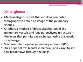
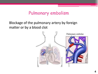
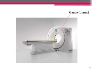
CT Pulmonary angiogram (CTPA)

1
At a glance …
Medical diagnostic test that employs computed
tomography to obtain an image of the pulmonary
arteries.
 CT offers a method of direct visualization of the
pulmonary vessels and lung parenchyma (structure in
the lungs that permits gas exchange) using diagnostic
x-ray images.
 Main use is to diagnose pulmonary embolism(PE).
 Uses a special dye (contrast material) and x-rays to see
how blood flows through the lungs.


2
Why the Test is Performed?
 The test is used to detect blood clots (pulmonary
embolism)
 May also be used to help your doctor diagnose:
• AV malformations of the lung
• Congenital (present from birth) narrowing of the
pulmonary vessels
• Pulmonary artery aneurysms
• Pulmonary hypertension - high blood pressure in the
arteries of the lungs
3
Pulmonary embolism
Blockage of the pulmonary artery by foreign
matter or by a blood clot

4
Arteriovenous malformation of the lung
Arteriovenous malformation or AVM is an abnormal
connection between arteries and veins, bypassing
the capillary system

5
Pulmonary artery aneurysm
An excessive localized swelling of the wall of pulmonary
artery.

6
7

How to Prepare for the Test?
 You should not eat or drink anything for 6
- 8 hours before the test.
 Health care provider need to be informed :
• If the patient is pregnant.
• If the patient ever had any allergic reactions to
x-ray contrast material or iodine substances.
• If the patient is allergic to any medications.
• Which medications patient is taking .
• If the patient ever had any bleeding problems.
How the Test is Performed ?
 Performed in the radiology department scanning room
 Patient lying flat (supine) on a CT table

 A timed intravenous injection of contrast media allows for
evaluation for pulmonary emboli.
 CT scanner gantry (donut) passes over and around the
patient to perform the scan. The gantry travels over the
patient from the neck to the abdomen to create the scan.
8
How the Test is Performed ?(cont..)
 Timing is essential and the radiographer will use
instructions to indicate when to breathe in, breath out
or hold breath.
 Intravenous contrast is used to highlight the pulmonary
vessels, and to determine the presence of a clot in the
lung. If the patient lies very still, and the chest is
stationary (breath hold) this optimizes the quality of the
scan.
9
Gantry(donut)

10
Example of a CTPA
 demonstrating a saddle embolus. The white area above the
center is the pulmonary artery, opacified by radio contrast.
 Inside it, the grey matter is blood clot.
 The black areas on either side are the lungs, with around it the
chest wall.

11
Risks
 Allergic reaction to the contrast dye.
 Damage to the blood vessel as the needle and catheter
are inserted.
 Blood clot traveling to the lungs, causing an embolism
 Excessive bleeding or a blood clot where the catheter is
inserted, which can reduce blood flow to the leg.
 Heart attack or stroke.
 Hematoma (a collection of blood at the site of the needle
puncture).
 Injury to the nerves at the puncture site
 Kidney damage from the dye.
12
Advantages





Sensitivity
Specificity
Lesser volume of contrast needed
Can be done in out patient basis

13
References
 http://www.nlm.nih.gov/medlineplus/ency/article/003813.htm
 http://healthengine.com.au/info/CT_Pulmonary_Angiogram
 http://en.wikipedia.org/wiki/CT_pulmonary_angiogram

14
15

Ct pulmonary angiogram (ctpa)

  • 1.
  • 2.
    At a glance… Medical diagnostic test that employs computed tomography to obtain an image of the pulmonary arteries.  CT offers a method of direct visualization of the pulmonary vessels and lung parenchyma (structure in the lungs that permits gas exchange) using diagnostic x-ray images.  Main use is to diagnose pulmonary embolism(PE).  Uses a special dye (contrast material) and x-rays to see how blood flows through the lungs.  2
  • 3.
    Why the Testis Performed?  The test is used to detect blood clots (pulmonary embolism)  May also be used to help your doctor diagnose: • AV malformations of the lung • Congenital (present from birth) narrowing of the pulmonary vessels • Pulmonary artery aneurysms • Pulmonary hypertension - high blood pressure in the arteries of the lungs 3
  • 4.
    Pulmonary embolism Blockage ofthe pulmonary artery by foreign matter or by a blood clot 4
  • 5.
    Arteriovenous malformation ofthe lung Arteriovenous malformation or AVM is an abnormal connection between arteries and veins, bypassing the capillary system 5
  • 6.
    Pulmonary artery aneurysm Anexcessive localized swelling of the wall of pulmonary artery. 6
  • 7.
    7 How to Preparefor the Test?  You should not eat or drink anything for 6 - 8 hours before the test.  Health care provider need to be informed : • If the patient is pregnant. • If the patient ever had any allergic reactions to x-ray contrast material or iodine substances. • If the patient is allergic to any medications. • Which medications patient is taking . • If the patient ever had any bleeding problems.
  • 8.
    How the Testis Performed ?  Performed in the radiology department scanning room  Patient lying flat (supine) on a CT table  A timed intravenous injection of contrast media allows for evaluation for pulmonary emboli.  CT scanner gantry (donut) passes over and around the patient to perform the scan. The gantry travels over the patient from the neck to the abdomen to create the scan. 8
  • 9.
    How the Testis Performed ?(cont..)  Timing is essential and the radiographer will use instructions to indicate when to breathe in, breath out or hold breath.  Intravenous contrast is used to highlight the pulmonary vessels, and to determine the presence of a clot in the lung. If the patient lies very still, and the chest is stationary (breath hold) this optimizes the quality of the scan. 9
  • 10.
  • 11.
    Example of aCTPA  demonstrating a saddle embolus. The white area above the center is the pulmonary artery, opacified by radio contrast.  Inside it, the grey matter is blood clot.  The black areas on either side are the lungs, with around it the chest wall. 11
  • 12.
    Risks  Allergic reactionto the contrast dye.  Damage to the blood vessel as the needle and catheter are inserted.  Blood clot traveling to the lungs, causing an embolism  Excessive bleeding or a blood clot where the catheter is inserted, which can reduce blood flow to the leg.  Heart attack or stroke.  Hematoma (a collection of blood at the site of the needle puncture).  Injury to the nerves at the puncture site  Kidney damage from the dye. 12
  • 13.
    Advantages     Sensitivity Specificity Lesser volume ofcontrast needed Can be done in out patient basis 13
  • 14.
  • 15.