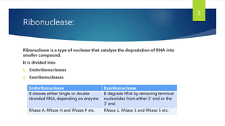
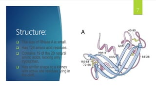
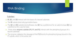
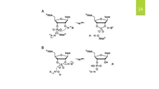
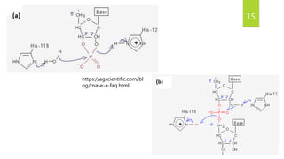
Structure and Mechanism ll:
Ribonuclease
REPRESENTENED BY:
SUFYAN ASHRAF (20012507-017)
AMEENA SHAHZADI (20012507-018)
1
Sr. No. Topic Slide
No.
1 Ribonuclease
2 Introduction and
History
3 Function and structure
4 Folding and stability
5 How RNase A binds to RNA
7 The reaction mechanism
8 Conclusion
9 Reference
Content Page: 2
Ribonuclease:
Ribonuclease is a type of nuclease that catalyse the degradation of RNA into
smaller compound.
It is divided into
1. Endoribonucleases
2. Exoribonucleases
Endoribonuclease Exoribonuclease
It cleaves either Single or double
stranded RNA, depending on enzyme
It degrade RNA by removing terminal
nucleotides from either 5’ end or the
3’ end
RNase A, RNase H and RNase P etc. RNase 1, RNase 3 and RNase 5 etc.
3
Ribonuclease A (RNase A):
Introduction:
 RNase A:
Ribonuclease A is a digestive enzyme secreted by the pancreas that specifically "digests" or
hydrolyzes RNA (but not DNA) polymers by endonuclease cleavage of the phosphodiester bonds
forming the covalent links between adjacent ribonucleotide residues in these molecules.
 What does “A” stand for in RNase A?
The "A" in its name refers not to its substrate specificity, but to the predominant form of the
enzyme produced by the bovine pancreas. RNase A is unmodified, whereas RNase B, RNase C,
and RNase D are mixtures of glycoforms. Because of its availability in large quantity and high
purity, RNase A has been the object of landmark work in protein chemistry and enzymology.
4
History:
A variety of methods were used to determine the structure of RNase-A
 Fast atom bombardment mass spectrometry (FABMS) - assigns disulfide
bonds of a protein with RNase A.
 Work on RNase A has yielded the first 3D structure of a protein containing an
isoaspartyl residue, derived from deamidation of an asparagine residue (Asn
67).
 NMR spectroscopy in elaborating both protein structure and protein folding
pathways.
5
Function:
 2 classes of enzyme that catalyze the synthesis or degradation of RNA:
1. RNA polymerase –synthesis
2. RNA depolymerases or “ribonucleases” – degradation.
 RNase A has been the object of landmark work on the folding stability and chemistry of
proteins in enzymology and in molecular evolution.
 RNA essential for life.
6
Structure:
 The size of RNase A is small.
 Has 124 amino acid residues.
 Contains 19 of the 20 natural
amino acids, lacking only
tryptophan.
 Has similar shape to a kidney
with active site residues lying in
the cleft.
7
Folding and stability:
 Four disulfide bonds:
1. Critical to stability on native enzyme.
2. more stability from Cys26-Cys84 and Cys58-Cys110 than from Cys65-
Cys72 and Cys40-Cys95 two proline residues with cis peptide bonds.
 The stability of RNase A is legendary.
 The two proline residues with cis peptide bonds, and the three
residues most important for catalysis is His12, His119, and Lys41.
8
RNA Binding:
Subsites:
 B1, B2, and B3 interact with the bases of a bound substrate.
 The B1 subsite bind only pyrimidine bases.
 The B2 and B3 subsites bind all bases, but B2 has a preference for an adenine base B3 has
a preference for a purine base.
 Three other enzymic subsites (P0, P1, and P2) interact with the phosphoryl groups of a
bound substrate.
 The enzyme catalyzes the cleavage of the P-O bond of a phosphoryl group bound in the
P1 subsite, which is the active site.
9
10
RNA Binding:
SUBSTRATE SPECIFICITY
 RNase A catalyzes the cleavage of the P-O bond of an RNA strand and the
hydrolysis of the P-O bond of a nucleoside 2',3'-cyclic phosphodiester on the 3'-
side of a pyrimidine residue
ONE-DIMENSIONAL DIFFUSION
 The ability to diffuse in one dimension can accelerate the formation of a site-
specific interaction within a linear biopolymer by up to 103 fold.
 Such facilitated diffusion is used by transcription factors and restriction
endonucleases to locate specific sites on double-stranded DNA
 Specifically, a uridine nucleotide is cleaved more quickly by RNase A if it is flanked
by a long stretch of poly(dA) than if it is flanked by a short stretch
11
12
Reaction Mechanism
IT IS A GENERAL ACID-BASE CATALYSIS
 The side chain of His 12 acts as a base that abstracts a proton from the 2’-
oxygen of a substrate molecule, and thereby facilitates its attack on the
phosphorus atom.
 This attack displace a nucleoside.
 His 119 acts as an acid that protonates the 5”-oxygen to facilitate its
displacement.
 Both products are released to solvent.
 The side chain of Lys41 and the main chain of Phe120 enhance catalysis by
stabilizing this transition state.
13
14
https://agscientific.com/bl
og/rnase-a-faq.html
15
Conclusion
 RNase A has been the most studied enzyme of the 20th century.
 Used to digest RNA produced by stomach microorganism.
 Methods now exist to produce unlimited quantities of RNase A and it’s
homologues.
 Can be used to exploit further use of RNase A in biotechnology and Medicine.
 RNase A will continue to be used as a model system.
16
Reference:
https://agscientific.com/blog/rnase-a-faq.html
17

Presentation (4).pptx

  • 1.
    Structure and Mechanismll: Ribonuclease REPRESENTENED BY: SUFYAN ASHRAF (20012507-017) AMEENA SHAHZADI (20012507-018) 1
  • 2.
    Sr. No. TopicSlide No. 1 Ribonuclease 2 Introduction and History 3 Function and structure 4 Folding and stability 5 How RNase A binds to RNA 7 The reaction mechanism 8 Conclusion 9 Reference Content Page: 2
  • 3.
    Ribonuclease: Ribonuclease is atype of nuclease that catalyse the degradation of RNA into smaller compound. It is divided into 1. Endoribonucleases 2. Exoribonucleases Endoribonuclease Exoribonuclease It cleaves either Single or double stranded RNA, depending on enzyme It degrade RNA by removing terminal nucleotides from either 5’ end or the 3’ end RNase A, RNase H and RNase P etc. RNase 1, RNase 3 and RNase 5 etc. 3
  • 4.
    Ribonuclease A (RNaseA): Introduction:  RNase A: Ribonuclease A is a digestive enzyme secreted by the pancreas that specifically "digests" or hydrolyzes RNA (but not DNA) polymers by endonuclease cleavage of the phosphodiester bonds forming the covalent links between adjacent ribonucleotide residues in these molecules.  What does “A” stand for in RNase A? The "A" in its name refers not to its substrate specificity, but to the predominant form of the enzyme produced by the bovine pancreas. RNase A is unmodified, whereas RNase B, RNase C, and RNase D are mixtures of glycoforms. Because of its availability in large quantity and high purity, RNase A has been the object of landmark work in protein chemistry and enzymology. 4
  • 5.
    History: A variety ofmethods were used to determine the structure of RNase-A  Fast atom bombardment mass spectrometry (FABMS) - assigns disulfide bonds of a protein with RNase A.  Work on RNase A has yielded the first 3D structure of a protein containing an isoaspartyl residue, derived from deamidation of an asparagine residue (Asn 67).  NMR spectroscopy in elaborating both protein structure and protein folding pathways. 5
  • 6.
    Function:  2 classesof enzyme that catalyze the synthesis or degradation of RNA: 1. RNA polymerase –synthesis 2. RNA depolymerases or “ribonucleases” – degradation.  RNase A has been the object of landmark work on the folding stability and chemistry of proteins in enzymology and in molecular evolution.  RNA essential for life. 6
  • 7.
    Structure:  The sizeof RNase A is small.  Has 124 amino acid residues.  Contains 19 of the 20 natural amino acids, lacking only tryptophan.  Has similar shape to a kidney with active site residues lying in the cleft. 7
  • 8.
    Folding and stability: Four disulfide bonds: 1. Critical to stability on native enzyme. 2. more stability from Cys26-Cys84 and Cys58-Cys110 than from Cys65- Cys72 and Cys40-Cys95 two proline residues with cis peptide bonds.  The stability of RNase A is legendary.  The two proline residues with cis peptide bonds, and the three residues most important for catalysis is His12, His119, and Lys41. 8
  • 9.
    RNA Binding: Subsites:  B1,B2, and B3 interact with the bases of a bound substrate.  The B1 subsite bind only pyrimidine bases.  The B2 and B3 subsites bind all bases, but B2 has a preference for an adenine base B3 has a preference for a purine base.  Three other enzymic subsites (P0, P1, and P2) interact with the phosphoryl groups of a bound substrate.  The enzyme catalyzes the cleavage of the P-O bond of a phosphoryl group bound in the P1 subsite, which is the active site. 9
  • 10.
  • 11.
    RNA Binding: SUBSTRATE SPECIFICITY RNase A catalyzes the cleavage of the P-O bond of an RNA strand and the hydrolysis of the P-O bond of a nucleoside 2',3'-cyclic phosphodiester on the 3'- side of a pyrimidine residue ONE-DIMENSIONAL DIFFUSION  The ability to diffuse in one dimension can accelerate the formation of a site- specific interaction within a linear biopolymer by up to 103 fold.  Such facilitated diffusion is used by transcription factors and restriction endonucleases to locate specific sites on double-stranded DNA  Specifically, a uridine nucleotide is cleaved more quickly by RNase A if it is flanked by a long stretch of poly(dA) than if it is flanked by a short stretch 11
  • 12.
  • 13.
    Reaction Mechanism IT ISA GENERAL ACID-BASE CATALYSIS  The side chain of His 12 acts as a base that abstracts a proton from the 2’- oxygen of a substrate molecule, and thereby facilitates its attack on the phosphorus atom.  This attack displace a nucleoside.  His 119 acts as an acid that protonates the 5”-oxygen to facilitate its displacement.  Both products are released to solvent.  The side chain of Lys41 and the main chain of Phe120 enhance catalysis by stabilizing this transition state. 13
  • 14.
  • 15.
  • 16.
    Conclusion  RNase Ahas been the most studied enzyme of the 20th century.  Used to digest RNA produced by stomach microorganism.  Methods now exist to produce unlimited quantities of RNase A and it’s homologues.  Can be used to exploit further use of RNase A in biotechnology and Medicine.  RNase A will continue to be used as a model system. 16
  • 17.