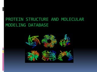
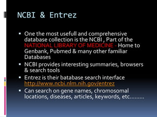
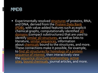
Secondary Structure Prediction of proteins Vijay Hemmadi
Secondary structure prediction has been around for almost a quarter of a century. The early methods suffered from a lack of data. Predictions were performed on single sequences rather than families of homologous sequences, and there were relatively few known 3D structures from which to derive parameters. Probably the most famous early methods are those of Chou & Fasman, Garnier, Osguthorbe & Robson (GOR) and Lim. Although the authors originally claimed quite high accuracies (70-80 %), under careful examination, the methods were shown to be only between 56 and 60% accurate (see Kabsch & Sander, 1984 given below). An early problem in secondary structure prediction had been the inclusion of structures used to derive parameters in the set of structures used to assess the accuracy of the method.
Some good references on the subject:
Sequence alig Sequence Alignment Pairwise alignment:-naveed ul mushtaq
Sequence Alignment Pairwise alignment:- Global Alignment and Local AlignmentTwo types of alignment Progressive Programs for multiple sequence alignment BLOSUM Point accepted mutation (PAM)PAM VS BLOSUM
protein structure prediction methods. homology modelling, fold recognition, threading, ab initio methods. in short and easy form slides. after one time read you can easily understand methods for protein structure prediction.
An integrated publicly accessible bioinformatics resource to support genomic/proteomic research and scientific discovery.
Established in 1984, by the National Biomedical Research Foundation (NBRF) Georgetown University Medial Center, Washington D.C., USA.
It is the source of annotated protein databases and analysis tools for the researchers.
Serve as primary resource for the exploration of protein information.
Accessible by text search for entry and list retrieval, and also BLAST search and peptide match.
INTRODUCTION
WHAT IS DATA AND DATABASE?
WHAT IS BIOLOGICAL DATABASE?
TYPES OF BIOLOGICAL DATABASE
PRIMARY DATABASE
Nucleic acid sequence database
Protein sequence database
SECONDARY DATABASE
COMPOSITE DATABASE
TERTIARY DATABASE
WHY NEED?
CONCLUSION
REFRENCES
UCSD Deans and Chairs Presentation - PDB & Drug DiscoveryPhilip Bourne
A presentation made to the Deans and Chairs of the UCSD Health Sciences on Jan. 25, 2011 concerning the role that the PDB might play in drug discovery going forward.
Lecture delivered by T. Ashok Kumar, Head, Department of Bioinformatics, Noorul Islam College of Arts and Science, Kumaracoil, Thuckalay, INDIA. UGC Sponsored National Workshop on BIOINFORMATICS AND GENOME ANALYSIS for College Teachers on August 11 & 12, 2014. Organized by Centre for Bioinformatics, Department of Zoology, NMCC.
Bioinformatics is the application of Information technology to store, organize and analyze the vast amount of biological data which is available in the form of sequences and structures of proteins and nucleic acids. The biological information of nucleic acids is available as sequences while the data of proteins is available as sequences and structures.
A biological database is a collection of data that is organized so that its contents can easily be accessed, managed, and updated. The activity of preparing a database can be divided in to:
Collection of data in a form which can be easily accessed
Making it available to a multi-user system (always available for the user)
Biological databases are libraries of life sciences information, collected from scientific experiments, published literature, high-throughput experiment technology, and computational analysis.
INTRODUCTION
DEFINITION OF BIOINFORMATICS
HISTORY
OBJECTIVE OF BIOINFORMATIC
TOOLS OF BIOINFORMATICS
PROCEDURE AND TOOLS OF BIOINFORMATIC
BIOLOGICAL DATABASES
HOMOLOGY AND SIMILARITY TOOLS (SEQUENCE ALIGNMENT)
PROTEIN FUNCTION ANALYSIS TOOLS
STRUCTURAL ANALYSIS TOOLS
SEQUENCE MANIPULATION TOOLS
SEQUENCE ANALYSIS TOOLS
APPLICATION
CONCLUSION
REFERENCES
Protein Sequence, Structure, and Functional Databases: UniProtKB, Swiss-Prot, TrEMBL, PIR, MIPS, PROSITE, PRINTS, BLOCKS, Pfam, NDRB, OWL, PDB, SCOP, CATH, NDB, PQS, SYSTERS, and Motif. Presented at UGC Sponsored National Workshop on Bioinformatics and Sequence Analysis conducted by Nesamony Memorial Christian College, Marthandam on 9th and 10th October, 2017 by Prof. T. Ashok Kumar
What is greenhouse gasses and how many gasses are there to affect the Earth.moosaasad1975
What are greenhouse gasses how they affect the earth and its environment what is the future of the environment and earth how the weather and the climate effects.
Slide 1: Title Slide
Extrachromosomal Inheritance
Slide 2: Introduction to Extrachromosomal Inheritance
Definition: Extrachromosomal inheritance refers to the transmission of genetic material that is not found within the nucleus.
Key Components: Involves genes located in mitochondria, chloroplasts, and plasmids.
Slide 3: Mitochondrial Inheritance
Mitochondria: Organelles responsible for energy production.
Mitochondrial DNA (mtDNA): Circular DNA molecule found in mitochondria.
Inheritance Pattern: Maternally inherited, meaning it is passed from mothers to all their offspring.
Diseases: Examples include Leber’s hereditary optic neuropathy (LHON) and mitochondrial myopathy.
Slide 4: Chloroplast Inheritance
Chloroplasts: Organelles responsible for photosynthesis in plants.
Chloroplast DNA (cpDNA): Circular DNA molecule found in chloroplasts.
Inheritance Pattern: Often maternally inherited in most plants, but can vary in some species.
Examples: Variegation in plants, where leaf color patterns are determined by chloroplast DNA.
Slide 5: Plasmid Inheritance
Plasmids: Small, circular DNA molecules found in bacteria and some eukaryotes.
Features: Can carry antibiotic resistance genes and can be transferred between cells through processes like conjugation.
Significance: Important in biotechnology for gene cloning and genetic engineering.
Slide 6: Mechanisms of Extrachromosomal Inheritance
Non-Mendelian Patterns: Do not follow Mendel’s laws of inheritance.
Cytoplasmic Segregation: During cell division, organelles like mitochondria and chloroplasts are randomly distributed to daughter cells.
Heteroplasmy: Presence of more than one type of organellar genome within a cell, leading to variation in expression.
Slide 7: Examples of Extrachromosomal Inheritance
Four O’clock Plant (Mirabilis jalapa): Shows variegated leaves due to different cpDNA in leaf cells.
Petite Mutants in Yeast: Result from mutations in mitochondrial DNA affecting respiration.
Slide 8: Importance of Extrachromosomal Inheritance
Evolution: Provides insight into the evolution of eukaryotic cells.
Medicine: Understanding mitochondrial inheritance helps in diagnosing and treating mitochondrial diseases.
Agriculture: Chloroplast inheritance can be used in plant breeding and genetic modification.
Slide 9: Recent Research and Advances
Gene Editing: Techniques like CRISPR-Cas9 are being used to edit mitochondrial and chloroplast DNA.
Therapies: Development of mitochondrial replacement therapy (MRT) for preventing mitochondrial diseases.
Slide 10: Conclusion
Summary: Extrachromosomal inheritance involves the transmission of genetic material outside the nucleus and plays a crucial role in genetics, medicine, and biotechnology.
Future Directions: Continued research and technological advancements hold promise for new treatments and applications.
Slide 11: Questions and Discussion
Invite Audience: Open the floor for any questions or further discussion on the topic.
Observation of Io’s Resurfacing via Plume Deposition Using Ground-based Adapt...Sérgio Sacani
Since volcanic activity was first discovered on Io from Voyager images in 1979, changes
on Io’s surface have been monitored from both spacecraft and ground-based telescopes.
Here, we present the highest spatial resolution images of Io ever obtained from a groundbased telescope. These images, acquired by the SHARK-VIS instrument on the Large
Binocular Telescope, show evidence of a major resurfacing event on Io’s trailing hemisphere. When compared to the most recent spacecraft images, the SHARK-VIS images
show that a plume deposit from a powerful eruption at Pillan Patera has covered part
of the long-lived Pele plume deposit. Although this type of resurfacing event may be common on Io, few have been detected due to the rarity of spacecraft visits and the previously low spatial resolution available from Earth-based telescopes. The SHARK-VIS instrument ushers in a new era of high resolution imaging of Io’s surface using adaptive
optics at visible wavelengths.
Richard's entangled aventures in wonderlandRichard Gill
Since the loophole-free Bell experiments of 2020 and the Nobel prizes in physics of 2022, critics of Bell's work have retreated to the fortress of super-determinism. Now, super-determinism is a derogatory word - it just means "determinism". Palmer, Hance and Hossenfelder argue that quantum mechanics and determinism are not incompatible, using a sophisticated mathematical construction based on a subtle thinning of allowed states and measurements in quantum mechanics, such that what is left appears to make Bell's argument fail, without altering the empirical predictions of quantum mechanics. I think however that it is a smoke screen, and the slogan "lost in math" comes to my mind. I will discuss some other recent disproofs of Bell's theorem using the language of causality based on causal graphs. Causal thinking is also central to law and justice. I will mention surprising connections to my work on serial killer nurse cases, in particular the Dutch case of Lucia de Berk and the current UK case of Lucy Letby.
Earliest Galaxies in the JADES Origins Field: Luminosity Function and Cosmic ...Sérgio Sacani
We characterize the earliest galaxy population in the JADES Origins Field (JOF), the deepest
imaging field observed with JWST. We make use of the ancillary Hubble optical images (5 filters
spanning 0.4−0.9µm) and novel JWST images with 14 filters spanning 0.8−5µm, including 7 mediumband filters, and reaching total exposure times of up to 46 hours per filter. We combine all our data
at > 2.3µm to construct an ultradeep image, reaching as deep as ≈ 31.4 AB mag in the stack and
30.3-31.0 AB mag (5σ, r = 0.1” circular aperture) in individual filters. We measure photometric
redshifts and use robust selection criteria to identify a sample of eight galaxy candidates at redshifts
z = 11.5 − 15. These objects show compact half-light radii of R1/2 ∼ 50 − 200pc, stellar masses of
M⋆ ∼ 107−108M⊙, and star-formation rates of SFR ∼ 0.1−1 M⊙ yr−1
. Our search finds no candidates
at 15 < z < 20, placing upper limits at these redshifts. We develop a forward modeling approach to
infer the properties of the evolving luminosity function without binning in redshift or luminosity that
marginalizes over the photometric redshift uncertainty of our candidate galaxies and incorporates the
impact of non-detections. We find a z = 12 luminosity function in good agreement with prior results,
and that the luminosity function normalization and UV luminosity density decline by a factor of ∼ 2.5
from z = 12 to z = 14. We discuss the possible implications of our results in the context of theoretical
models for evolution of the dark matter halo mass function.
Seminar of U.V. Spectroscopy by SAMIR PANDASAMIR PANDA
Spectroscopy is a branch of science dealing the study of interaction of electromagnetic radiation with matter.
Ultraviolet-visible spectroscopy refers to absorption spectroscopy or reflect spectroscopy in the UV-VIS spectral region.
Ultraviolet-visible spectroscopy is an analytical method that can measure the amount of light received by the analyte.
1. Bioinformatics is about using mathematics,
statistics and information technology to extract
useful information from large and complex
biological datasets.
SUB-TOPIC:- PROTEIN STRUCTURE &
MOLECULAR MODELING DATABASE
6. Levels of protein structure
Primary structure-The primary structure of a protein refers to
the linear sequence of amino acids in the polypeptide chain.
The primary structure is held together by covalent bonds such
as peptide bonds, which are made during the process of protein
biosynthesis or translation.
Secondary structure-Secondary structure refers to highly
regular local sub-structures.Two main types of secondary
structure, the alpha helix and the beta strand orbeta sheets
Tertiary structure-Tertiary structure refers to the three-
dimensional structure of a single, double, or triple
bonded protein molecule.The alpha-helix and beta pleated-
sheets are folded into a compact globular structure.
Quaternary structure-Quaternary structure is the three-
dimensional structure of a multi-subunit protein and how the
subunits fit together.
8. NCBI & Entrez
One the most usefull and comprehensive
database collection is the NCBI , Part of the
NATIONAL LIBRARY OF MEDICINE - Home to
Genbank, Pubmed & many other familiar
Databases
NCBI provides interesting summaries, browsers
& search tools
Entrez is their batabase search interface
http://www.ncbi.nlm.nih.gov/entrez
Can search on gene names, chromosomal
locations, diseases, articles, keywords, etc.........
12. MMDB
Experimentally resolved structures of proteins, RNA,
and DNA, derived from the Protein Data Bank
(PDB), with value-added features such as explicit
chemical graphs, computationally identified 3D
domains (compact substructures) that are used to
identify similar 3D structures, as well as links to
literature, similar sequences, information
about chemicals bound to the structures, and more.
These connections make it possible, for example,
to find 3D structures for homologs of a protein
sequence of interest, then interactively view
the sequence-structure relationships, active
sites, bound chemicals, journal articles, and more.
18. ExPASy(PROTPARAM)
ProtParam (References / Documentation) is a
tool which allows the computation of various
physical and chemical parameters for a given
protein stored in Swiss-Prot orTrEMBL or for a
user entered sequence.
The computed parameters include the molecular
weight, theoretical pI, amino acid composition,
atomic composition, extinction coefficient,
estimated half-life, instability index, aliphatic
index and grand average of hydropathicity
22. Uniprot
The mission of UniProt is to provide the
scientific community with a comprehensive,
high-quality and freely accessible resource of
protein sequence and functional information.
25. Basic Local Alignment Search
Tool(blast)
Finds regions of local similarity
between sequences.
Compares nucleotide or protein
sequences to sequence databases.
Calculates statistical significance from
matches.
Can be used to infer functional and
evolutionary relationships between
sequences.
It can also help in identifying members
of the gene family.
30. Protein data bank
Protein Data Bank (PDB) is a repository for
the 3-D structural data of large biological
molecules, such as proteins and nucleic
acids.
PDB is overseen by an organization called
theWorldwide Protein Data Bank, wwPDB.
PDB is a key resource in areas of structural
biology, such as structural genomics.
31.
32.
33.
34. Useful Features of the Molecular Modeling
Database
Facilitate computation on 3D structure data
Analysis of individual structures and relationships
among them
biological and geometrical features within 3D
structures
conserved protein domain annotations
evolutionary relationships among 3D structures
functional relationships among 3D structures
Interactive views of sequence-structure relationships
Connections between 3D structure records and
associated literature, molecular, and chemical data