Protein Microarrays Methods and Protocols 2011th Edition Ulrike Korf
Protein Microarrays Methods and Protocols 2011th Edition Ulrike Korf
Protein Microarrays Methods and Protocols 2011th Edition Ulrike Korf
Protein Microarrays Methods and Protocols 2011th Edition Ulrike Korf
Protein Microarrays Methods and Protocols 2011th Edition Ulrike Korf
1.
Visit https://ebookultra.com todownload the full version and
explore more ebooks or textbooks
Protein Microarrays Methods and Protocols 2011th
Edition Ulrike Korf
_____ Click the link below to download _____
https://ebookultra.com/download/protein-microarrays-methods-
and-protocols-2011th-edition-ulrike-korf/
Explore and download more ebooks or textbooks at ebookultra.com
2.
Here are somerecommended products that we believe you will be
interested in. You can click the link to download.
Protein Arrays Methods and Protocols 1st Edition Eric Fung
https://ebookultra.com/download/protein-arrays-methods-and-
protocols-1st-edition-eric-fung/
Protein Chromatography Methods and Protocols 2nd Edition
Dermot Walls
https://ebookultra.com/download/protein-chromatography-methods-and-
protocols-2nd-edition-dermot-walls/
Molecular Chaperones Methods and Protocols Methods in
Molecular Biology v787 2011th Edition Stuart K. Calderwood
https://ebookultra.com/download/molecular-chaperones-methods-and-
protocols-methods-in-molecular-biology-v787-2011th-edition-stuart-k-
calderwood/
Plant Protein Secretion Methods and Protocols 2nd Edition
Liwen Jiang
https://ebookultra.com/download/plant-protein-secretion-methods-and-
protocols-2nd-edition-liwen-jiang/
3.
Protein Dynamics Methodsand Protocols 2014th Edition
Dennis R. Livesay
https://ebookultra.com/download/protein-dynamics-methods-and-
protocols-2014th-edition-dennis-r-livesay/
G Protein Signaling Methods and Protocols 1st Edition
Wendy K. Greentree
https://ebookultra.com/download/g-protein-signaling-methods-and-
protocols-1st-edition-wendy-k-greentree-2/
G Protein Signaling Methods and Protocols 1st Edition
Wendy K. Greentree
https://ebookultra.com/download/g-protein-signaling-methods-and-
protocols-1st-edition-wendy-k-greentree/
BIB Protein Supersecondary Structure Methods and Protocols
2nd ed. Edition Kister
https://ebookultra.com/download/bib-protein-supersecondary-structure-
methods-and-protocols-2nd-ed-edition-kister/
Nuclear G Protein Coupled Receptors Methods and Protocols
1st Edition Bruce G. Allen
https://ebookultra.com/download/nuclear-g-protein-coupled-receptors-
methods-and-protocols-1st-edition-bruce-g-allen/
5.
Protein Microarrays Methodsand Protocols 2011th
Edition Ulrike Korf Digital Instant Download
Author(s): Ulrike Korf
ISBN(s): 9781617792854, 1617792853
Edition: 2011
File Details: PDF, 8.47 MB
Year: 2011
Language: english
7.
Me t ho d s i n Mo l e c u l a r Bi o l o g y ™
Series Editor
John M. Walker
School of Life Sciences
University of Hertfordshire
Hatfield, Hertfordshire, AL10 9AB, UK
For further volumes:
http://www.springer.com/series/7651
v
Preface
Proteins are involvedin almost any aspect of cellular function. The cellular proteome is
subjected to a steady flow of dynamic changes, and therefore is a very suitable readout for
the functional properties of a cell or an organism. Proteins, for example, build the cellular
architecture, and are essential components of membranous compartments confining a cell,
as well as subcellular organelles. Networks of tightly regulated enzymes are in command of
the energy supply, and provide molecular building blocks, such as carbohydrates, lipids, and
nucleic acids. Other proteins are involved in replication and transcriptional processes, and
assist in the translation of new proteins. Proteins in extracellular fluids maintain the com-
munication between cells of a tissue as well as within an organism and may serve as disease
biomarkers. The number of different proteins encoded by the genome is increased by at
least an order of magnitude, due to the introduction of posttranslational modifications,
such as glycosylation, lipid-modifications, acetylation, and by protein phosphorylation
which is the best-studied mode of cellular regulation.
Understanding protein function and the regulation of signaling networks requires
large-scale efforts which enable the dynamic analysis of numerous samples in parallel.
Progress in functional proteomics has been limited for a long time, partially because of limi-
tations in assay sensitivity and sample capacity. Protein microarrays have the ability to over-
come these limitations so that a highly parallel analysis of hundreds of proteins in thousands
of samples is attainable. Advancements in the field of robotics and signal detection have
facilitated an increase in sensitivity and sample capacity and, therefore, have contributed to
the evolution of an increasing number of robust protein microarray applications. Thus, due
to the robustness and flexibility of this experimental platform, diverse applications can now
be implemented in principles of different types of biochemical assays.
This volume presents an up-to-date collection of robust strategies in the field of protein
microarrays, and summarizes recent advantages in the field of printing technologies, the
development of suitable surface materials, as well as detection and quantification technolo-
gies. Parallel to the advancement of wet-lab techniques, new software tools were developed
for data analysis in order to deal with large data sets generated by protein microarray
applications.
Thanks to all article authors for taking the time to prepare a chapter for this book, the
series editor for shaping the idea for this volume, people at Springer for their uncomplicated
and helpful advice, and special thanks to my family for their patience and cooperation while
I edited the articles in this book to their completion.
I am confident that this book will stimulate the application and further advancement of
this powerful technology in labs worldwide. I am very much looking forward to the future
of protein microarray-based applications.
Heidelberg, Germany Ulrike Korf
vii
Contents
Preface . .. . . . . . . . . . . . . . . . . . . . . . . . . . . . . . . . . . . . . . . . . . . . . . . . . . . . . . . . . . . v
Contributors . . . . . . . . . . . . . . . . . . . . . . . . . . . . . . . . . . . . . . . . . . . . . . . . . . . . . . . . . ix
Part I Reverse Phase Protein Arrays
1 Reverse Phase Protein Microarrays for Clinical Applications . . . . . . . . . . . . . . . . . . 3
Mariaelena Pierobon, Claudio Belluco, Lance A. Liotta,
and Emanuel F. Petricoin III
2 Impact of Blocking and Detection Chemistries on Antibody Performance
for Reverse Phase Protein Arrays . . . . . . . . . . . . . . . . . . . . . . . . . . . . . . . . . . . . . . 13
Kristi Ambroz
3 Phosphoprotein Stability in Clinical Tissue and Its Relevance
for Reverse Phase Protein Microarray Technology . . . . . . . . . . . . . . . . . . . . . . . . . 23
Virginia Espina, Claudius Mueller, and Lance A. Liotta
4 Utilization of RNAi to Validate Antibodies for Reverse Phase Protein Arrays . . . . . 45
Heiko Mannsperger, Stefan Uhlmann, Ulrike Korf, and Özgür Sahin
5 Antibody-Mediated Signal Amplification for Reverse Phase Protein
Array-Based Protein Quantification . . . . . . . . . . . . . . . . . . . . . . . . . . . . . . . . . . . . 55
Jan C. Brase, Heiko Mannsperger, Holger Sültmann, and Ulrike Korf
6 Reverse-Phase Protein Lysate Microarray (RPA) for the Experimental Validation
of Quantitative Protein Network Models . . . . . . . . . . . . . . . . . . . . . . . . . . . . . . . . 65
Satoshi S. Nishizuka
7 Characterization of Kinase Inhibitors Using Reverse Phase Protein Arrays . . . . . . . 79
Georg Martiny-Baron, Dorothea Haasen, Daniel D’Dorazio,
Johannes Voshol, and Doriano Fabbro
8 Use of Formalin-Fixed and Paraffin-Embedded Tissues for Diagnosis
and Therapy in Routine Clinical Settings . . . . . . . . . . . . . . . . . . . . . . . . . . . . . . . . 109
Daniela Berg, Katharina Malinowsky, Bilge Reischauer, Claudia Wolff,
and Karl-Friedrich Becker
9 Producing Reverse Phase Protein Microarrays from Formalin-Fixed Tissues . . . . . . 123
Claudia Wolff, Christina Schott, Katharina Malinowsky, Daniela Berg,
and Karl-Friedrich Becker
10 Use of Reverse Phase Protein Microarrays to Study Protein Expression
in Leukemia: Technical and Methodological Lessons Learned . . . . . . . . . . . . . . . . 141
Steven M. Kornblau and Kevin R. Coombes
14.
viii Contents
Part II AntibodyMicroarrays
11 Antibody Microarrays as Tools for Biomarker Discovery . . . . . . . . . . . . . . . . . . . . . 159
Marta Sanchez-Carbayo
12 Assessment of Antibody Specificity Using Suspension Bead Arrays . . . . . . . . . . . . . 183
Jochen M. Schwenk and Peter Nilsson
13 Quantitative Analysis of Phosphoproteins Using Microspot Immunoassays . . . . . . . 191
Frauke Henjes, Frank Götschel, Anika Jöcker, and Ulrike Korf
14 Robust Protein Profiling with Complex Antibody Microarrays
in a Dual-Colour Mode . . . . . . . . . . . . . . . . . . . . . . . . . . . . . . . . . . . . . . . . . . . . . 203
Christoph Schröder, Mohamed S.S. Alhamdani, Kurt Fellenberg,
Andrea Bauer, Anette Jacob, and Jörg D. Hoheisel
15 High-Throughput Studies of Protein Glycoforms Using Antibody–Lectin
Sandwich Arrays . . . . . . . . . . . . . . . . . . . . . . . . . . . . . . . . . . . . . . . . . . . . . . . . . . 223
Brian B. Haab and Tingting Yue
16 Microspot Immunoassay-Based Analysis of Plasma Protein Profiles
for Biomarker Discovery Strategies . . . . . . . . . . . . . . . . . . . . . . . . . . . . . . . . . . . . 237
Johanna Sonntag, Heiko Mannsperger, Anika Jöcker, and Ulrike Korf
17 Recombinant Antibodies for the Generation of Antibody Arrays . . . . . . . . . . . . . . 247
Carl A.K. Borrebaeck and Christer Wingren
Part III Protein Microarrays
18 Producing Protein Microarrays from DNA Microarrays . . . . . . . . . . . . . . . . . . . . . 265
Oda Stoevesandt, Michael J. Taussig, and Mingyue He
19 Cell Arrays and High-Content Screening . . . . . . . . . . . . . . . . . . . . . . . . . . . . . . . . 277
Holger Erfle, Anastasia Eskova, Jürgen Reymann,
and Vytaute Starkuviene
20 Probing Calmodulin Protein–Protein Interactions Using
High-Content Protein Arrays . . . . . . . . . . . . . . . . . . . . . . . . . . . . . . . . . . . . . . . . . 289
David J. O’Connell, Mikael Bauer, Sara Linse,
and Dolores J. Cahill
21 Protein Function Microarrays for Customised Systems-Oriented
Proteome Analysis . . . . . . . . . . . . . . . . . . . . . . . . . . . . . . . . . . . . . . . . . . . . . . . . . 305
Jonathan M. Blackburn and Aubrey Shoko
22 Optimized Autoantibody Profiling on Protein Arrays . . . . . . . . . . . . . . . . . . . . . . . 331
Sara L. O’Kane, John K. O’Brien, and Dolores J. Cahill
Part IV Sample Immobilization Strategies
23 Inkjet Printing for the Production of Protein Microarrays . . . . . . . . . . . . . . . . . . . 345
Iain McWilliam, Marisa Chong Kwan, and Duncan Hall
24 Impact of Substrates for Probe Immobilization . . . . . . . . . . . . . . . . . . . . . . . . . . . 363
Ursula Sauer
25 Contact Printing of Protein Microarrays . . . . . . . . . . . . . . . . . . . . . . . . . . . . . . . . 379
John Austin and Antonia H. Holway
Index . . . . . . . . . . . . . . . . . . . . . . . . . . . . . . . . . . . . . . . . . . . . . . . . . . . . . . . . . . . . . . 395
15.
ix
Contributors
Mohamed S.S. Alhamdani• Functional Genome Analysis,
German Cancer Research Center (DKFZ), Heidelberg, Germany
Kristi Ambroz • Director of Biotechnology Reagent Operations and Technical Support,
LI-COR, Lincoln, NE, USA
John Austin • Aushon BioSystems Inc., Concord, MA, USA
Andrea Bauer • Functional Genome Analysis, German Cancer
Research Center (DKFZ), Heidelberg, Germany
Mikael Bauer • Department of Biophysical Chemistry, Lund University, Lund, Sweden
Karl-Friedrich Becker • Institut für Pathologie, Technische Universität München,
Munich, Germany
Claudio Belluco • CRO-IRCCS, National Cancer Institute, Aviano, Italy
Daniela Berg • Institut für Pathologie, Technische Universität München, Munich,
Germany
Jonathan M. Blackburn • Division of Medical Biochemistry & Institute for Infectious
Disease & Molecular Medicine, University of Cape Town, Cape Town, South Africa
Carl A.K. Borrebaeck • Department of Immunotechnology, Lund University,
Lund, Sweden; CREATE Health, Lund University, Lund, Sweden
Jan C. Brase • Division of Molecular Genome Analysis, German Cancer Research
Center (DKFZ), Heidelberg, Germany
Dolores J. Cahill • Translational Science, School of Medicine and Medical Sciences,
UCD Conway Institute, University College Dublin, Dublin, Ireland
Kevin R. Coombes • Departments of Bioinformatics and Computational Biology,
The University of Texas M.D. Anderson Cancer Center, Houston, TX, USA
Daniel D’Dorazio • Center for Proteomic Chemistry, Novartis Pharma AG,
Basel, Switzerland
Holger Erfle • BioQuant, University of Heidelberg, Heidelberg, Germany
Anastasia Eskova • BioQuant, University of Heidelberg, Heidelberg, Germany
Virginia Espina • Center for Applied Proteomics and Molecular Medicine,
George Mason University, Manassas, VA, USA
Doriano Fabbro • Center for Proteomic Chemistry, Novartis Pharma AG,
Basel, Switzerland
Kurt Fellenberg • Chair of Proteomics and Bioanalytics, Technical University
Munich, Freising, Germany
Frank Götschel • Division of Molecular Genome Analysis, German Cancer
Research Center (DKFZ), Heidelberg, Germany
Brian B. Haab • Van Andel Research Institute, Grand Rapids, MI, USA
Dorothea Haasen • Center for Proteomic Chemistry, Novartis Pharma AG,
Basel, Switzerland
Duncan Hall • Arrayjet Ltd., MIC, Roslin, UK
Mingyue He • The Babraham Institute, Cambridge, UK
16.
x Contributors
Frauke Henjes• Division of Molecular Genome Analysis, German Cancer
Research Center (DKFZ), Heidelberg, Germany
Jörg D. Hoheisel • Functional Genome Analysis, German Cancer
Research Center (DKFZ), Heidelberg, Germany
Antonia H. Holway • Associate Director, Translational Research, Lahey Clinic,
Burlington, MA, USA
Anette Jacob • Functional Genome Analysis, German Cancer
Research Center (DKFZ), Heidelberg, Germany
Anika Jöcker • Division of Molecular Genome Analysis, German Cancer
Research Center (DKFZ), Heidelberg, Germany
Ulrike Korf • Division of Molecular Genome Analysis, German Cancer
Research Center (DKFZ), Heidelberg, Germany
Steven M. Kornblau • Departments of Stem Cell Transplantation
and Cellular Therapy, The University of Texas M.D. Anderson Cancer Center,
Houston, TX, USA
Marisa Chong Kwan • Arrayjet Ltd., MIC, Roslin, UK
Sara Linse • Department of Biophysical Chemistry, Lund University, Lund, Sweden
Lance A. Liotta • Center for Applied Proteomics and Molecular Medicine,
George Mason University, Manassas, VA, USA
Katharina Malinowsky • Institut für Pathologie, Technische Universität München,
Munich, Germany
Heiko Mannsperger • Division of Molecular Genome Analysis, German Cancer
Research Center (DKFZ), Heidelberg, Germany
Georg Martiny-Baron • Center for Proteomic Chemistry, Novartis Pharma AG,
Basel, Switzerland
Iain McWilliam • Arrayjet Ltd., MIC, Roslin, UK
Claudius Mueller • Center for Applied Proteomics and Molecular Medicine,
George Mason University, Manassas, VA, USA
Peter Nilsson • SciLifeLab Stockholm, KTH – Royal Institute of Technology,
Tomtebodav, Sweden
Satoshi S. Nishizuka • Molecular Therapeutics Laboratory, Department of Surgery,
Iwate Medical University School of Medicine, Uchimura, Japan
John K. O’Brien • Wellcome Trust Genome Campus, Cambridge, UK
David J. O’Connell • Conway Institute of Biomolecular & Biomedical Research,
University College Dublin, Dublin, Ireland
Sara L. O’Kane • Conway Institute of Biomolecular & Biomedical Research,
University College Dublin, Dublin, Ireland
Emanuel F. Petricoin III • Center for Applied Proteomics and Molecular Medicine,
George Mason University, Manassas, VA, USA
Mariaelena Pierobon • Center for Applied Proteomics and Molecular Medicine,
George Mason University, Manassas, VA, USA
Bilge Reischauer • Institut für Pathologie, Technische Universität München,
Munich, Germany
Jürgen Reymann • BioQuant, University of Heidelberg, Heidelberg, Germany
Özgür Sahin • Division of Molecular Genome Analysis, German Cancer
Research Center (DKFZ), Heidelberg, Germany
17.
xi
Contributors
Marta Sanchez-Carbayo •Tumor Markers Group, Spanish National Cancer
Research Center, Madrid, Spain
Ursula Sauer • Health & Environment Department, Biosensor Technologies,
AIT Austrian Institute of Technology GmbH, Seibersdorf, Austria
Christina Schott • Institut für Pathologie, Technische Universität München,
Munich, Germany
Christoph Schröder • Functional Genome Analysis, German Cancer
Research Center (DKFZ), Heidelberg, Germany
Jochen M. Schwenk • SciLifeLab Stockholm, KTH – Royal Institute
of Technology, Tomtebodav, Sweden
Aubrey Shoko • Centre for Proteomic & Genomic Research, University of Cape Town,
Cape Town, South Africa
Johanna Sonntag • Division of Molecular Genome Analysis, German Cancer Research
Center (DKFZ), Heidelberg, Germany
Vytaute Starkuviene • BioQuant, University of Heidelberg, Heidelberg, Germany
Oda Stoevesandt • Protein Technology Group, Babraham Bioscience Technologies Ltd,
Cambridge, UK
Holger Sültmann • Division of Molecular Genome Analysis, German Cancer
Research Center (DKFZ), Heidelberg, Germany
Michael J. Taussig • Protein Technology Group, Babraham Bioscience
Technologies Ltd, Cambridge, UK
Stefan Uhlmann • Division of Molecular Genome Analysis, German Cancer
Research Center (DKFZ), Heidelberg, Germany
Johannes Voshol • Center for Proteomic Chemistry, Novartis Pharma AG,
Basel, Switzerland
Christer Wingren • Department of Immunotechnology, Lund University,
Lund, Sweden; CREATE Health, Lund University, Lund, Sweden
Claudia Wolff • Institut für Pathologie, Technische Universität München,
Munich, Germany
Tingting Yue • Van Andel Research Institute, Grand Rapids, MI, USA
4 M. Pierobonet al.
decisions will shift from the therapy itself to the biomarkers that are
used to stratify and personalize the therapy. These biomarkers will
serve as “gatekeepers” for therapeutic decision-making processes
as a companion diagnostic and provide the physician with critical
missing information on helping to guide which targeted therapies
to consider. Consequently, the discovery of biomarkers that provide
predictive and prognostic ability for patient stratification/therapy
selection, that is the companion diagnostics of the future, is taking
on an increasingly intense focus in all areas of translational research.
Because of the central, causal role that alterations in cell signaling
and aberrant cell signaling have in tumorigenesis (1–8), phospho-
protein pathway biomarkers may be among the most important
class of biomarkers for prediction, prognosis, and patient-tailored
therapy (4, 8–10). The hope that gene expression analysis will
provide a direct route to unraveling and elucidating ongoing protein
signaling events and provide an effective molecular surrogate for
protein pathway biomarkers has largely dissipated as recent studies
have revealed little correlation between gene expression and protein
expression (11, 12). Moreover, protein expression levels themselves
are not able to predict the phosphorylation levels of signaling
activation, which points to the need for technologies that can
directly assess and measure the activation state of the cellular
“circuitry” and generate the pathway biomarker information that
is critically needed.
Post-translational protein modifications (PTM), mainly phospho-
rylation, are now known to control the kinase-driven signaling net-
works that are abarrently activated in human cancers (13–27). The
vast majority of protein phosphorylation occurs on serine and thre-
onine residues with the remainder (approximately 10%) occurring
on tyrosine. Many growth factor receptor (e.g., vascular endothe-
lial growth factor receptor (VEGFR), epidermal growth factor
receptor (EGFR), c-erbB2)-mediated signaling are based on recep-
tors that are themselves kinase enzymes, and mainly utilize tyrosine
phosphorylation-based PTM. Upon ligand binding, the receptors
dimerize, self-phosphorylate, which then form structural altera-
tions and new binding sites for downstream protein kinase interac-
tions (13–27). Downstream signaling cascades are comprised of
enzymatic networks of kinases and phosphatases and their sub-
strates, linking together based on defined phosphorylation events
that then provide the necessary substrates for structural interac-
tions such as through SH2 and SH3 domains (13–27). How the
cell orchestrates coordinate control of these signaling networks is
also under intense investigation, and new approaches using math-
ematical modeling of the networks are now being explored in
2. Cell Signaling
Activation
Alterations in
Human Cancer
23.
5
1 Reverse PhaseProtein Microarrays for Clinical Applications
order to both reconstruct signaling networks de novo and/or
exploit the pathway architecture to identify optimal therapeutic
strategies (28–35). While cancer, at a functional level, is a disease
of the signaling pathway network, the complexity of the human
“kinome,” comprised of less than a thousand proteins (36) is of
relatively low-dimensional space compared to the genome or the
entire proteome. Recent extensive genomic analysis of individual
human tumor specimens has revealed a complex heterogeneous
portrait of hundreds of independent somatic genetic mutations
(5–7). Which of these specific mutations represent the tipping
points for transition into different stages of tumorigenesis and
metastasis remains unknown. While the mutational portraits of
cancer appear complex and highly heterogeneous, the cells con-
taining mutations that ultimately and functionally provide a sur-
vival advantage are selected out. This functional selection is manifest
in cell signaling pathway changes that are responsible for altered
cell growth, death, motility, differentiation, and metabolism. As
complex as signaling networks may be in the myriad of possible
connections and permutations of protein–protein linkages, cell sig-
naling ultimately must abide by chemistry and physical heuristics.
Based on this, one would predict that disparate tumor types,
defined in the past by location and histology, would share common
signaling alteration “themes” regardless of the apparent differences
at the somatic mutational backdrop within each patient. Indeed,
this appears to be the case as a growing cadre of data points to an
entirely new categorization of human cancer, based on functional
protein pathway activation themes, and not on mutational status,
location, tumor grading, and gene expression. An example of this
is the ubiquitous nature of AKT/mTOR pathway derangements,
growth factor receptor-mediated signal pathway activation, and
ras–raf–ERK network activation in a large number of human can-
cers, regardless of location and organ microenvironment (37–42).
Protein microarrays represent a technology platform that could
address the limitations of previous platforms through the analysis
and quantitative measurement of many phosphoprotein biomark-
ers at once from a clinical biopsy specimen. In particular, the reverse
phase protein microarray (RPMA) is proving to be a powerful
enabling technology for the analysis of clinical material for pathway
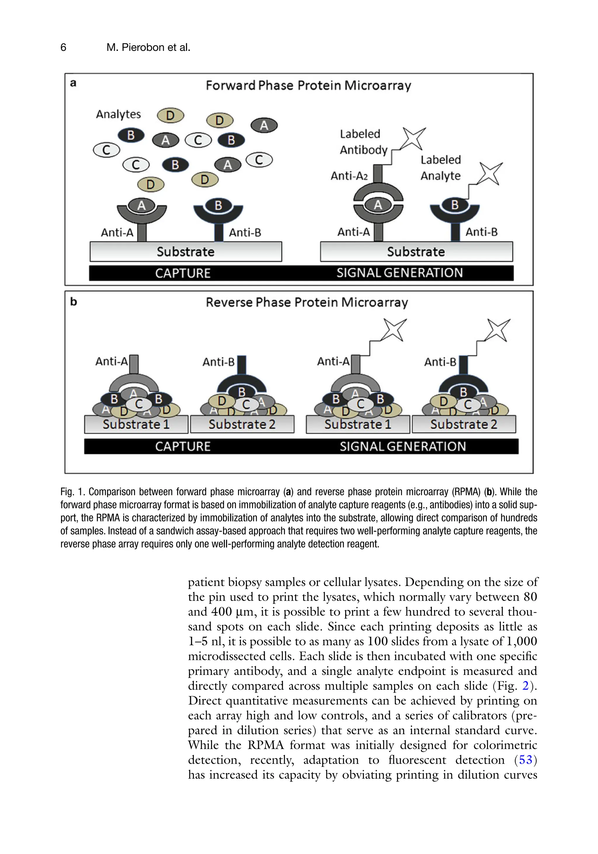
phosphoprotein biomarker profiling (43–52) (Fig. 1).
In contrast to a forward phase format (e.g., antibody array)
where the analyte detecting molecule is immobilized, with the
RPMA format, cellular lysates from individual test samples are
printed directly and immobilized on the array surface such that a
finished array could be comprised of lysates from cells from different
3. Reverse Phase
Protein
Microarrays:
Enabling
Technology for
Patient-Tailored
Therapeutics
24.
6 M. Pierobonet al.
patient biopsy samples or cellular lysates. Depending on the size of
the pin used to print the lysates, which normally vary between 80
and 400 mm, it is possible to print a few hundred to several thou-
sand spots on each slide. Since each printing deposits as little as
1–5 nl, it is possible to as many as 100 slides from a lysate of 1,000
microdissected cells. Each slide is then incubated with one specific
primary antibody, and a single analyte endpoint is measured and
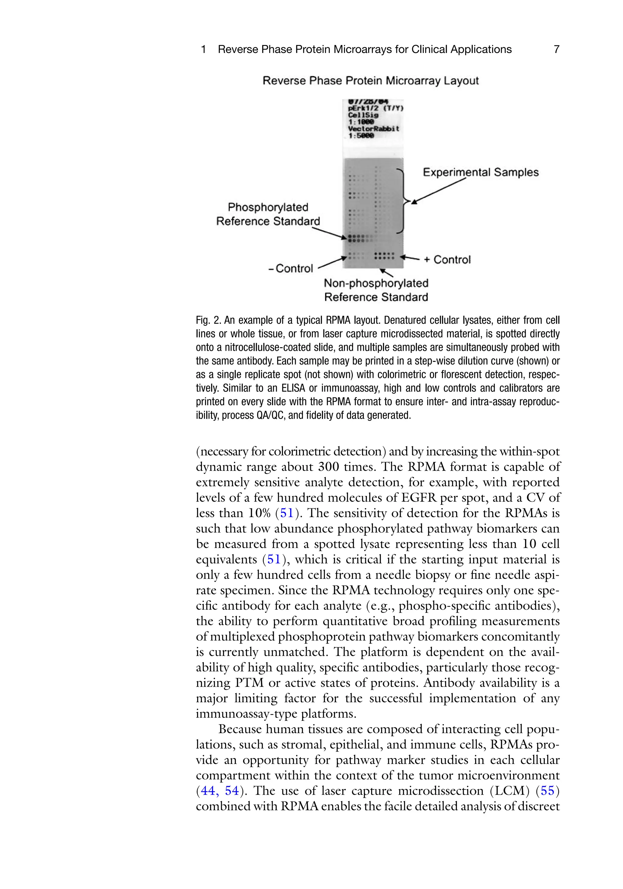
directly compared across multiple samples on each slide (Fig. 2).
Direct quantitative measurements can be achieved by printing on
each array high and low controls, and a series of calibrators (pre-
pared in dilution series) that serve as an internal standard curve.
While the RPMA format was initially designed for colorimetric
detection, recently, adaptation to fluorescent detection (53)
has increased its capacity by obviating printing in dilution curves
Fig. 1. Comparison between forward phase microarray (a) and reverse phase protein microarray (RPMA) (b). While the
forward phase microarray format is based on immobilization of analyte capture reagents (e.g., antibodies) into a solid sup-
port, the RPMA is characterized by immobilization of analytes into the substrate, allowing direct comparison of hundreds
of samples. Instead of a sandwich assay-based approach that requires two well-performing analyte capture reagents, the
reverse phase array requires only one well-performing analyte detection reagent.
25.
7
1 Reverse PhaseProtein Microarrays for Clinical Applications
(necessary for colorimetric detection) and by increasing the within-spot
dynamic range about 300 times. The RPMA format is capable of
extremely sensitive analyte detection, for example, with reported
levels of a few hundred molecules of EGFR per spot, and a CV of
less than 10% (51). The sensitivity of detection for the RPMAs is
such that low abundance phosphorylated pathway biomarkers can
be measured from a spotted lysate representing less than 10 cell
equivalents (51), which is critical if the starting input material is
only a few hundred cells from a needle biopsy or fine needle aspi-
rate specimen. Since the RPMA technology requires only one spe-
cific antibody for each analyte (e.g., phospho-specific antibodies),
the ability to perform quantitative broad profiling measurements
of multiplexed phosphoprotein pathway biomarkers concomitantly
is currently unmatched. The platform is dependent on the avail-
ability of high quality, specific antibodies, particularly those recog-
nizing PTM or active states of proteins. Antibody availability is a
major limiting factor for the successful implementation of any
immunoassay-type platforms.
Because human tissues are composed of interacting cell popu-
lations, such as stromal, epithelial, and immune cells, RPMAs pro-
vide an opportunity for pathway marker studies in each cellular
compartment within the context of the tumor microenvironment
(44, 54). The use of laser capture microdissection (LCM) (55)
combined with RPMA enables the facile detailed analysis of
discreet
Fig. 2. An example of a typical RPMA layout. Denatured cellular lysates, either from cell
lines or whole tissue, or from laser capture microdissected material, is spotted directly
onto a nitrocellulose-coated slide, and multiple samples are simultaneously probed with
the same antibody. Each sample may be printed in a step-wise dilution curve (shown) or
as a single replicate spot (not shown) with colorimetric or florescent detection, respec-
tively. Similar to an ELISA or immunoassay, high and low controls and calibrators are
printed on every slide with the RPMA format to ensure inter- and intra-assay reproduc-
ibility, process QA/QC, and fidelity of data generated.
Gutenberg” appears, orwith which the phrase “Project
Gutenberg” is associated) is accessed, displayed, performed,
viewed, copied or distributed:
This eBook is for the use of anyone anywhere in the United
States and most other parts of the world at no cost and
with almost no restrictions whatsoever. You may copy it,
give it away or re-use it under the terms of the Project
Gutenberg License included with this eBook or online at
www.gutenberg.org. If you are not located in the United
States, you will have to check the laws of the country
where you are located before using this eBook.
1.E.2. If an individual Project Gutenberg™ electronic work is
derived from texts not protected by U.S. copyright law (does not
contain a notice indicating that it is posted with permission of
the copyright holder), the work can be copied and distributed to
anyone in the United States without paying any fees or charges.
If you are redistributing or providing access to a work with the
phrase “Project Gutenberg” associated with or appearing on the
work, you must comply either with the requirements of
paragraphs 1.E.1 through 1.E.7 or obtain permission for the use
of the work and the Project Gutenberg™ trademark as set forth
in paragraphs 1.E.8 or 1.E.9.
1.E.3. If an individual Project Gutenberg™ electronic work is
posted with the permission of the copyright holder, your use and
distribution must comply with both paragraphs 1.E.1 through
1.E.7 and any additional terms imposed by the copyright holder.
Additional terms will be linked to the Project Gutenberg™
License for all works posted with the permission of the copyright
holder found at the beginning of this work.
1.E.4. Do not unlink or detach or remove the full Project
Gutenberg™ License terms from this work, or any files
28.
containing a partof this work or any other work associated with
Project Gutenberg™.
1.E.5. Do not copy, display, perform, distribute or redistribute
this electronic work, or any part of this electronic work, without
prominently displaying the sentence set forth in paragraph 1.E.1
with active links or immediate access to the full terms of the
Project Gutenberg™ License.
1.E.6. You may convert to and distribute this work in any binary,
compressed, marked up, nonproprietary or proprietary form,
including any word processing or hypertext form. However, if
you provide access to or distribute copies of a Project
Gutenberg™ work in a format other than “Plain Vanilla ASCII” or
other format used in the official version posted on the official
Project Gutenberg™ website (www.gutenberg.org), you must,
at no additional cost, fee or expense to the user, provide a copy,
a means of exporting a copy, or a means of obtaining a copy
upon request, of the work in its original “Plain Vanilla ASCII” or
other form. Any alternate format must include the full Project
Gutenberg™ License as specified in paragraph 1.E.1.
1.E.7. Do not charge a fee for access to, viewing, displaying,
performing, copying or distributing any Project Gutenberg™
works unless you comply with paragraph 1.E.8 or 1.E.9.
1.E.8. You may charge a reasonable fee for copies of or
providing access to or distributing Project Gutenberg™
electronic works provided that:
• You pay a royalty fee of 20% of the gross profits you derive
from the use of Project Gutenberg™ works calculated using the
method you already use to calculate your applicable taxes. The
fee is owed to the owner of the Project Gutenberg™ trademark,
but he has agreed to donate royalties under this paragraph to
the Project Gutenberg Literary Archive Foundation. Royalty
29.
payments must bepaid within 60 days following each date on
which you prepare (or are legally required to prepare) your
periodic tax returns. Royalty payments should be clearly marked
as such and sent to the Project Gutenberg Literary Archive
Foundation at the address specified in Section 4, “Information
about donations to the Project Gutenberg Literary Archive
Foundation.”
• You provide a full refund of any money paid by a user who
notifies you in writing (or by e-mail) within 30 days of receipt
that s/he does not agree to the terms of the full Project
Gutenberg™ License. You must require such a user to return or
destroy all copies of the works possessed in a physical medium
and discontinue all use of and all access to other copies of
Project Gutenberg™ works.
• You provide, in accordance with paragraph 1.F.3, a full refund of
any money paid for a work or a replacement copy, if a defect in
the electronic work is discovered and reported to you within 90
days of receipt of the work.
• You comply with all other terms of this agreement for free
distribution of Project Gutenberg™ works.
1.E.9. If you wish to charge a fee or distribute a Project
Gutenberg™ electronic work or group of works on different
terms than are set forth in this agreement, you must obtain
permission in writing from the Project Gutenberg Literary
Archive Foundation, the manager of the Project Gutenberg™
trademark. Contact the Foundation as set forth in Section 3
below.
1.F.
1.F.1. Project Gutenberg volunteers and employees expend
considerable effort to identify, do copyright research on,
transcribe and proofread works not protected by U.S. copyright
30.
law in creatingthe Project Gutenberg™ collection. Despite these
efforts, Project Gutenberg™ electronic works, and the medium
on which they may be stored, may contain “Defects,” such as,
but not limited to, incomplete, inaccurate or corrupt data,
transcription errors, a copyright or other intellectual property
infringement, a defective or damaged disk or other medium, a
computer virus, or computer codes that damage or cannot be
read by your equipment.
1.F.2. LIMITED WARRANTY, DISCLAIMER OF DAMAGES - Except
for the “Right of Replacement or Refund” described in
paragraph 1.F.3, the Project Gutenberg Literary Archive
Foundation, the owner of the Project Gutenberg™ trademark,
and any other party distributing a Project Gutenberg™ electronic
work under this agreement, disclaim all liability to you for
damages, costs and expenses, including legal fees. YOU AGREE
THAT YOU HAVE NO REMEDIES FOR NEGLIGENCE, STRICT
LIABILITY, BREACH OF WARRANTY OR BREACH OF CONTRACT
EXCEPT THOSE PROVIDED IN PARAGRAPH 1.F.3. YOU AGREE
THAT THE FOUNDATION, THE TRADEMARK OWNER, AND ANY
DISTRIBUTOR UNDER THIS AGREEMENT WILL NOT BE LIABLE
TO YOU FOR ACTUAL, DIRECT, INDIRECT, CONSEQUENTIAL,
PUNITIVE OR INCIDENTAL DAMAGES EVEN IF YOU GIVE
NOTICE OF THE POSSIBILITY OF SUCH DAMAGE.
1.F.3. LIMITED RIGHT OF REPLACEMENT OR REFUND - If you
discover a defect in this electronic work within 90 days of
receiving it, you can receive a refund of the money (if any) you
paid for it by sending a written explanation to the person you
received the work from. If you received the work on a physical
medium, you must return the medium with your written
explanation. The person or entity that provided you with the
defective work may elect to provide a replacement copy in lieu
of a refund. If you received the work electronically, the person
or entity providing it to you may choose to give you a second
opportunity to receive the work electronically in lieu of a refund.
31.
If the secondcopy is also defective, you may demand a refund
in writing without further opportunities to fix the problem.
1.F.4. Except for the limited right of replacement or refund set
forth in paragraph 1.F.3, this work is provided to you ‘AS-IS’,
WITH NO OTHER WARRANTIES OF ANY KIND, EXPRESS OR
IMPLIED, INCLUDING BUT NOT LIMITED TO WARRANTIES OF
MERCHANTABILITY OR FITNESS FOR ANY PURPOSE.
1.F.5. Some states do not allow disclaimers of certain implied
warranties or the exclusion or limitation of certain types of
damages. If any disclaimer or limitation set forth in this
agreement violates the law of the state applicable to this
agreement, the agreement shall be interpreted to make the
maximum disclaimer or limitation permitted by the applicable
state law. The invalidity or unenforceability of any provision of
this agreement shall not void the remaining provisions.
1.F.6. INDEMNITY - You agree to indemnify and hold the
Foundation, the trademark owner, any agent or employee of the
Foundation, anyone providing copies of Project Gutenberg™
electronic works in accordance with this agreement, and any
volunteers associated with the production, promotion and
distribution of Project Gutenberg™ electronic works, harmless
from all liability, costs and expenses, including legal fees, that
arise directly or indirectly from any of the following which you
do or cause to occur: (a) distribution of this or any Project
Gutenberg™ work, (b) alteration, modification, or additions or
deletions to any Project Gutenberg™ work, and (c) any Defect
you cause.
Section 2. Information about the Mission
of Project Gutenberg™
32.
Project Gutenberg™ issynonymous with the free distribution of
electronic works in formats readable by the widest variety of
computers including obsolete, old, middle-aged and new
computers. It exists because of the efforts of hundreds of
volunteers and donations from people in all walks of life.
Volunteers and financial support to provide volunteers with the
assistance they need are critical to reaching Project
Gutenberg™’s goals and ensuring that the Project Gutenberg™
collection will remain freely available for generations to come. In
2001, the Project Gutenberg Literary Archive Foundation was
created to provide a secure and permanent future for Project
Gutenberg™ and future generations. To learn more about the
Project Gutenberg Literary Archive Foundation and how your
efforts and donations can help, see Sections 3 and 4 and the
Foundation information page at www.gutenberg.org.
Section 3. Information about the Project
Gutenberg Literary Archive Foundation
The Project Gutenberg Literary Archive Foundation is a non-
profit 501(c)(3) educational corporation organized under the
laws of the state of Mississippi and granted tax exempt status
by the Internal Revenue Service. The Foundation’s EIN or
federal tax identification number is 64-6221541. Contributions
to the Project Gutenberg Literary Archive Foundation are tax
deductible to the full extent permitted by U.S. federal laws and
your state’s laws.
The Foundation’s business office is located at 809 North 1500
West, Salt Lake City, UT 84116, (801) 596-1887. Email contact
links and up to date contact information can be found at the
Foundation’s website and official page at
www.gutenberg.org/contact
33.
Section 4. Informationabout Donations to
the Project Gutenberg Literary Archive
Foundation
Project Gutenberg™ depends upon and cannot survive without
widespread public support and donations to carry out its mission
of increasing the number of public domain and licensed works
that can be freely distributed in machine-readable form
accessible by the widest array of equipment including outdated
equipment. Many small donations ($1 to $5,000) are particularly
important to maintaining tax exempt status with the IRS.
The Foundation is committed to complying with the laws
regulating charities and charitable donations in all 50 states of
the United States. Compliance requirements are not uniform
and it takes a considerable effort, much paperwork and many
fees to meet and keep up with these requirements. We do not
solicit donations in locations where we have not received written
confirmation of compliance. To SEND DONATIONS or determine
the status of compliance for any particular state visit
www.gutenberg.org/donate.
While we cannot and do not solicit contributions from states
where we have not met the solicitation requirements, we know
of no prohibition against accepting unsolicited donations from
donors in such states who approach us with offers to donate.
International donations are gratefully accepted, but we cannot
make any statements concerning tax treatment of donations
received from outside the United States. U.S. laws alone swamp
our small staff.
Please check the Project Gutenberg web pages for current
donation methods and addresses. Donations are accepted in a
number of other ways including checks, online payments and
34.
credit card donations.To donate, please visit:
www.gutenberg.org/donate.
Section 5. General Information About
Project Gutenberg™ electronic works
Professor Michael S. Hart was the originator of the Project
Gutenberg™ concept of a library of electronic works that could
be freely shared with anyone. For forty years, he produced and
distributed Project Gutenberg™ eBooks with only a loose
network of volunteer support.
Project Gutenberg™ eBooks are often created from several
printed editions, all of which are confirmed as not protected by
copyright in the U.S. unless a copyright notice is included. Thus,
we do not necessarily keep eBooks in compliance with any
particular paper edition.
Most people start at our website which has the main PG search
facility: www.gutenberg.org.
This website includes information about Project Gutenberg™,
including how to make donations to the Project Gutenberg
Literary Archive Foundation, how to help produce our new
eBooks, and how to subscribe to our email newsletter to hear
about new eBooks.
35.
Welcome to ourwebsite – the ideal destination for book lovers and
knowledge seekers. With a mission to inspire endlessly, we offer a
vast collection of books, ranging from classic literary works to
specialized publications, self-development books, and children's
literature. Each book is a new journey of discovery, expanding
knowledge and enriching the soul of the reade
Our website is not just a platform for buying books, but a bridge
connecting readers to the timeless values of culture and wisdom. With
an elegant, user-friendly interface and an intelligent search system,
we are committed to providing a quick and convenient shopping
experience. Additionally, our special promotions and home delivery
services ensure that you save time and fully enjoy the joy of reading.
Let us accompany you on the journey of exploring knowledge and
personal growth!
ebookultra.com