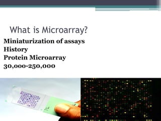
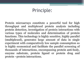
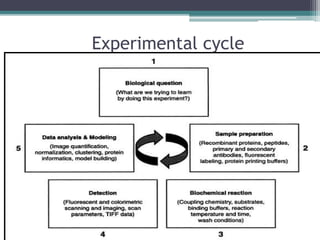
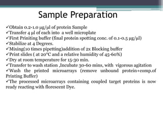
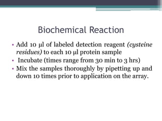
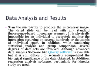
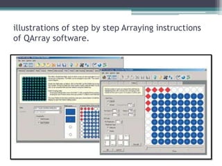
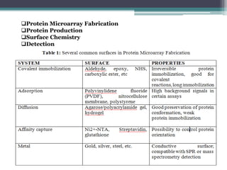
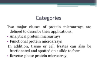
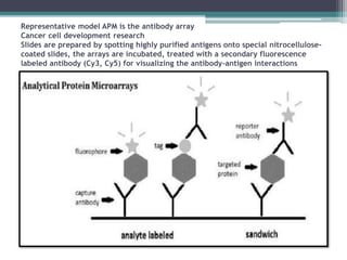
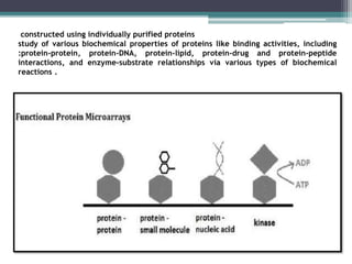
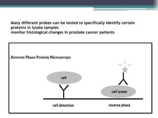
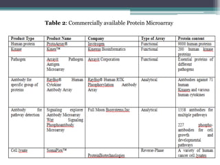
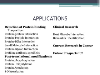
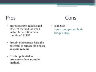
PROTEIN MICROARRAY
Ghalia Nawal
Roll#33
MS I Biotechnology
2015-2017
What is Microarray?
Miniaturization of assays
History
Protein Microarray
30,ooo-250,000
Principle:
Protein microarrays constitute a powerful tool for high
throughput and multiplexed protein analysis including
protein detection, investigation of protein interactions with
various types of molecules and determination of protein
functions. This technology is highly sensitive, highly parallel
(multiplexed), generates large amount of data in single
experiment with comparatively low sample consumption so
is highly economical and facilitate the parallel screening of
thousands of interactions, encompassing protein anti-body,
enzyme substrate, protein ligand or protein drug and
protein –protein interactions.
Experimental cycle
Sample Preparation
Obtain 0.2-1.0 μg/μl of protein Sample
Transfer 4 μl of each into a well microplate
First Priniting buffer (final protein spotting conc. of 0.1-0.5 μg/μl)
Stabilize at 4 Degrees.
Mixing(10 times pipetting)addition of 2x Blocking buffer
Print slides ( at 20°C and a relative humidity of 45-60%)
Dry at room temperature for 15-30 min.
Transfer to wash station ,Incubate 30-60 mins, with vigorous agitation
Wash the printed microarrays (remove unbound protein+comp.of
Printing Buffer)
The processed microarrays containing coupled target proteins is now
ready reacting with florescent Dye.
Biochemical Reaction
• Add 10 μl of labeled detection reagent (cysteine
residues) to each 10 μl protein sample
• Incubate (times range from 30 min to 3 hrs)
• Mix the samples thoroughly by pipetting up and
down 10 times prior to application on the array.
Data Analysis and Results
• Scan the microarray to produce the microarray image.
The dried slide can be read with any standard
fluorescence-based microarray scanner . It is physically
impossible for an individual to accurately monitor the
interaction occurring on several hundreds or thousands
of individual spots. In addition, while conducting
statistical analysis and group comparison, several
degrees of data sets are involved. Although advanced
data analysis Software like QArray software is available
but it is still difficult to accurately comprehend the
biological significance of the data obtained. In Addition,
regression analysis software, particularly for kinetics
study are used.
illustrations of step by step Arraying instructions
of QArray software.
Protein Microarray Fabrication
Protein Production
Surface Chemistry
Detection
Categories
Two major classes of protein microarrays are
defined to describe their applications:
• Analytical protein microarrays
• Functional protein microarrays
In addition, tissue or cell lysates can also be
fractionated and spotted on a slide to form
• Reverse-phase protein microarray.
Representative model APM is the antibody array
Cancer cell development research
Slides are prepared by spotting highly purified antigens onto special nitrocellulose-
coated slides, the arrays are incubated, treated with a secondary fluorescence
labeled antibody (Cy3, Cy5) for visualizing the antibody-antigen interactions
constructed using individually purified proteins
study of various biochemical properties of proteins like binding activities, including
:protein-protein, protein-DNA, protein-lipid, protein-drug and protein-peptide
interactions, and enzyme-substrate relationships via various types of biochemical
reactions .
Many different probes can be tested to specifically identify certain
proteins in lysate samples
monitor histological changes in prostate cancer patients
Table 2: Commercially available Protein Microarray
APPLICATIONS
Detection of Protein Binding
Properties:
Protein-protein interaction
Protein-Peptide Interaction
Protein-DNA Interaction
Small Molecule Interaction
Protein-Glycan Interaction
Profiling antibody specificity
Post-translational modifications:
Protein phosphorylation
Protein Ubiquitylation
Protein Acetylation
S-Nitrosylation
Clinical Research
Host Microbe Interaction
Biomarker Identification
Current Research in Cancer
Future Prospects!!!!!
Pros Cons
• more sensitive, reliable and
efficient method for small
molecule detection than
traditional ELISA
• Protein microarrays have the
potential to replace singleplex
analysis systems
• Greater potential in
proteomics than any other
method .
• High Cost
$500-1000 per antibody
$10 per oligo
ANY
QUESTION

Protein microarray

  • 1.
  • 2.
    What is Microarray? Miniaturizationof assays History Protein Microarray 30,ooo-250,000
  • 3.
    Principle: Protein microarrays constitutea powerful tool for high throughput and multiplexed protein analysis including protein detection, investigation of protein interactions with various types of molecules and determination of protein functions. This technology is highly sensitive, highly parallel (multiplexed), generates large amount of data in single experiment with comparatively low sample consumption so is highly economical and facilitate the parallel screening of thousands of interactions, encompassing protein anti-body, enzyme substrate, protein ligand or protein drug and protein –protein interactions.
  • 4.
  • 5.
    Sample Preparation Obtain 0.2-1.0μg/μl of protein Sample Transfer 4 μl of each into a well microplate First Priniting buffer (final protein spotting conc. of 0.1-0.5 μg/μl) Stabilize at 4 Degrees. Mixing(10 times pipetting)addition of 2x Blocking buffer Print slides ( at 20°C and a relative humidity of 45-60%) Dry at room temperature for 15-30 min. Transfer to wash station ,Incubate 30-60 mins, with vigorous agitation Wash the printed microarrays (remove unbound protein+comp.of Printing Buffer) The processed microarrays containing coupled target proteins is now ready reacting with florescent Dye.
  • 6.
    Biochemical Reaction • Add10 μl of labeled detection reagent (cysteine residues) to each 10 μl protein sample • Incubate (times range from 30 min to 3 hrs) • Mix the samples thoroughly by pipetting up and down 10 times prior to application on the array.
  • 7.
    Data Analysis andResults • Scan the microarray to produce the microarray image. The dried slide can be read with any standard fluorescence-based microarray scanner . It is physically impossible for an individual to accurately monitor the interaction occurring on several hundreds or thousands of individual spots. In addition, while conducting statistical analysis and group comparison, several degrees of data sets are involved. Although advanced data analysis Software like QArray software is available but it is still difficult to accurately comprehend the biological significance of the data obtained. In Addition, regression analysis software, particularly for kinetics study are used.
  • 8.
    illustrations of stepby step Arraying instructions of QArray software.
  • 9.
    Protein Microarray Fabrication ProteinProduction Surface Chemistry Detection
  • 10.
    Categories Two major classesof protein microarrays are defined to describe their applications: • Analytical protein microarrays • Functional protein microarrays In addition, tissue or cell lysates can also be fractionated and spotted on a slide to form • Reverse-phase protein microarray.
  • 11.
    Representative model APMis the antibody array Cancer cell development research Slides are prepared by spotting highly purified antigens onto special nitrocellulose- coated slides, the arrays are incubated, treated with a secondary fluorescence labeled antibody (Cy3, Cy5) for visualizing the antibody-antigen interactions
  • 12.
    constructed using individuallypurified proteins study of various biochemical properties of proteins like binding activities, including :protein-protein, protein-DNA, protein-lipid, protein-drug and protein-peptide interactions, and enzyme-substrate relationships via various types of biochemical reactions .
  • 13.
    Many different probescan be tested to specifically identify certain proteins in lysate samples monitor histological changes in prostate cancer patients
  • 14.
    Table 2: Commerciallyavailable Protein Microarray
  • 15.
    APPLICATIONS Detection of ProteinBinding Properties: Protein-protein interaction Protein-Peptide Interaction Protein-DNA Interaction Small Molecule Interaction Protein-Glycan Interaction Profiling antibody specificity Post-translational modifications: Protein phosphorylation Protein Ubiquitylation Protein Acetylation S-Nitrosylation Clinical Research Host Microbe Interaction Biomarker Identification Current Research in Cancer Future Prospects!!!!!
  • 16.
    Pros Cons • moresensitive, reliable and efficient method for small molecule detection than traditional ELISA • Protein microarrays have the potential to replace singleplex analysis systems • Greater potential in proteomics than any other method . • High Cost $500-1000 per antibody $10 per oligo
  • 17.

Editor's Notes

  • #12 proteins are detected after antibody capture using direct protein labeling