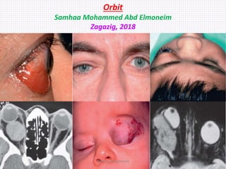
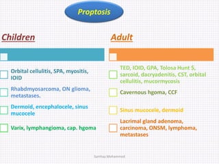
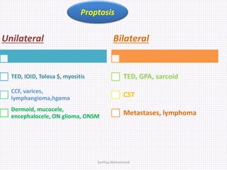
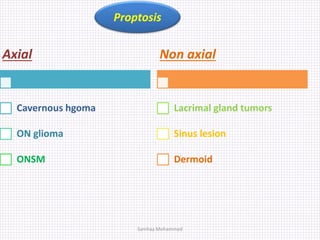
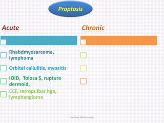
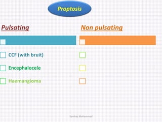
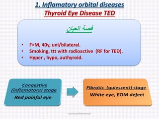
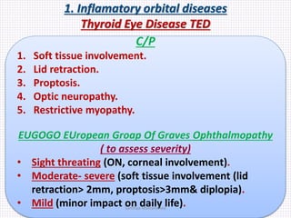
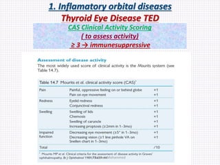
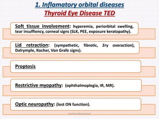
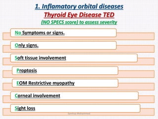
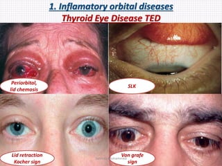
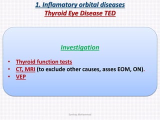
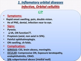
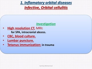
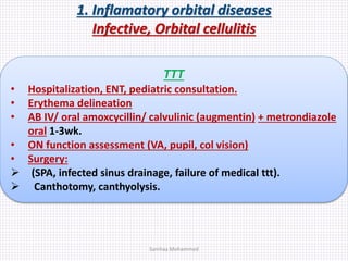
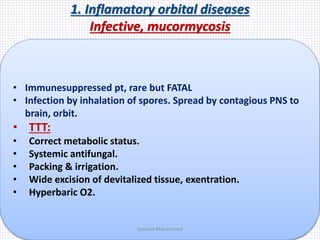
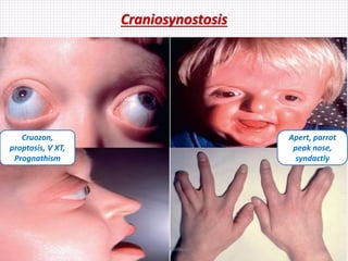
Orbit
Samhaa Mohammed Abd Elmoneim
Zagazig, 2018
Samhaa Mohammed
Orbital
anatomy
2, 3, 3, 4
Maxilla
Palatine
Samhaa Mohammed
Orbit anatomy
Lesser & greater
shpenoid wings
Sup & inf orbital
fissures
Zygomatic
Maxillary
Infraorbital
groove
Zygomatico
maxillary suture
Infraorbital
foramen
palatine
Ant lacrimal crest
Ethmoid
Lacrimal
Supra orbital notch
Trochlea
Frontal
Optic foramen
Samhaa Mohammed
Orbital dis. symptoms
• Eyelid and conjunctival swelling.
• Redness, watering.
• Pain (sometimes exacerbated by eye movement).
• Increasing ocular prominence, displacement or a
sunken impression of the eye.
• Double vision and blurring.
• Pulsing sensation or audible bruit.
Samhaa Mohammed
Orbital dis. signs
1. Soft tissue involvement (ptosis, chemosis, skin discolouration, ebibulbar injection).
2. Proptosis, exophthalmos (axial with conal lesions, dystopia with extraconal
lesions).
• Horizontal & vertical measurement using exophthalmometer.
• > 20mm from corneal apex.
• Difference > 2mm between both eyes.
3. Lagophthalos, palbebral fissure.
• Pseudoproptosis ( high myopia, lid retraction, contralat ptosis,enophthalmos,
microphthalmos, shallow orbit).
4. Enophthalmos ( congenital, trauma, post radiation, oculodigital sign in bind eye,
sclerosis as schirrous carcinoma or inflamatory)
• Pseudo enophthalmos (microophthalmos, phthisis pulbi, ptosis contralateral
proptosis)
5. Ophthalmoplegia (FDT, differential IOP, saccadic mov.).
6. Pulsation (CCF, encephalocele), bruit (CCF).
7. OA, OD odema.opticociliary shunt, choroidal folds.Samhaa Mohammed
Orbital dis. Exam.
• Proptosis from side & behind + measurement (Hertel
or ruler).
• Dystopia vertical/ horizontal (ruler).
• Specific
• lid signs in TED (Dalrymple, Von Gafe, Kocher, Stellwag)
• EOM motility
• Pupil & fundus
• Lagophthalmos
• Orbital rim
• Pulsation
• Retropulsion
• Infraorbital sensation
• Pain (malignancy)
Samhaa Mohammed
Orbital dis. investigation
• CT (bone)
• MRI (soft tissue)
• FNAB
Samhaa Mohammed
Causes
Inflamatory non infectious (TED,
IOID, Sarcoid, Wegner
granulomatosis, Ig4)
Infectious (orbital cellulitis, SPA)
Neoplastic (1ry or 2ry, B, M or
metastases
Traumatic (orbital fracture, CCF, RB
hge)
Vascular (CCF, varix,
lymphangioma)
Malformation (congenital
syndrome, synostosis)
Proptosis
Samhaa Mohammed
Children
Orbital cellulitis, SPA, myositis,
IOID
Rhabdmyosarcoma, ON glioma,
metastases.
Dermoid, encephalocele, sinus
mucocele
Varix, lymphangioma, cap. hgoma
Adult
TED, IOID, GPA, Tolosa Hunt $,
sarcoid, dacryadenitis, CST, orbital
cellulitis, mucormycosis
Cavernous hgoma, CCF
Sinus mucocele, dermoid
Lacrimal gland adenoma,
carcinoma, ONSM, lymphoma,
metastases
Proptosis
Samhaa Mohammed
Unilateral
TED, IOID, Tolosa $, myositis
CCF, varices,
lymphangioma,hgoma
Dermoid, mucocele,
encephalocele, ON glioma, ONSM
Bilateral
TED, GPA, sarcoid
CST
Metastases, lymphoma
Proptosis
Samhaa Mohammed
Axial
Cavernous hgoma
ON glioma
ONSM
Non axial
Lacrimal gland tumors
Sinus lesion
Dermoid
Proptosis
Samhaa Mohammed
Acute
Rhabdmyosarcoma,
lymphoma
Orbital cellulitis, myositis
IOID, Tolosa $, rupture
dermoid,
CCF, retropulbar hge,
lymphangioma
Chronic
Proptosis
Samhaa Mohammed
Pulsating
CCF (with bruit)
Encephalocele
Haemangioma
Non pulsating
Proptosis
Samhaa Mohammed
Orbital diseases
1. inflamatory
1. Non infective
 TED
 IOID
 Myositis
 GPA
 Tolosa Hunt $
2. Infective
 Orbital cellulitis
 SPA
 Mucormycosis
Samhaa Mohammed
1. Inflamatory orbital diseases
Thyroid Eye Disease TED
‫العيان‬ ‫قصة‬
• F>M, 40y, uni/bilateral.
• Smoking, ttt with radioactive (RF for TED).
• Hyper , hypo, authyroid.
Samhaa Mohammed
1. Inflamatory orbital diseases
Thyroid Eye Disease TED
C/P
1. Soft tissue involvement.
2. Lid retraction.
3. Proptosis.
4. Optic neuropathy.
5. Restrictive myopathy.
EUGOGO EUropean Groap Of Graves Ophthalmopathy
( to assess severity)
• Sight threating (ON, corneal involvement).
• Moderate- severe (soft tissue involvement (lid
retraction> 2mm, proptosis>3mm& diplopia).
• Mild (minor impact on daily life).Samhaa Mohammed
1. Inflamatory orbital diseases
Thyroid Eye Disease TED
CAS Clinical Activity Scoring
( to assess activity)
≥ 3 → immunesuppressive
Samhaa Mohammed
1. Inflamatory orbital diseases
Thyroid Eye Disease TED
Soft tissue involvement: hyperemia, periorbital swelling,
tear insuffiency, corneal signs (SLK, PEE, exposure keratopathy).
Lid retraction: (sympathetic, fibrotic, 2ry overaction),
Dalrymple, Kocher, Von Grafe signs).
Proptosis
Restrictive myopathy: (ophthalmoplegia, IR, MR).
Optic neuropathy: (lost ON function).
Samhaa Mohammed
1. Inflamatory orbital diseases
Thyroid Eye Disease TED
(NO SPECS score) to assess severity
No Symptoms or signs.
Only signs.
Soft tissue involvement
Proptosis
EOM Restrictive myopathy
Corneal involvement
Sight loss
Samhaa Mohammed
1. Inflamatory orbital diseases
Thyroid Eye Disease TED
Periorbital,
lid chemosis
SLK
Lid retraction
Kocher sign
Von grafe
sign
Samhaa Mohammed
1. Inflamatory orbital diseases
Thyroid Eye Disease TED
Investigation
• Thyroid function tests
• CT, MRI (to exclude other causes, asses EOM, ON).
• VEP
Samhaa Mohammed
1. Inflamatory orbital diseases
Thyroid Eye Disease TED
Defect lt
elevation
Defect rt
depression
Axial CT
EOM ++
Coronal CT
Spared LR
Restrictive
myopathy
Samhaa Mohammed
1. Inflamatory orbital diseases
Thyroid Eye Disease TED
TTT
• Stop smoking.
• Control thyroid function.
• Steroid in case of ttt with radioactive iodine.
Steroid doses
• Moderate to severe: oral 1mg/kg/d then tapering acc. to
response, or IV 0.5g/w for 6w then 0.25g/w for 6w then oral
for several months.
• Sight threating ON, exposure keratopathy:
 IV 0.5-1g for 3d then convert to oral 40mg/d.
 Shift to surgical decompression in steroid no response or
contraindication.
 LFT, GIT prophylaxis, osteoporosis protection.
Samhaa Mohammed
1. Inflamatory orbital diseases
Thyroid Eye Disease TED
Mild disease
• Lubricants.
• NSAIDS, steroids,
ciclosporin.
• Head elevation.
• Taping in exposure
keratopathy.
Moderate to severe
• TTT score 3 of 7:
•1. Spontaneous orbital pain.
•2. Gaze-evoked orbital pain.
•3. Eyelid swelling
•4. Eyelid erythema.
•5. Conjunctival redness
considered to be due to
active (inflammatory phase)
TED.
•6. Chemosis.
•7. Inflammation of caruncle
or plica.
• Syst steroid iv oral &
orbital,
immunosuppressive,
radiotherapy
Post inflamatory
complication
• Orbit → EOM → lid
•1. Proptosis (surgical
decompression:
•Lateral wall (↓ 4-5mm, less
diplopia)
•+ medial wall (diplopia)
•+ floor (hypoglobus,
infraorbital n damage, ↓6-
9mm)
•+ part of roof.
•2. Restrictive myopathy
•Prism then surgery (diplopia
in pp & reading) .
•Stable angle, infl subsides.
•3. Lid retraction
•Mild no ttt
•Botox injection then
mullerectomy (mild) or UL, LL
recession (severe)
Samhaa Mohammed
Orbital diseases
1. inflamatory
1. Non infective
 TED
 IOID
 Myositis
 GPA
 Tolosa Hunt $
2. Infective
 Orbital cellulitis
 SPA
 Mucormycosis
Samhaa Mohammed
1. Inflamatory orbital diseases
Infective, Orbital cellulitis
 Preseptal cellulitis (from SC tissue till septum, staph
areus, strept pyogenes, No proptosis, EOM affection)
 Orbital cellulitis (behind orbital septum, strept
pneumonia, staph areus, strept pyogenes, H influenza).
Infection spread from preseptal cellulitis, PNS,
dacryocystitis, midfacial, dental infection, trauma, surgery,
blood borne.
Samhaa Mohammed
1. Inflamatory orbital diseases
Infective, Orbital cellulitis
C/P
• Symptoms:
• Rapid onset swelling, pain, double vision.
• Hx of PNS, dental, infection near to eye.
• Signs:
• Pyrexia.
• ↓VA, ON function??.
• Proptosis (axial, non axial in SPA).
• Painful ophthalmoplegia.
• ON swelling, ch folds.
• Complication:
• SERIOUS: CSS, brain abcess, meningitis.
• OCULAR: Compressive ON, Exposure keratopathy,
endophthalmitis, CRAO.
• SPA subperiosteal abcess (medial wall)
Samhaa Mohammed
1. Inflamatory orbital diseases
Infective, Orbital cellulitis
Investigation
• High resolution CT, MRI:
for SPA, intracranial abcess.
• CBC, blood culture.
• Lumbar puncture.
• Tetanus immunization: in trauma
Samhaa Mohammed
1. Inflamatory orbital diseases
Infective, Orbital cellulitis
TTT
• Hospitalization, ENT, pediatric consultation.
• Erythema delineation
• AB IV/ oral amoxcycillin/ calvulinic (augmentin) + metrondiazole
oral 1-3wk.
• ON function assessment (VA, pupil, col vision)
• Surgery:
 (SPA, infected sinus drainage, failure of medical ttt).
 Canthotomy, canthyolysis.
Samhaa Mohammed
1. Inflamatory orbital diseases
Infective, mucormycosis
• Immunesuppressed pt, rare but FATAL
• Infection by inhalation of spores. Spread by contagious PNS to
brain, orbit.
• TTT:
• Correct metabolic status.
• Systemic antifungal.
• Packing & irrigation.
• Wide excision of devitalized tissue, exentration.
• Hyperbaric O2.
Samhaa Mohammed
Orbital diseases
1. inflamatory
1. Non infective
 TED
 IOID
 Myositis
 GPA
 Tolosa Hunt $
2. Infective
 Orbital cellulitis
 SPA
 Mucormycosis
Samhaa Mohammed
1. Inflamatory orbital diseases
Idiopathic Orbital Inflamatory Disease IOID
• Orbital pseudotumour, Space occupying orbital inflamation.
• Non specific, non infective, non neoplastic pleomorphic
inflamation → reactive fibrosis.
• Unilateral in adults, Bilateral in children
C/P
 Dx: by exclusion (infection, lymphoma, GPA, sarcoidosis)
 Symptoms: Acute/ subacute painful swelling, periocular
redness, pyrexia in children.
 Signs:
• Congestive proptosis.
• Ophthalmoplegia
• ON dysfunction.
• Choroidal folds.
Samhaa Mohammed
1. Inflamatory orbital diseases
Idiopathic Orbital Inflamatory Disease IOID
Course
Severe
prolonged
(frozen orbit)
Spontaneous
(resolution
within wks)
Intermittent
(with
remission)
Samhaa Mohammed
1. Inflamatory orbital diseases
Idiopathic Orbital Inflamatory Disease IOID
Investigation
• CT (Ill defined contents, involved EOM tendons).
• Biopsy (rule out malignancy) in persistent cases.
• Wide range investigation to exclude DD
(lymphoma, sarcoid, GPA, infection).
Samhaa Mohammed
1. Inflamatory orbital diseases
Idiopathic Orbital Inflamatory Disease IOID
TTT
 Observation: in mild, spontaneous.
 NSAIDs alone (e.g. ibuprofen) in mild disease + proton
pump inhibitor
 Systemic steroids after Dx has been confirmed, as
they may mask other pathology such as infection and
Wegener granulomatosis.
 Oral prednisolone is initially given at a dose of 1.0–1.5
mg/ kg/day, subsequently being tapered and
discontinued over a number of weeks.
 Further treatment may be needed in the event of
recurrence.
 Orbital depot steroid
 Radiotherapy, Immunesuppressive
 Surgical resection in highly resistant cases.Samhaa Mohammed
Orbital diseases
1. inflamatory
1. Non infective
 TED
 IOID
 Myositis
 GPA
 Tolosa Hunt $
2. Infective
 Orbital cellulitis
 SPA
 Mucormycosis
Samhaa Mohammed
1. Inflamatory orbital diseases
Orbital myositis
• Idiopathic inflamation of one or more EOM
• TTT like mild IOID.
Samhaa Mohammed
1. Inflamatory orbital diseases
Acute dacryoadenitis
• Idiopathic lacrimal gland inflamation.
• Viral, sarcoidosis & sjogren $ (chronic inf), IOID.
C/P
 Signs:
• S shaped ptosis, dystopia.
• Perauricular LN
 Investigation:
• CT: Lacrimal gland ++, no bony erosion.
• Biopsy: exclude neoplasm (resistant to ttt).
Samhaa Mohammed
1. Inflamatory orbital diseases
Tolosa Hunt syndrome
Wegner granulomatosis
Tolosa Hunt Syndrome
• Unilateral non granulomatous inflamation of orbital apex, SOF,
cavernous sinus.
• Dx by exclusion other neoplastic, inflamatory causes.
• Ocular: CN palsies (2,3,4,5₁,₂,6)
Wegner granulomatosis
• Bilateral multisystem inflamation.
• PNS, nasopharynx, Renal involvement.
• +ve ANCA C.
• Ocular: PUK, scleritis, retinal vascular occlusion.
• TTT with sterois, cyclophosphamide, surgical decompression.
Samhaa Mohammed
Orbital diseases
1. inflamatory
1. Non infective
 TED
 IOID
 Myositis
 GPA
 Tolosa Hunt $
2. Infective
 Orbital cellulitis
 SPA
 Mucormycosis
Samhaa Mohammed
Orbital diseases
2. Vascular Orbital diseases
 Cavernous sinus thrombosis:
Clotting inside CS due to 2ry infection leads to all orbital signs, MRI, MRV.
 CCF carotid cavernous fistula.
 Varix.
 Lymphangioma
 Capillary haemangioma.
 Cavernous haemangioma.
Samhaa Mohammed
2. Vascular orbital diseases
Carotid Cavernous Fistula CCF
• ICA/ECA & cavernous sinus communication.
• Ocular signs due to venous stasis, ↑episcleral pr, ↓arterial
supply to CN.
Direct CCF
-ICA & CS
-high flow
-Trauma, rupture aneurysm in middle
age female HTN
Indirect CCF
-dural branches of ICA/ECA & CS
-low flow
-Atherosclerosis, congenital + minor
trauma/ CT dis.Samhaa Mohammed
2. Vascular orbital diseases
Carotid Cavernous Fistula CCF
• Symptoms:
Direct CCF
-classic Δ (pulsatile proptosis,
whooshing noise, conj. chemosis)
-Within days after head trauma
Indirect CCF
-Conjunctival vascular engorgement
-Gradual onset of one or both eyes
Samhaa Mohammed
2. Vascular orbital diseases
Carotid Cavernous Fistula CCF
• Signs: Direct CCF
-immediate vision loss due to (ON),
long term (glaucoma, exposure k).
- ipsilat., bil., contra. Proptosis,
ophthalmoplegia
-Pulsatile with bruit proptosis
cancelled with pressure on carotid.
-IOP ↑(↑episcleral pr, NVG), stasis.
Indirect CCF
Milder than direct, corkscrew b v
-Gradual onset of one or both eyes
Samhaa Mohammed
2. Vascular orbital diseases
CCF
Investigation
• CT, MRI, MRV/A (SOV , EOM ++)
• Orbital doppler (abnormal flow).
• Catheter digital subtraction angiography (definite Dx)
• CT, MRI angiography
Samhaa Mohammed
2. Vascular orbital diseases
CCF
Corkscrew
conj. bv
Vascular
tortousity
CT axial,
SOV ++
CT coronal,
EOM ++
Samhaa Mohammed
2. Vascular orbital diseases
CCF
TTT
• CCF NOT life threating. Protect eye, consult neurosurgeon.
• Spontaneous closure in majority of cases.
• Post traumatic CCF is less likely for spontaneous closure
• Direct CCF→ transarterial embolization (repair a, occlude sinus) or
craniotomy.
• Indirect CCF→ spontaneous occlusion, transvenous sinus occlusion
Samhaa Mohammed
Orbital diseases
2. Vascular causes
 Cavernous sinus thrombosis:
Clotting inside CS due to 2ry infection, blood dis. leads to all orbital signs, MRI,
MRV.
CN (3,4,6,5₁,₂,₃), Horner
 CCF carotid cavernous fistula.
 Varix.
 Lymphangioma
 Capillary haemangioma.
 Cavernous haemangioma.
Samhaa Mohammed
2. Vascular orbital diseases
Varices
lymphangioma
Varix
• Hamartoma. Thin wall vein like connected to b v (orbit, conj,
lid), from childhood to middle age.
• Superonasal, no pulsation or bruit, proptosis ↑with valsalva.
• Complication: acute hge, thrombosis, phlebolith (MRI with
contrast), enophthalmos (fat atrophy).
• Surgery in ON, recurrent thrombosis. Surgery is difficult.
Lymphangioma
• Hamartoma. Venous lymphatic malformation. Early childhood.
• Not connected to b v, but rupture → chocolate cysts.
• Association (intracranial, lid, conj, oropharynx)
• Superonasal, ant or post (proptosis) orbital.
• Surgery in persistent/sight threating chocolate cyst.Samhaa Mohammed
2. Vascular orbital diseases
Varix
Lid
varices
Conj
varices
Orbital
varices
With
vasalva
Samhaa Mohammed
2. Vascular orbital diseases
Varix
Enophthalmos
Phlebolith
radiology?
Samhaa Mohammed
2. Vascular orbital diseases
lymphangioma, chocolate cyst
Samhaa Mohammed
2. Vascular orbital diseases
Capillary haemangioma
Capillary Haemangioma
• Hamartoma, most common tumour in orbit & periorbital
area in childhood.
• No true capsule. Small vessels anastamosis.
• Systemic association: high COP, Kasbach Merritt $(50% fatal
dt thrombocytopenia), PHACE $
• Preseptal . (strawberry), orbital. Exclude large extensive post orbital
lesions.
• Invest.: US (medium internal reflectivity),CT, MRI (soft finger like
expansion with No bony erosion).
• TTT: its course is progression over 1st 6m then resolves gradually within
3-7y.
• Treat in anisometropia, squint, amblyopia, ON, exposure keratopathy.
• Oral BB (2mg/kg/d) by pediatrician/ topical.
• Local steroid 1–2 ml total of 40 mg/ml in 1-3 session of triamicinolone.
• Laser to superficial part, alfa interferon, local resection.Samhaa Mohammed
2. Vascular orbital diseases
capillary haemangioma
Preseptal Forniceal
CT intraconal
homogenous mass
Preseptal
strawbelrry
Samhaa Mohammed
2. Vascular orbital diseases
cavernous haemangioma
Cavernous haemangioma
• Middle age female. Progression in pregnancy, lateral wall.
• Encapsulated endothelial lined vascular channels seperated
by septae.
• C/P:
• unilateral slowly progressive axial proptosis.
• Lesion at orbital apex cause ON without proptosis.
• Invest.: US, CT, MRI (well circumscribed oval mass in ms cone).
• TTT:
• Discovered accidentaly with no symptoms = no ttt.
• Symptomatic cases with progression need surgery.
Samhaa Mohammed
2. Vascular orbital diseases
capillary haemangioma
Axial
proptosis
Retropulbal well
circumscribed
EncapsulatedSamhaa Mohammed
Orbital diseases
2. Vascular Orbital diseases
 Cavernous sinus thrombosis:
Clotting inside CS due to 2ry infection leads to all orbital signs, MRI, MRV.
 CCF carotid cavernous fistula.
 Varix.
 Lymphangioma
 Capillary haemangioma.
 Cavernous haemangioma.
Samhaa Mohammed
Causes
Inflamatory non infectious (TED,
IOID, Sarcoid, Wegner
granulomatosis, Ig4)
Infectious (orbital cellulitis, SPA)
Neoplastic (1ry or 2ry, B, M or
metastases
Traumatic (orbital fracture, CCF, RB
hge)
Vascular (CCF, varix,
lymphangioma)
Malformation (congenital
syndrome, synostosis)
Proptosis
Samhaa Mohammed
Orbital diseases
3. Neoplastic causes
 Lacrimal gland tumours:
• Pleomorphic adenoma.
• Lacrimal gland adenocarcinoma.
 Neurnal tumours:
• Optic n. glioma.
• Optic n. sheath meinigioma ONSM.
• Plexiform neurofibroma
• Isolated neurofibroma.
 Lymphoma.
 Rhabdmyosarcoma.
 Metastases.
Samhaa Mohammed
3. Neoplastic orbital diseases
lacrimal gland pleomorphic adenoma
 Arise from myoepithelial cells in ducts & secretory elements.
 Benign mixed cell tumour (myoeithelial). Outer layer
(metaplasia to myxoid tissue) & inner layer (glanular tissue to
keratin and squamous differentiation).
 Most common benign epithelial tumour of lacrimal gland.
 Young middle age.
C/P
• Symptoms:
• Slowly progressive painless proptosis.
• Signs:
• Orbital lobe ++ causes inferonasal dystopia, post extension
causes proptosis
• Palpebral lobe ++ causes UL swelling S shaped ptosis.
Samhaa Mohammed
3. Neoplastic orbital diseases
lacrimal gland pleomorphic adenoma
Investigation
• CT:
• Rounded lesion with smooth surface with no destruction of
bony orbit.
• Biopsy: XXX contraindicated in suspicious cases to avoid
seeding.
TTT
• Anterior portionthrough anterior transseptal approach.
• Orbital portion excision with intact capsule through lateral
orbitotomy.
• Incomplete excision leads to seeding, recurrence, malignant
transformation
Samhaa Mohammed
3. Neoplastic orbital diseases
lacrimal gland carcinoma
 Middle age. High mortality rate.
 Adenoidcystic (50%), pleomorphic adenocarcinoma,
mucoepidermoid, squamous cell carcinoma
 Shorter Hx than benign.
 Course (incomplete excision of B, B→M, denovo M)
C/P
• Symptoms:
• Pain (lacrimal n infiltration in malignancy or inflamation)
• Signs:
• Inferonasal dystopia.
• Post extension causes proptosis, epibulbar congestion,
ophthalmoplegia.
• Hypothesia, choroidal folds, OD swelling.
Samhaa Mohammed
3. Neoplastic orbital diseases
lacrimal gland carcinoma
Investigation
• CT:
• Irregular mass with bony erosion.
• Biopsy:
• Necessary for Dx
TTT
• Neurology consultation (cavernous sinus spread)
• Tumour excision ± chemotherapy/ brachytherapy.
• Exentration
• Incomplete excision leads to seeding, recurrence, malignant
transformation
Samhaa Mohammed
3. Neoplastic orbital diseases
lacrimal adenoma/ carcinoma
Lacrimal gland
adenocarcinoma
Pleomorphic
adenoma
Samhaa Mohammed
Orbital diseases
3. Neoplastic causes
 Lacrimal gland tumours:
• Pleomorphic adenoma.
• Lacrimal gland adenocarcinoma.
 Neurnal tumours:
• Optic n. glioma.
• Optic n. sheath meinigioma ONSM.
• Plexiform neurofibroma
• Isolated neurofibroma.
 Lymphoma.
 Rhabdmyosarcoma.
 Metastases.
Samhaa Mohammed
3. Neoplastic orbital tumours
neurnal, ON glioma
 Slowly progressive astrocytoma.
 Childhood .
 Variable prognosis.
 NF1 association.
C/P
• Symptoms:
• Slowly progressive visual loss..
• Signs:
• Inferotemporal dystopia, non axial proptosis
• ON swelling, atrophy, intracranial spread.
Investigation
• CT (intracranial chiasm extension), MRI (fusiform ON ++).
Samhaa Mohammed
3. Neoplastic orbital diseases
neurnal ON glioma
TTT
Surgery means lost vision ¡¡
• Observation
• Surgiical excision with preserved globe
• Poor vision, significant proptosis.
• To prevent intracranial extension.
• ± radio or chemotherapy (incomplete excision cases)
Samhaa Mohammed
3. Neoplastic orbital tumours
ON glioma
Fusiform ON ++
Axial CT
ON Glioma
with dystopia
Samhaa Mohammed
3. Neoplastic orbital tumours
ON sheath meningioma ONSM
 Arises from arachnoid meningoepithelial cells of intraorbital
part of ON
 Affect middle age females.
 Good prognosis in adult, poor in children
 NF2 association.
 Histology menigothelial cells, psammoma bodies
C/P
• Symptoms:
• Transient, gradual VA decrease
• Signs:
• Hoyett Spencer Δ (progressive vision loss, OA, OC shunts)
• Reverse sequence (ON dysfunction&atrophy → OC shunts→
EOM restriction → proptosis)
Samhaa Mohammed
3. Neoplastic orbital tumours
ON sheath meningioma ONSM
Investigation
• MRI (investigation of choice)
• CT: ON thickening & calcification.
TTT
• No need in middle age
• Stereoactic radiotherapy with surgery
Samhaa Mohammed
3. Neoplastic orbital tumours
ONSM
MRI, ONSM
ONSM
calcification,
CT
Samhaa Mohammed
3. Neoplastic orbital tumours
plexiform neurofibroma
isolated neurofibroma
Plexiform NF
• Most common peripheral neurnal tumour of orbit.
• NF1, presenntation in childhood with S shaped ptosis (bag of
worms)
• May turn malignant
• Surgery not recommended (attached to important structure,
difficult complete excision)
Isolated neurofibroma
• Less common, 10% with NF1.
• Adult age.
• Straightforward surgery (well circumscribed, relative avascular)
Samhaa Mohammed
Orbital diseases
3. Neoplastic causes
 Lacrimal gland tumours:
• Pleomorphic adenoma.
• Lacrimal gland adenocarcinoma.
 Neurnal tumours:
• Optic n. glioma.
• Optic n. sheath meinigioma ONSM.
• Plexiform neurofibroma
• Isolated neurofibroma.
 Lymphoma.
 Rhabdmyosarcoma.
 Metastases.
Samhaa Mohammed
3. Neoplastic orbital tumours
Rhadmyosarcoma
 The most common soft tissue sarcoma of childhood.
 The most common 1ry orbit malignancy in children.
 Fom undifferentiated mesenchymal cells that have ability to
differentiate to striated ms.
 Genetic RB1
Types
1. Embryonal:
• Good prognosis, most common
2. Alveolar:
• Most Aggressive
3. Batyroid
4. Pleomorphic
Samhaa Mohammed
3. Neoplastic orbital tumours
Rhadmyosarcoma
C/P
• Symptoms:
• Rapidly progressive unilateral proptosis (mimic orbital cellulitis)
• Signs:
• Proptosis, diplopia, swelling, redness (NOT warm).
• Invest.:
• MRI, CT: poorly defined mass, bony destruction
• Incisional biopsy to confirm Dx.
• Systemic for metastases (lung, bone).
TTT
• Radiotherapy, chemotherapy, surgical debulking.
• Confined orbital lesion has good prognosis.Samhaa Mohammed
3. Neoplastic orbital tumours
Rhabdomyosarcoma
MRI T2, globe
indentation, proptosis
CT, Orbital, intracranial
invasionSamhaa Mohammed
Orbital diseases
3. Neoplastic causes
 Lacrimal gland tumours:
• Pleomorphic adenoma.
• Lacrimal gland adenocarcinoma.
 Neurnal tumours:
• Optic n. glioma.
• Optic n. sheath meinigioma ONSM.
• Plexiform neurofibroma
• Isolated neurofibroma.
 Lymphoma.
 Rhabdmyosarcoma.
 Metastases.
Samhaa Mohammed
3. Neoplastic orbital tumours
Lymphoma
 Majority of orbital lymphoma of non-Hodgkin (B cell origin)
 Older patients.
 1ry or 2ry metastases.
 Variable unpredictable course.
C/P
• Symptoms:
• May be no symptoms with insidious onset.
• Signs:
• Orbital lymphoma at any site with rubbery consistency.
• May be confined to conj, lacrimal gland. Masquerade uveitis.
• LN ++, systemic assessment.
• Invest.:
• MRI orbit
• Biopsy to establish Dx
• Systemic work up
• TTT: radiotherapy, chemotherapy, immuntherapy, local resection.
Samhaa Mohammed
3. Neoplastic orbital tumours
Lymphoma
Orbital lymphoma
• Benign reactive hyperplasia
• Hyper cellular lesion. Mature lymphocyte, 20% M.
• Atypical lymphoid hyperplasia
• Hyper cellular lesioon. borderline maturity. 30% M.
• Malignant lymphoma
• Hyper cellular lesion. Immature malignant B cells.
Definite Dx by biopsy
Samhaa Mohammed
Orbital diseases
3. Neoplastic causes
 Lacrimal gland tumours:
• Pleomorphic adenoma.
• Lacrimal gland adenocarcinoma.
 Neurnal tumours:
• Optic n. glioma.
• Optic n. sheath meinigioma ONSM.
• Plexiform neurofibroma
• Isolated neurofibroma.
 Lymphoma.
 Rhabdmyosarcoma.
 Metastases.
Samhaa Mohammed
3. Neoplastic orbital tumours
adult metastases
 Choroid is more common site than orbit.
 1ry site from breast (70%), lung, prostate, skin, GIT.
C/P
Any orbital sign
• Proptosis, dystopia, opthalmoplegia, enophthalmos, chronic inflamation.
Invest.
• CT, MRI, FNAB, for 1y tumour.
TTT
Save
life
Save
vision
Save
globeSamhaa Mohammed
3. Neoplastic orbital tumours
childhood metastases
orbital invasion
Neuroblastoma
• One of the most common M in childhood
Myeloid sarcoma
• Bilateral orbital involvement. May preceed lsystemic leukemia
Langerhans cell histocytosis
• Histyocyte proliferation lead to bone destruction.
Orbital invasion from adjacent tumours
• Sinus tumour (ethmoid, maxillary, nasopharyngeal carcinoma).
• Bony invasion (fibrous dysplasia, intracranial meningioma).
• Invasion from lid, conjunctiva, IO tumours.
Samhaa Mohammed
3. Neoplastic orbital tumours
Metastases
Non encapsulated mass in
renal carcinoma metastases
Orbital secondaries in
neuroblastoma
Samhaa Mohammed
Causes
Inflamatory non infectious (TED,
IOID, Sarcoid, Wegner
granulomatosis, Ig4)
Infectious (orbital cellulitis, SPA)
Neoplastic (1ry or 2ry, B, M or
metastases
Traumatic (orbital fracture, CCF, RB
hge)
Vascular (CCF, varix,
lymphangioma)
Malformation (congenital,
syndrome, synostosis)
Proptosis
Samhaa Mohammed
4. Congenital orbital tumours
Dermoid
Superficial
• Superotemporal well circumscribed smooth firm lesion.
• Posterior border can be palpable.
• Imaging: well circumscribed heterogenous cystic lesion.
• Ttt: observation if small. Systemic steroid if rupture. Excision in toto
Deep
• More in adult life, with gradual proptosis or leaky inflamation.
• Dystopia, proptosis, indistinct post margin.
• Imaging: well circumscribed heterogenous cystic lesion with bony defect /
intracranial extension.
• Ttt: excision in toto.
Samhaa Mohammed
Orbital dermoid
Samhaa Mohammed
Deep demoid
with slight
dystopia
Superficial
superotemporal
dermoid
Orbital dermoid
Samhaa Mohammed
Superficial dermoid
at surgery
4. Congenital orbital defects
Encephalocele
• Congenital defect at the base of the skull with herniation of cranial tissue.
• Pulsatile proptosis without bruit.
Meningeocele: meninges herniation.
Meningeoencephalocele: + brain tissue
Anterior encephalocele:
• Superomedial defect .
• Globe displacement forward and laterally.
Posterior encephalocele:
• Shift the globe forward and inferior.
• Associated with NF1
CT: bony defect
Samhaa Mohammed
Encephalocele
Samhaa Mohammed
Post
encephalocele
Ant
encephaloocele
Large bony
defect (CT)
DD
Dermoid
Capillary hgoma
Microphthalmos
with cyst
4. Congenital orbital malformation
craniosynostosis
 Premature cranial suture closure leads to abnormal shaped
skull.
Crouzon syndrome (AD)
• Proptosis (shallow orbit), prominent lower jaw (prognathism).
• Hypertolerism, short anteropost diameter, V exotropia.
• Exposure keratopathy, ON.
Apert syndrome (AD, sporadic)
• Most severe craniosynostosis, Oxycephaly (conical shape).
• Peaked nose, low set ears, midfacial hypolplasia.
• Hypertelorism, proptosis less than crouzon.
Pfeiffer syndrome (AD)
• As Apert, downslanting palpebral fissure.
Samhaa Mohammed
Craniosynostosis
Apert, parrot
peak nose,
syndactly
Cruozon,
proptosis, V XT,
Prognathism
Samhaa Mohammed
Anophthalmic socket
Surgeries:
• Enucleation
• Removal of globe in primary IO malignancy, blind painful eye or after
severe trauma.
• Evisceration
• Removal of entire content of globe with sparing sclera, EOM (better
movement
• Not in IO malignancy
• Exentration
• Removal of globe, soft tissue of orbit, part of bony part.
• In orbital malignancy, mucormycosis.
• After surgery, prothesis attached to surrounding skin, mounted in
glasses
Samhaa Mohammed
Anophthalmic socket
Indication:
• Intraocular, orbital malignancy
• Blind painful eye
• Severe destructive penetrating trauma
• Non treatable endophthalmitis
• Symapthetic ophthalmia
Samhaa Mohammed
Anophthalmic socket
Rehabilitation
• Cosmotic shell
• Cover shrunken unsightly eye.
• Over implant after maintaining fornices by conformer.
• Orbital implant
• Non integrated (silicon, acrylic), integrated (polythelene,
hydroxyapatite) → provide better motility.
• Subperiosteal floor implant.
• Filler material orbital injection.
• Dermis fat graft (failure or other implants to be implanted).
Choose the largest
implant for the pt socket
Samhaa Mohammed
Anophthalmic socket
Complication
• Post enucleation socket syndrome PESS
• UL ptosis, LL laxity, sulcus deformity, lost volume.
• Ttt: implant (orbital, subperiosteal, filler, dermis fat), levator resection, LTS
• Poor mobility
• Type of implant, size of shell
• Ttt: prothesis fitting, increase implant size.
• Implant extrusion, migration/ exposure.
• Ttt: resuturing , implant exchange, if failed dermis fat graft
• Fornices shortening
• Trauma, infection, radiotherapy, no conformer to maintain fornix.
• Ttt: AMG, scleral or hard palate graft
• Prothesis discharge/ infection.
• Ttt: cleaning, polishing, treat exposed suture, AB, steroid, lubricant.Samhaa Mohammed
Anophthalmic socket
PESS
Samhaa Mohammed
Anophthalmic socket
Post
evisceration
Conformer
Post exentration with
mounted glasses
Samhaa Mohammed

Orbit

  • 1.
    Orbit Samhaa Mohammed AbdElmoneim Zagazig, 2018 Samhaa Mohammed
  • 2.
    Orbital anatomy 2, 3, 3,4 Maxilla Palatine Samhaa Mohammed
  • 3.
    Orbit anatomy Lesser &greater shpenoid wings Sup & inf orbital fissures Zygomatic Maxillary Infraorbital groove Zygomatico maxillary suture Infraorbital foramen palatine Ant lacrimal crest Ethmoid Lacrimal Supra orbital notch Trochlea Frontal Optic foramen Samhaa Mohammed
  • 4.
    Orbital dis. symptoms •Eyelid and conjunctival swelling. • Redness, watering. • Pain (sometimes exacerbated by eye movement). • Increasing ocular prominence, displacement or a sunken impression of the eye. • Double vision and blurring. • Pulsing sensation or audible bruit. Samhaa Mohammed
  • 5.
    Orbital dis. signs 1.Soft tissue involvement (ptosis, chemosis, skin discolouration, ebibulbar injection). 2. Proptosis, exophthalmos (axial with conal lesions, dystopia with extraconal lesions). • Horizontal & vertical measurement using exophthalmometer. • > 20mm from corneal apex. • Difference > 2mm between both eyes. 3. Lagophthalos, palbebral fissure. • Pseudoproptosis ( high myopia, lid retraction, contralat ptosis,enophthalmos, microphthalmos, shallow orbit). 4. Enophthalmos ( congenital, trauma, post radiation, oculodigital sign in bind eye, sclerosis as schirrous carcinoma or inflamatory) • Pseudo enophthalmos (microophthalmos, phthisis pulbi, ptosis contralateral proptosis) 5. Ophthalmoplegia (FDT, differential IOP, saccadic mov.). 6. Pulsation (CCF, encephalocele), bruit (CCF). 7. OA, OD odema.opticociliary shunt, choroidal folds.Samhaa Mohammed
  • 6.
    Orbital dis. Exam. •Proptosis from side & behind + measurement (Hertel or ruler). • Dystopia vertical/ horizontal (ruler). • Specific • lid signs in TED (Dalrymple, Von Gafe, Kocher, Stellwag) • EOM motility • Pupil & fundus • Lagophthalmos • Orbital rim • Pulsation • Retropulsion • Infraorbital sensation • Pain (malignancy) Samhaa Mohammed
  • 7.
    Orbital dis. investigation •CT (bone) • MRI (soft tissue) • FNAB Samhaa Mohammed
  • 8.
    Causes Inflamatory non infectious(TED, IOID, Sarcoid, Wegner granulomatosis, Ig4) Infectious (orbital cellulitis, SPA) Neoplastic (1ry or 2ry, B, M or metastases Traumatic (orbital fracture, CCF, RB hge) Vascular (CCF, varix, lymphangioma) Malformation (congenital syndrome, synostosis) Proptosis Samhaa Mohammed
  • 9.
    Children Orbital cellulitis, SPA,myositis, IOID Rhabdmyosarcoma, ON glioma, metastases. Dermoid, encephalocele, sinus mucocele Varix, lymphangioma, cap. hgoma Adult TED, IOID, GPA, Tolosa Hunt $, sarcoid, dacryadenitis, CST, orbital cellulitis, mucormycosis Cavernous hgoma, CCF Sinus mucocele, dermoid Lacrimal gland adenoma, carcinoma, ONSM, lymphoma, metastases Proptosis Samhaa Mohammed
  • 10.
    Unilateral TED, IOID, Tolosa$, myositis CCF, varices, lymphangioma,hgoma Dermoid, mucocele, encephalocele, ON glioma, ONSM Bilateral TED, GPA, sarcoid CST Metastases, lymphoma Proptosis Samhaa Mohammed
  • 11.
    Axial Cavernous hgoma ON glioma ONSM Nonaxial Lacrimal gland tumors Sinus lesion Dermoid Proptosis Samhaa Mohammed
  • 12.
    Acute Rhabdmyosarcoma, lymphoma Orbital cellulitis, myositis IOID,Tolosa $, rupture dermoid, CCF, retropulbar hge, lymphangioma Chronic Proptosis Samhaa Mohammed
  • 13.
  • 14.
    Orbital diseases 1. inflamatory 1.Non infective  TED  IOID  Myositis  GPA  Tolosa Hunt $ 2. Infective  Orbital cellulitis  SPA  Mucormycosis Samhaa Mohammed
  • 15.
    1. Inflamatory orbitaldiseases Thyroid Eye Disease TED ‫العيان‬ ‫قصة‬ • F>M, 40y, uni/bilateral. • Smoking, ttt with radioactive (RF for TED). • Hyper , hypo, authyroid. Samhaa Mohammed
  • 16.
    1. Inflamatory orbitaldiseases Thyroid Eye Disease TED C/P 1. Soft tissue involvement. 2. Lid retraction. 3. Proptosis. 4. Optic neuropathy. 5. Restrictive myopathy. EUGOGO EUropean Groap Of Graves Ophthalmopathy ( to assess severity) • Sight threating (ON, corneal involvement). • Moderate- severe (soft tissue involvement (lid retraction> 2mm, proptosis>3mm& diplopia). • Mild (minor impact on daily life).Samhaa Mohammed
  • 17.
    1. Inflamatory orbitaldiseases Thyroid Eye Disease TED CAS Clinical Activity Scoring ( to assess activity) ≥ 3 → immunesuppressive Samhaa Mohammed
  • 18.
    1. Inflamatory orbitaldiseases Thyroid Eye Disease TED Soft tissue involvement: hyperemia, periorbital swelling, tear insuffiency, corneal signs (SLK, PEE, exposure keratopathy). Lid retraction: (sympathetic, fibrotic, 2ry overaction), Dalrymple, Kocher, Von Grafe signs). Proptosis Restrictive myopathy: (ophthalmoplegia, IR, MR). Optic neuropathy: (lost ON function). Samhaa Mohammed
  • 19.
    1. Inflamatory orbitaldiseases Thyroid Eye Disease TED (NO SPECS score) to assess severity No Symptoms or signs. Only signs. Soft tissue involvement Proptosis EOM Restrictive myopathy Corneal involvement Sight loss Samhaa Mohammed
  • 20.
    1. Inflamatory orbitaldiseases Thyroid Eye Disease TED Periorbital, lid chemosis SLK Lid retraction Kocher sign Von grafe sign Samhaa Mohammed
  • 21.
    1. Inflamatory orbitaldiseases Thyroid Eye Disease TED Investigation • Thyroid function tests • CT, MRI (to exclude other causes, asses EOM, ON). • VEP Samhaa Mohammed
  • 22.
    1. Inflamatory orbitaldiseases Thyroid Eye Disease TED Defect lt elevation Defect rt depression Axial CT EOM ++ Coronal CT Spared LR Restrictive myopathy Samhaa Mohammed
  • 23.
    1. Inflamatory orbitaldiseases Thyroid Eye Disease TED TTT • Stop smoking. • Control thyroid function. • Steroid in case of ttt with radioactive iodine. Steroid doses • Moderate to severe: oral 1mg/kg/d then tapering acc. to response, or IV 0.5g/w for 6w then 0.25g/w for 6w then oral for several months. • Sight threating ON, exposure keratopathy:  IV 0.5-1g for 3d then convert to oral 40mg/d.  Shift to surgical decompression in steroid no response or contraindication.  LFT, GIT prophylaxis, osteoporosis protection. Samhaa Mohammed
  • 24.
    1. Inflamatory orbitaldiseases Thyroid Eye Disease TED Mild disease • Lubricants. • NSAIDS, steroids, ciclosporin. • Head elevation. • Taping in exposure keratopathy. Moderate to severe • TTT score 3 of 7: •1. Spontaneous orbital pain. •2. Gaze-evoked orbital pain. •3. Eyelid swelling •4. Eyelid erythema. •5. Conjunctival redness considered to be due to active (inflammatory phase) TED. •6. Chemosis. •7. Inflammation of caruncle or plica. • Syst steroid iv oral & orbital, immunosuppressive, radiotherapy Post inflamatory complication • Orbit → EOM → lid •1. Proptosis (surgical decompression: •Lateral wall (↓ 4-5mm, less diplopia) •+ medial wall (diplopia) •+ floor (hypoglobus, infraorbital n damage, ↓6- 9mm) •+ part of roof. •2. Restrictive myopathy •Prism then surgery (diplopia in pp & reading) . •Stable angle, infl subsides. •3. Lid retraction •Mild no ttt •Botox injection then mullerectomy (mild) or UL, LL recession (severe) Samhaa Mohammed
  • 25.
    Orbital diseases 1. inflamatory 1.Non infective  TED  IOID  Myositis  GPA  Tolosa Hunt $ 2. Infective  Orbital cellulitis  SPA  Mucormycosis Samhaa Mohammed
  • 26.
    1. Inflamatory orbitaldiseases Infective, Orbital cellulitis  Preseptal cellulitis (from SC tissue till septum, staph areus, strept pyogenes, No proptosis, EOM affection)  Orbital cellulitis (behind orbital septum, strept pneumonia, staph areus, strept pyogenes, H influenza). Infection spread from preseptal cellulitis, PNS, dacryocystitis, midfacial, dental infection, trauma, surgery, blood borne. Samhaa Mohammed
  • 27.
    1. Inflamatory orbitaldiseases Infective, Orbital cellulitis C/P • Symptoms: • Rapid onset swelling, pain, double vision. • Hx of PNS, dental, infection near to eye. • Signs: • Pyrexia. • ↓VA, ON function??. • Proptosis (axial, non axial in SPA). • Painful ophthalmoplegia. • ON swelling, ch folds. • Complication: • SERIOUS: CSS, brain abcess, meningitis. • OCULAR: Compressive ON, Exposure keratopathy, endophthalmitis, CRAO. • SPA subperiosteal abcess (medial wall) Samhaa Mohammed
  • 28.
    1. Inflamatory orbitaldiseases Infective, Orbital cellulitis Investigation • High resolution CT, MRI: for SPA, intracranial abcess. • CBC, blood culture. • Lumbar puncture. • Tetanus immunization: in trauma Samhaa Mohammed
  • 29.
    1. Inflamatory orbitaldiseases Infective, Orbital cellulitis TTT • Hospitalization, ENT, pediatric consultation. • Erythema delineation • AB IV/ oral amoxcycillin/ calvulinic (augmentin) + metrondiazole oral 1-3wk. • ON function assessment (VA, pupil, col vision) • Surgery:  (SPA, infected sinus drainage, failure of medical ttt).  Canthotomy, canthyolysis. Samhaa Mohammed
  • 30.
    1. Inflamatory orbitaldiseases Infective, mucormycosis • Immunesuppressed pt, rare but FATAL • Infection by inhalation of spores. Spread by contagious PNS to brain, orbit. • TTT: • Correct metabolic status. • Systemic antifungal. • Packing & irrigation. • Wide excision of devitalized tissue, exentration. • Hyperbaric O2. Samhaa Mohammed
  • 31.
    Orbital diseases 1. inflamatory 1.Non infective  TED  IOID  Myositis  GPA  Tolosa Hunt $ 2. Infective  Orbital cellulitis  SPA  Mucormycosis Samhaa Mohammed
  • 32.
    1. Inflamatory orbitaldiseases Idiopathic Orbital Inflamatory Disease IOID • Orbital pseudotumour, Space occupying orbital inflamation. • Non specific, non infective, non neoplastic pleomorphic inflamation → reactive fibrosis. • Unilateral in adults, Bilateral in children C/P  Dx: by exclusion (infection, lymphoma, GPA, sarcoidosis)  Symptoms: Acute/ subacute painful swelling, periocular redness, pyrexia in children.  Signs: • Congestive proptosis. • Ophthalmoplegia • ON dysfunction. • Choroidal folds. Samhaa Mohammed
  • 33.
    1. Inflamatory orbitaldiseases Idiopathic Orbital Inflamatory Disease IOID Course Severe prolonged (frozen orbit) Spontaneous (resolution within wks) Intermittent (with remission) Samhaa Mohammed
  • 34.
    1. Inflamatory orbitaldiseases Idiopathic Orbital Inflamatory Disease IOID Investigation • CT (Ill defined contents, involved EOM tendons). • Biopsy (rule out malignancy) in persistent cases. • Wide range investigation to exclude DD (lymphoma, sarcoid, GPA, infection). Samhaa Mohammed
  • 35.
    1. Inflamatory orbitaldiseases Idiopathic Orbital Inflamatory Disease IOID TTT  Observation: in mild, spontaneous.  NSAIDs alone (e.g. ibuprofen) in mild disease + proton pump inhibitor  Systemic steroids after Dx has been confirmed, as they may mask other pathology such as infection and Wegener granulomatosis.  Oral prednisolone is initially given at a dose of 1.0–1.5 mg/ kg/day, subsequently being tapered and discontinued over a number of weeks.  Further treatment may be needed in the event of recurrence.  Orbital depot steroid  Radiotherapy, Immunesuppressive  Surgical resection in highly resistant cases.Samhaa Mohammed
  • 36.
    Orbital diseases 1. inflamatory 1.Non infective  TED  IOID  Myositis  GPA  Tolosa Hunt $ 2. Infective  Orbital cellulitis  SPA  Mucormycosis Samhaa Mohammed
  • 37.
    1. Inflamatory orbitaldiseases Orbital myositis • Idiopathic inflamation of one or more EOM • TTT like mild IOID. Samhaa Mohammed
  • 38.
    1. Inflamatory orbitaldiseases Acute dacryoadenitis • Idiopathic lacrimal gland inflamation. • Viral, sarcoidosis & sjogren $ (chronic inf), IOID. C/P  Signs: • S shaped ptosis, dystopia. • Perauricular LN  Investigation: • CT: Lacrimal gland ++, no bony erosion. • Biopsy: exclude neoplasm (resistant to ttt). Samhaa Mohammed
  • 39.
    1. Inflamatory orbitaldiseases Tolosa Hunt syndrome Wegner granulomatosis Tolosa Hunt Syndrome • Unilateral non granulomatous inflamation of orbital apex, SOF, cavernous sinus. • Dx by exclusion other neoplastic, inflamatory causes. • Ocular: CN palsies (2,3,4,5₁,₂,6) Wegner granulomatosis • Bilateral multisystem inflamation. • PNS, nasopharynx, Renal involvement. • +ve ANCA C. • Ocular: PUK, scleritis, retinal vascular occlusion. • TTT with sterois, cyclophosphamide, surgical decompression. Samhaa Mohammed
  • 40.
    Orbital diseases 1. inflamatory 1.Non infective  TED  IOID  Myositis  GPA  Tolosa Hunt $ 2. Infective  Orbital cellulitis  SPA  Mucormycosis Samhaa Mohammed
  • 41.
    Orbital diseases 2. VascularOrbital diseases  Cavernous sinus thrombosis: Clotting inside CS due to 2ry infection leads to all orbital signs, MRI, MRV.  CCF carotid cavernous fistula.  Varix.  Lymphangioma  Capillary haemangioma.  Cavernous haemangioma. Samhaa Mohammed
  • 42.
    2. Vascular orbitaldiseases Carotid Cavernous Fistula CCF • ICA/ECA & cavernous sinus communication. • Ocular signs due to venous stasis, ↑episcleral pr, ↓arterial supply to CN. Direct CCF -ICA & CS -high flow -Trauma, rupture aneurysm in middle age female HTN Indirect CCF -dural branches of ICA/ECA & CS -low flow -Atherosclerosis, congenital + minor trauma/ CT dis.Samhaa Mohammed
  • 43.
    2. Vascular orbitaldiseases Carotid Cavernous Fistula CCF • Symptoms: Direct CCF -classic Δ (pulsatile proptosis, whooshing noise, conj. chemosis) -Within days after head trauma Indirect CCF -Conjunctival vascular engorgement -Gradual onset of one or both eyes Samhaa Mohammed
  • 44.
    2. Vascular orbitaldiseases Carotid Cavernous Fistula CCF • Signs: Direct CCF -immediate vision loss due to (ON), long term (glaucoma, exposure k). - ipsilat., bil., contra. Proptosis, ophthalmoplegia -Pulsatile with bruit proptosis cancelled with pressure on carotid. -IOP ↑(↑episcleral pr, NVG), stasis. Indirect CCF Milder than direct, corkscrew b v -Gradual onset of one or both eyes Samhaa Mohammed
  • 45.
    2. Vascular orbitaldiseases CCF Investigation • CT, MRI, MRV/A (SOV , EOM ++) • Orbital doppler (abnormal flow). • Catheter digital subtraction angiography (definite Dx) • CT, MRI angiography Samhaa Mohammed
  • 46.
    2. Vascular orbitaldiseases CCF Corkscrew conj. bv Vascular tortousity CT axial, SOV ++ CT coronal, EOM ++ Samhaa Mohammed
  • 47.
    2. Vascular orbitaldiseases CCF TTT • CCF NOT life threating. Protect eye, consult neurosurgeon. • Spontaneous closure in majority of cases. • Post traumatic CCF is less likely for spontaneous closure • Direct CCF→ transarterial embolization (repair a, occlude sinus) or craniotomy. • Indirect CCF→ spontaneous occlusion, transvenous sinus occlusion Samhaa Mohammed
  • 48.
    Orbital diseases 2. Vascularcauses  Cavernous sinus thrombosis: Clotting inside CS due to 2ry infection, blood dis. leads to all orbital signs, MRI, MRV. CN (3,4,6,5₁,₂,₃), Horner  CCF carotid cavernous fistula.  Varix.  Lymphangioma  Capillary haemangioma.  Cavernous haemangioma. Samhaa Mohammed
  • 49.
    2. Vascular orbitaldiseases Varices lymphangioma Varix • Hamartoma. Thin wall vein like connected to b v (orbit, conj, lid), from childhood to middle age. • Superonasal, no pulsation or bruit, proptosis ↑with valsalva. • Complication: acute hge, thrombosis, phlebolith (MRI with contrast), enophthalmos (fat atrophy). • Surgery in ON, recurrent thrombosis. Surgery is difficult. Lymphangioma • Hamartoma. Venous lymphatic malformation. Early childhood. • Not connected to b v, but rupture → chocolate cysts. • Association (intracranial, lid, conj, oropharynx) • Superonasal, ant or post (proptosis) orbital. • Surgery in persistent/sight threating chocolate cyst.Samhaa Mohammed
  • 50.
    2. Vascular orbitaldiseases Varix Lid varices Conj varices Orbital varices With vasalva Samhaa Mohammed
  • 51.
    2. Vascular orbitaldiseases Varix Enophthalmos Phlebolith radiology? Samhaa Mohammed
  • 52.
    2. Vascular orbitaldiseases lymphangioma, chocolate cyst Samhaa Mohammed
  • 53.
    2. Vascular orbitaldiseases Capillary haemangioma Capillary Haemangioma • Hamartoma, most common tumour in orbit & periorbital area in childhood. • No true capsule. Small vessels anastamosis. • Systemic association: high COP, Kasbach Merritt $(50% fatal dt thrombocytopenia), PHACE $ • Preseptal . (strawberry), orbital. Exclude large extensive post orbital lesions. • Invest.: US (medium internal reflectivity),CT, MRI (soft finger like expansion with No bony erosion). • TTT: its course is progression over 1st 6m then resolves gradually within 3-7y. • Treat in anisometropia, squint, amblyopia, ON, exposure keratopathy. • Oral BB (2mg/kg/d) by pediatrician/ topical. • Local steroid 1–2 ml total of 40 mg/ml in 1-3 session of triamicinolone. • Laser to superficial part, alfa interferon, local resection.Samhaa Mohammed
  • 54.
    2. Vascular orbitaldiseases capillary haemangioma Preseptal Forniceal CT intraconal homogenous mass Preseptal strawbelrry Samhaa Mohammed
  • 55.
    2. Vascular orbitaldiseases cavernous haemangioma Cavernous haemangioma • Middle age female. Progression in pregnancy, lateral wall. • Encapsulated endothelial lined vascular channels seperated by septae. • C/P: • unilateral slowly progressive axial proptosis. • Lesion at orbital apex cause ON without proptosis. • Invest.: US, CT, MRI (well circumscribed oval mass in ms cone). • TTT: • Discovered accidentaly with no symptoms = no ttt. • Symptomatic cases with progression need surgery. Samhaa Mohammed
  • 56.
    2. Vascular orbitaldiseases capillary haemangioma Axial proptosis Retropulbal well circumscribed EncapsulatedSamhaa Mohammed
  • 57.
    Orbital diseases 2. VascularOrbital diseases  Cavernous sinus thrombosis: Clotting inside CS due to 2ry infection leads to all orbital signs, MRI, MRV.  CCF carotid cavernous fistula.  Varix.  Lymphangioma  Capillary haemangioma.  Cavernous haemangioma. Samhaa Mohammed
  • 58.
    Causes Inflamatory non infectious(TED, IOID, Sarcoid, Wegner granulomatosis, Ig4) Infectious (orbital cellulitis, SPA) Neoplastic (1ry or 2ry, B, M or metastases Traumatic (orbital fracture, CCF, RB hge) Vascular (CCF, varix, lymphangioma) Malformation (congenital syndrome, synostosis) Proptosis Samhaa Mohammed
  • 59.
    Orbital diseases 3. Neoplasticcauses  Lacrimal gland tumours: • Pleomorphic adenoma. • Lacrimal gland adenocarcinoma.  Neurnal tumours: • Optic n. glioma. • Optic n. sheath meinigioma ONSM. • Plexiform neurofibroma • Isolated neurofibroma.  Lymphoma.  Rhabdmyosarcoma.  Metastases. Samhaa Mohammed
  • 60.
    3. Neoplastic orbitaldiseases lacrimal gland pleomorphic adenoma  Arise from myoepithelial cells in ducts & secretory elements.  Benign mixed cell tumour (myoeithelial). Outer layer (metaplasia to myxoid tissue) & inner layer (glanular tissue to keratin and squamous differentiation).  Most common benign epithelial tumour of lacrimal gland.  Young middle age. C/P • Symptoms: • Slowly progressive painless proptosis. • Signs: • Orbital lobe ++ causes inferonasal dystopia, post extension causes proptosis • Palpebral lobe ++ causes UL swelling S shaped ptosis. Samhaa Mohammed
  • 61.
    3. Neoplastic orbitaldiseases lacrimal gland pleomorphic adenoma Investigation • CT: • Rounded lesion with smooth surface with no destruction of bony orbit. • Biopsy: XXX contraindicated in suspicious cases to avoid seeding. TTT • Anterior portionthrough anterior transseptal approach. • Orbital portion excision with intact capsule through lateral orbitotomy. • Incomplete excision leads to seeding, recurrence, malignant transformation Samhaa Mohammed
  • 62.
    3. Neoplastic orbitaldiseases lacrimal gland carcinoma  Middle age. High mortality rate.  Adenoidcystic (50%), pleomorphic adenocarcinoma, mucoepidermoid, squamous cell carcinoma  Shorter Hx than benign.  Course (incomplete excision of B, B→M, denovo M) C/P • Symptoms: • Pain (lacrimal n infiltration in malignancy or inflamation) • Signs: • Inferonasal dystopia. • Post extension causes proptosis, epibulbar congestion, ophthalmoplegia. • Hypothesia, choroidal folds, OD swelling. Samhaa Mohammed
  • 63.
    3. Neoplastic orbitaldiseases lacrimal gland carcinoma Investigation • CT: • Irregular mass with bony erosion. • Biopsy: • Necessary for Dx TTT • Neurology consultation (cavernous sinus spread) • Tumour excision ± chemotherapy/ brachytherapy. • Exentration • Incomplete excision leads to seeding, recurrence, malignant transformation Samhaa Mohammed
  • 64.
    3. Neoplastic orbitaldiseases lacrimal adenoma/ carcinoma Lacrimal gland adenocarcinoma Pleomorphic adenoma Samhaa Mohammed
  • 65.
    Orbital diseases 3. Neoplasticcauses  Lacrimal gland tumours: • Pleomorphic adenoma. • Lacrimal gland adenocarcinoma.  Neurnal tumours: • Optic n. glioma. • Optic n. sheath meinigioma ONSM. • Plexiform neurofibroma • Isolated neurofibroma.  Lymphoma.  Rhabdmyosarcoma.  Metastases. Samhaa Mohammed
  • 66.
    3. Neoplastic orbitaltumours neurnal, ON glioma  Slowly progressive astrocytoma.  Childhood .  Variable prognosis.  NF1 association. C/P • Symptoms: • Slowly progressive visual loss.. • Signs: • Inferotemporal dystopia, non axial proptosis • ON swelling, atrophy, intracranial spread. Investigation • CT (intracranial chiasm extension), MRI (fusiform ON ++). Samhaa Mohammed
  • 67.
    3. Neoplastic orbitaldiseases neurnal ON glioma TTT Surgery means lost vision ¡¡ • Observation • Surgiical excision with preserved globe • Poor vision, significant proptosis. • To prevent intracranial extension. • ± radio or chemotherapy (incomplete excision cases) Samhaa Mohammed
  • 68.
    3. Neoplastic orbitaltumours ON glioma Fusiform ON ++ Axial CT ON Glioma with dystopia Samhaa Mohammed
  • 69.
    3. Neoplastic orbitaltumours ON sheath meningioma ONSM  Arises from arachnoid meningoepithelial cells of intraorbital part of ON  Affect middle age females.  Good prognosis in adult, poor in children  NF2 association.  Histology menigothelial cells, psammoma bodies C/P • Symptoms: • Transient, gradual VA decrease • Signs: • Hoyett Spencer Δ (progressive vision loss, OA, OC shunts) • Reverse sequence (ON dysfunction&atrophy → OC shunts→ EOM restriction → proptosis) Samhaa Mohammed
  • 70.
    3. Neoplastic orbitaltumours ON sheath meningioma ONSM Investigation • MRI (investigation of choice) • CT: ON thickening & calcification. TTT • No need in middle age • Stereoactic radiotherapy with surgery Samhaa Mohammed
  • 71.
    3. Neoplastic orbitaltumours ONSM MRI, ONSM ONSM calcification, CT Samhaa Mohammed
  • 72.
    3. Neoplastic orbitaltumours plexiform neurofibroma isolated neurofibroma Plexiform NF • Most common peripheral neurnal tumour of orbit. • NF1, presenntation in childhood with S shaped ptosis (bag of worms) • May turn malignant • Surgery not recommended (attached to important structure, difficult complete excision) Isolated neurofibroma • Less common, 10% with NF1. • Adult age. • Straightforward surgery (well circumscribed, relative avascular) Samhaa Mohammed
  • 73.
    Orbital diseases 3. Neoplasticcauses  Lacrimal gland tumours: • Pleomorphic adenoma. • Lacrimal gland adenocarcinoma.  Neurnal tumours: • Optic n. glioma. • Optic n. sheath meinigioma ONSM. • Plexiform neurofibroma • Isolated neurofibroma.  Lymphoma.  Rhabdmyosarcoma.  Metastases. Samhaa Mohammed
  • 74.
    3. Neoplastic orbitaltumours Rhadmyosarcoma  The most common soft tissue sarcoma of childhood.  The most common 1ry orbit malignancy in children.  Fom undifferentiated mesenchymal cells that have ability to differentiate to striated ms.  Genetic RB1 Types 1. Embryonal: • Good prognosis, most common 2. Alveolar: • Most Aggressive 3. Batyroid 4. Pleomorphic Samhaa Mohammed
  • 75.
    3. Neoplastic orbitaltumours Rhadmyosarcoma C/P • Symptoms: • Rapidly progressive unilateral proptosis (mimic orbital cellulitis) • Signs: • Proptosis, diplopia, swelling, redness (NOT warm). • Invest.: • MRI, CT: poorly defined mass, bony destruction • Incisional biopsy to confirm Dx. • Systemic for metastases (lung, bone). TTT • Radiotherapy, chemotherapy, surgical debulking. • Confined orbital lesion has good prognosis.Samhaa Mohammed
  • 76.
    3. Neoplastic orbitaltumours Rhabdomyosarcoma MRI T2, globe indentation, proptosis CT, Orbital, intracranial invasionSamhaa Mohammed
  • 77.
    Orbital diseases 3. Neoplasticcauses  Lacrimal gland tumours: • Pleomorphic adenoma. • Lacrimal gland adenocarcinoma.  Neurnal tumours: • Optic n. glioma. • Optic n. sheath meinigioma ONSM. • Plexiform neurofibroma • Isolated neurofibroma.  Lymphoma.  Rhabdmyosarcoma.  Metastases. Samhaa Mohammed
  • 78.
    3. Neoplastic orbitaltumours Lymphoma  Majority of orbital lymphoma of non-Hodgkin (B cell origin)  Older patients.  1ry or 2ry metastases.  Variable unpredictable course. C/P • Symptoms: • May be no symptoms with insidious onset. • Signs: • Orbital lymphoma at any site with rubbery consistency. • May be confined to conj, lacrimal gland. Masquerade uveitis. • LN ++, systemic assessment. • Invest.: • MRI orbit • Biopsy to establish Dx • Systemic work up • TTT: radiotherapy, chemotherapy, immuntherapy, local resection. Samhaa Mohammed
  • 79.
    3. Neoplastic orbitaltumours Lymphoma Orbital lymphoma • Benign reactive hyperplasia • Hyper cellular lesion. Mature lymphocyte, 20% M. • Atypical lymphoid hyperplasia • Hyper cellular lesioon. borderline maturity. 30% M. • Malignant lymphoma • Hyper cellular lesion. Immature malignant B cells. Definite Dx by biopsy Samhaa Mohammed
  • 80.
    Orbital diseases 3. Neoplasticcauses  Lacrimal gland tumours: • Pleomorphic adenoma. • Lacrimal gland adenocarcinoma.  Neurnal tumours: • Optic n. glioma. • Optic n. sheath meinigioma ONSM. • Plexiform neurofibroma • Isolated neurofibroma.  Lymphoma.  Rhabdmyosarcoma.  Metastases. Samhaa Mohammed
  • 81.
    3. Neoplastic orbitaltumours adult metastases  Choroid is more common site than orbit.  1ry site from breast (70%), lung, prostate, skin, GIT. C/P Any orbital sign • Proptosis, dystopia, opthalmoplegia, enophthalmos, chronic inflamation. Invest. • CT, MRI, FNAB, for 1y tumour. TTT Save life Save vision Save globeSamhaa Mohammed
  • 82.
    3. Neoplastic orbitaltumours childhood metastases orbital invasion Neuroblastoma • One of the most common M in childhood Myeloid sarcoma • Bilateral orbital involvement. May preceed lsystemic leukemia Langerhans cell histocytosis • Histyocyte proliferation lead to bone destruction. Orbital invasion from adjacent tumours • Sinus tumour (ethmoid, maxillary, nasopharyngeal carcinoma). • Bony invasion (fibrous dysplasia, intracranial meningioma). • Invasion from lid, conjunctiva, IO tumours. Samhaa Mohammed
  • 83.
    3. Neoplastic orbitaltumours Metastases Non encapsulated mass in renal carcinoma metastases Orbital secondaries in neuroblastoma Samhaa Mohammed
  • 84.
    Causes Inflamatory non infectious(TED, IOID, Sarcoid, Wegner granulomatosis, Ig4) Infectious (orbital cellulitis, SPA) Neoplastic (1ry or 2ry, B, M or metastases Traumatic (orbital fracture, CCF, RB hge) Vascular (CCF, varix, lymphangioma) Malformation (congenital, syndrome, synostosis) Proptosis Samhaa Mohammed
  • 85.
    4. Congenital orbitaltumours Dermoid Superficial • Superotemporal well circumscribed smooth firm lesion. • Posterior border can be palpable. • Imaging: well circumscribed heterogenous cystic lesion. • Ttt: observation if small. Systemic steroid if rupture. Excision in toto Deep • More in adult life, with gradual proptosis or leaky inflamation. • Dystopia, proptosis, indistinct post margin. • Imaging: well circumscribed heterogenous cystic lesion with bony defect / intracranial extension. • Ttt: excision in toto. Samhaa Mohammed
  • 86.
    Orbital dermoid Samhaa Mohammed Deepdemoid with slight dystopia Superficial superotemporal dermoid
  • 87.
  • 88.
    4. Congenital orbitaldefects Encephalocele • Congenital defect at the base of the skull with herniation of cranial tissue. • Pulsatile proptosis without bruit. Meningeocele: meninges herniation. Meningeoencephalocele: + brain tissue Anterior encephalocele: • Superomedial defect . • Globe displacement forward and laterally. Posterior encephalocele: • Shift the globe forward and inferior. • Associated with NF1 CT: bony defect Samhaa Mohammed
  • 89.
    Encephalocele Samhaa Mohammed Post encephalocele Ant encephaloocele Large bony defect(CT) DD Dermoid Capillary hgoma Microphthalmos with cyst
  • 90.
    4. Congenital orbitalmalformation craniosynostosis  Premature cranial suture closure leads to abnormal shaped skull. Crouzon syndrome (AD) • Proptosis (shallow orbit), prominent lower jaw (prognathism). • Hypertolerism, short anteropost diameter, V exotropia. • Exposure keratopathy, ON. Apert syndrome (AD, sporadic) • Most severe craniosynostosis, Oxycephaly (conical shape). • Peaked nose, low set ears, midfacial hypolplasia. • Hypertelorism, proptosis less than crouzon. Pfeiffer syndrome (AD) • As Apert, downslanting palpebral fissure. Samhaa Mohammed
  • 91.
  • 92.
    Anophthalmic socket Surgeries: • Enucleation •Removal of globe in primary IO malignancy, blind painful eye or after severe trauma. • Evisceration • Removal of entire content of globe with sparing sclera, EOM (better movement • Not in IO malignancy • Exentration • Removal of globe, soft tissue of orbit, part of bony part. • In orbital malignancy, mucormycosis. • After surgery, prothesis attached to surrounding skin, mounted in glasses Samhaa Mohammed
  • 93.
    Anophthalmic socket Indication: • Intraocular,orbital malignancy • Blind painful eye • Severe destructive penetrating trauma • Non treatable endophthalmitis • Symapthetic ophthalmia Samhaa Mohammed
  • 94.
    Anophthalmic socket Rehabilitation • Cosmoticshell • Cover shrunken unsightly eye. • Over implant after maintaining fornices by conformer. • Orbital implant • Non integrated (silicon, acrylic), integrated (polythelene, hydroxyapatite) → provide better motility. • Subperiosteal floor implant. • Filler material orbital injection. • Dermis fat graft (failure or other implants to be implanted). Choose the largest implant for the pt socket Samhaa Mohammed
  • 95.
    Anophthalmic socket Complication • Postenucleation socket syndrome PESS • UL ptosis, LL laxity, sulcus deformity, lost volume. • Ttt: implant (orbital, subperiosteal, filler, dermis fat), levator resection, LTS • Poor mobility • Type of implant, size of shell • Ttt: prothesis fitting, increase implant size. • Implant extrusion, migration/ exposure. • Ttt: resuturing , implant exchange, if failed dermis fat graft • Fornices shortening • Trauma, infection, radiotherapy, no conformer to maintain fornix. • Ttt: AMG, scleral or hard palate graft • Prothesis discharge/ infection. • Ttt: cleaning, polishing, treat exposed suture, AB, steroid, lubricant.Samhaa Mohammed
  • 96.
  • 97.