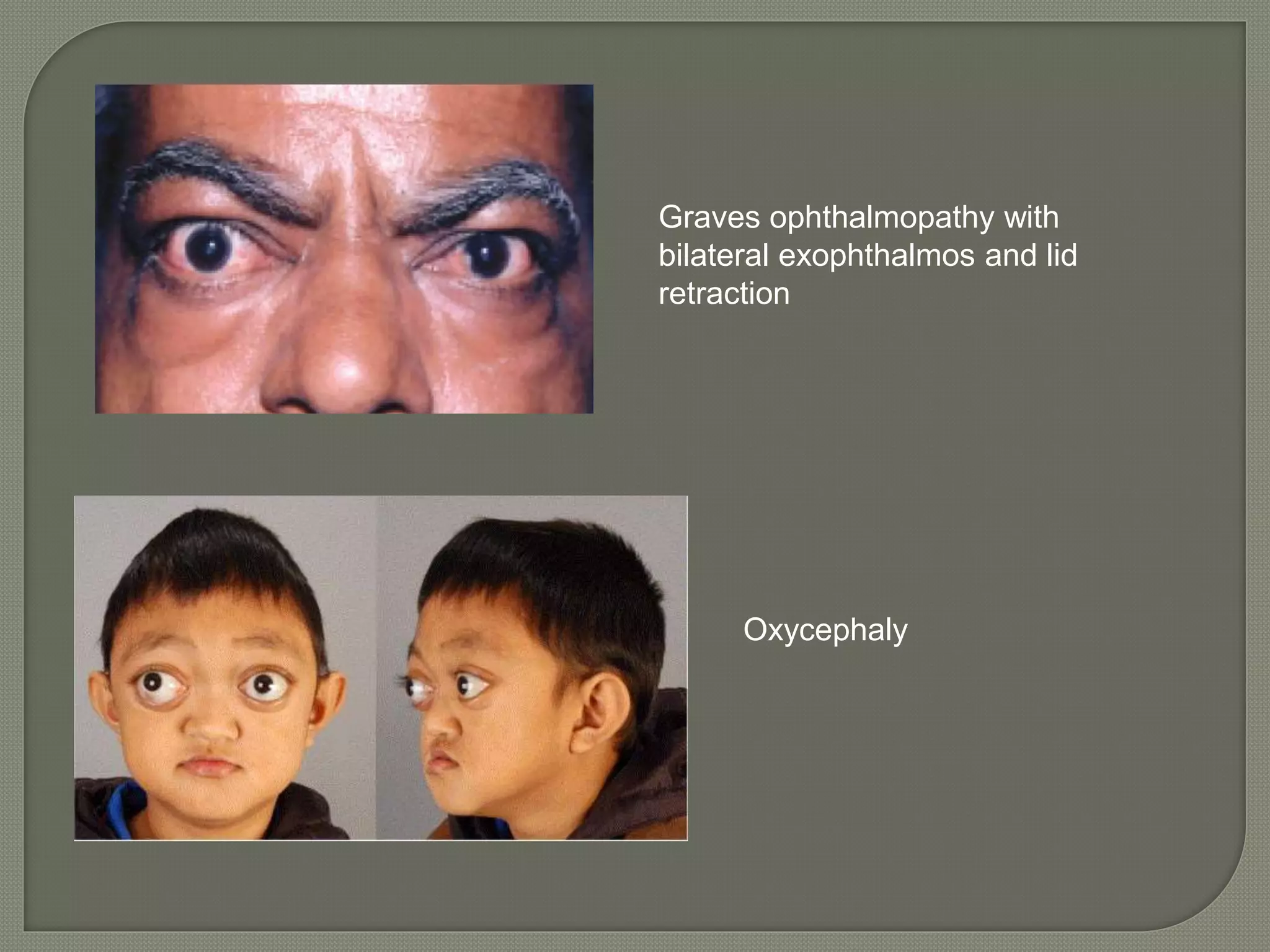
2. Forward displacement of eyeball beyond
the orbital margins.
Exophthalmos is synonymous, but is
usually used for the displacement
associated with thyroid disease.
11. Develops with extremely sudden onset
Common causes are
• Orbital emphysema
• Fracture of medial orbital wall
• Orbital hemorrhage
• Rupture of ethmoidal mucocele
12. Appears and disappears on its own.
Common causes are
• Periodic orbital oedema
• Orbital varix
• Reccurent orbital haemorrhage
• Highly vascular tumours
13. Causes:
• Pulsating vascular lesions like Caroticocavernous
fistula and saccular aneurysms of ophthalmic
artery.
• Also by transmitted cerebral pulsations
associated with deficiency of orbital roof. These
include congenital meningocele or
meningoencephalocele, neurofibromatosis,
traumatic or operative hiatus
14. One eye may look to be larger than the
other eye
Causes
• Unilateral high axial myopia
• Unilateral buphthalmos
• Pseudocornea or anterior staphyloma
• Retraction of eyelid of one eye
• Enophthalmos of opposite eye
16. Local Examination
Inspection - to diff proptosis and
pseudoproptosis, if proptosis is unilateral or
bilateral, to note the shape of the skull
Palpation – swellings, regional lymph nodes,
orbital rim
Auscultation – to know abnormal vascular
communications like caroticocavernous fistula
Transillumination
Visual acuity – may decrease due to pressure
in the back of the eyeball, optic nerve
compression, exposure keratopathy
17. Pupil reactions
Fundoscopy – venous engorgement,
hemorrage, disc edema
Occular motility – decreased in thyroid
ophthalmopathy
Exophthalmometry –
• Measures the protrusion of the apex of the cornea
from the from outer orbital margin
• Normal – 10 to 21 mm, symmetrical in both eyes
• Difference of more than 2 mm is significant
19. Systemic Examination
Laboratory investigations
• thyroid function test
• haematological studies
• Casoni’s test
• Stool examination for ova and cysts
• Urine analysis for bence jones proteins
Imaging techniques
• Non-invasive – Plain X rays, Compted
tomography scanning, Ultrasonography, MRI
• Invasive – Not done