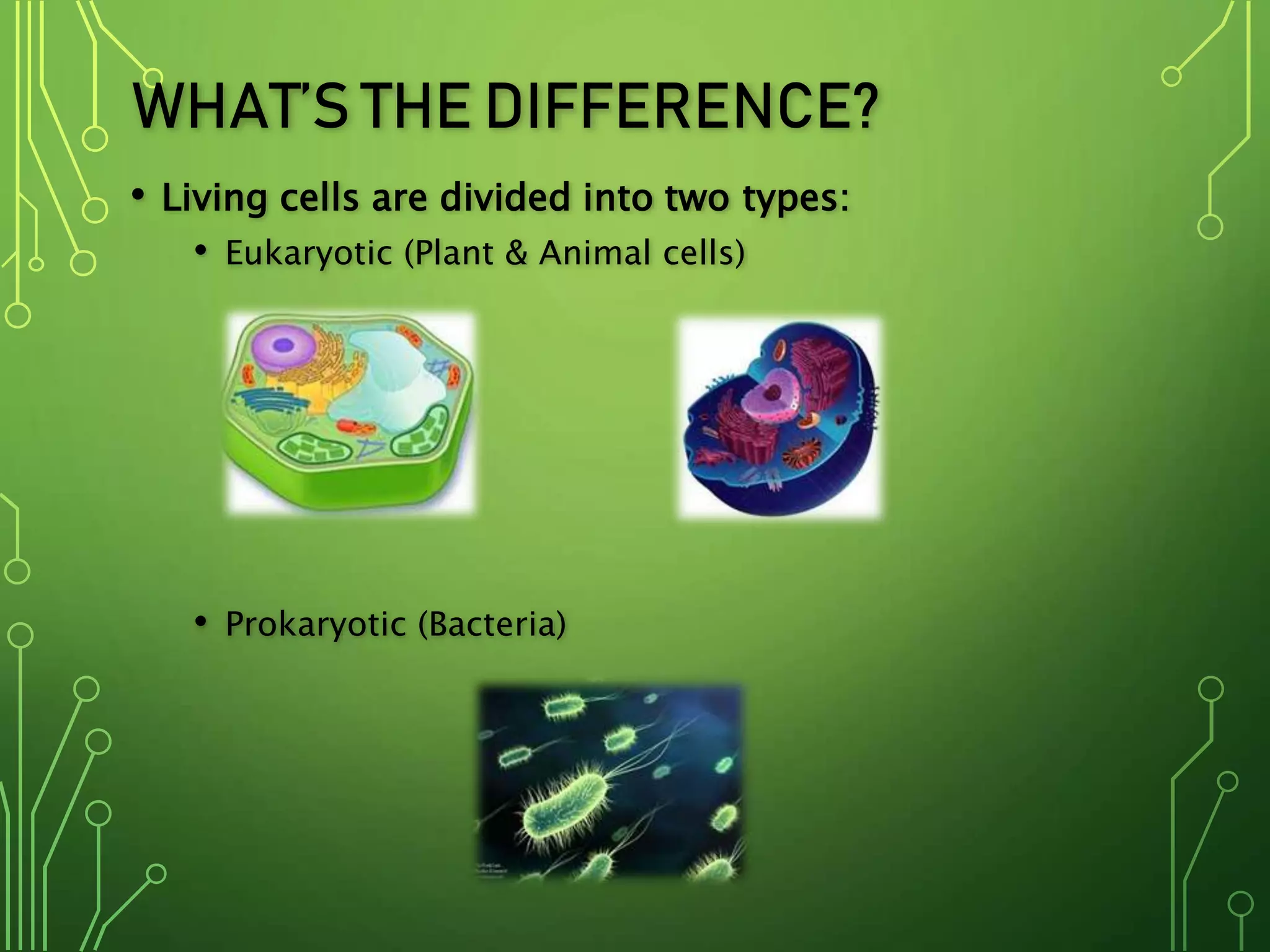
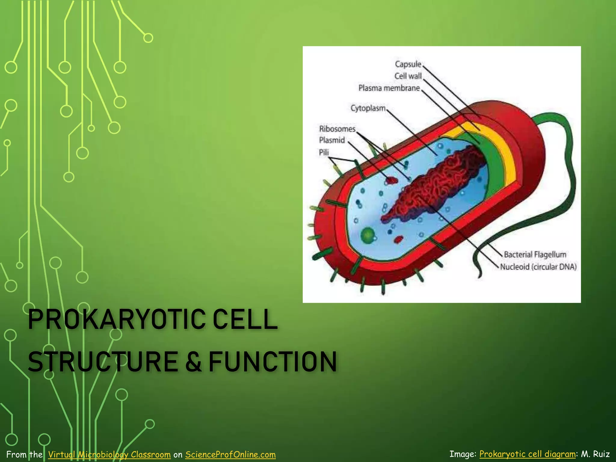
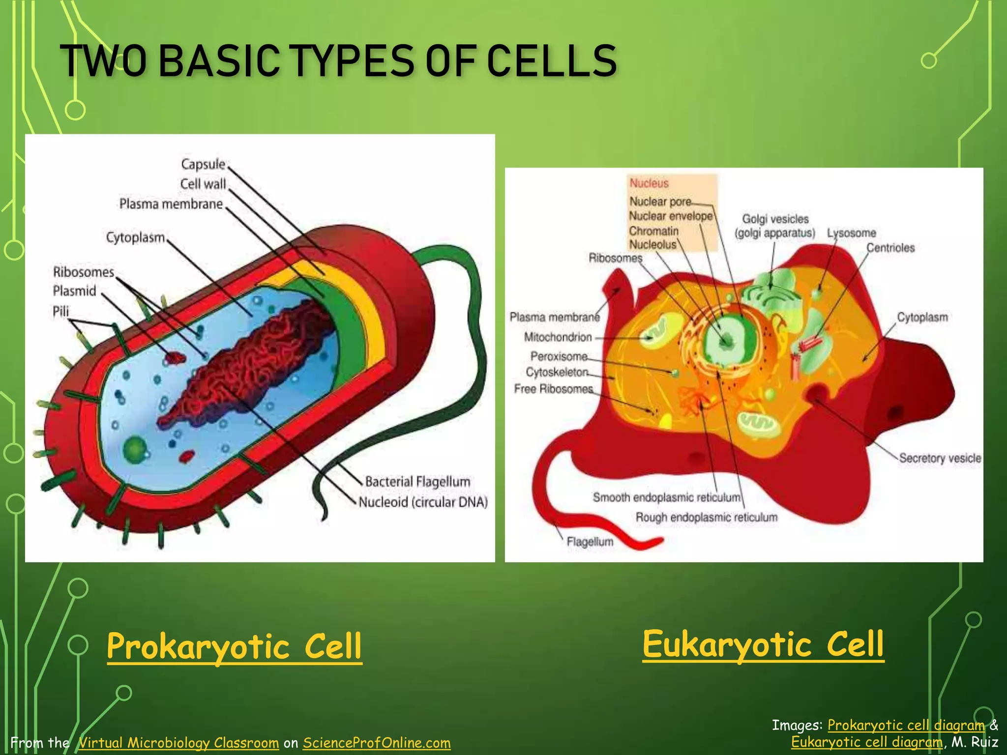
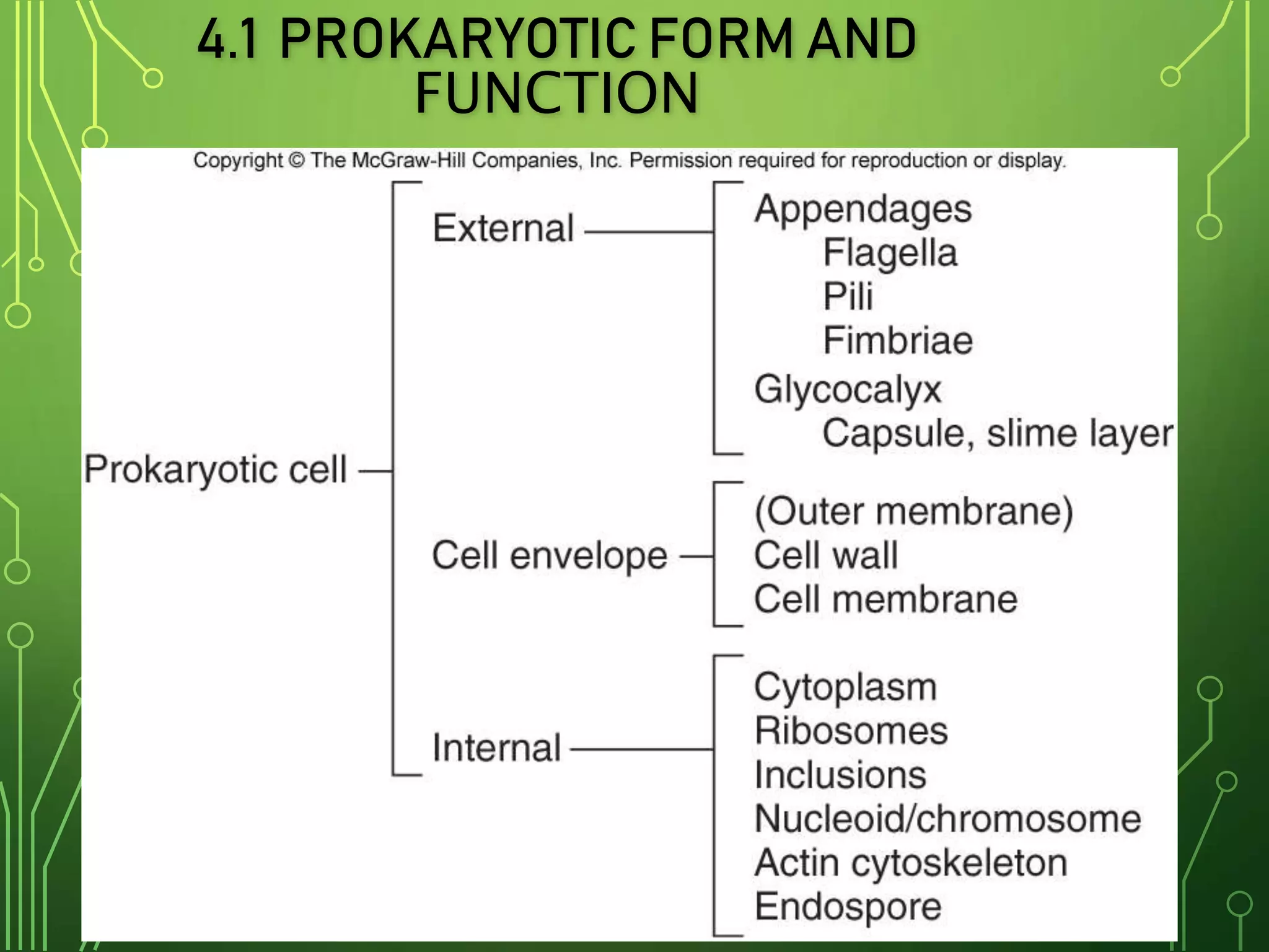
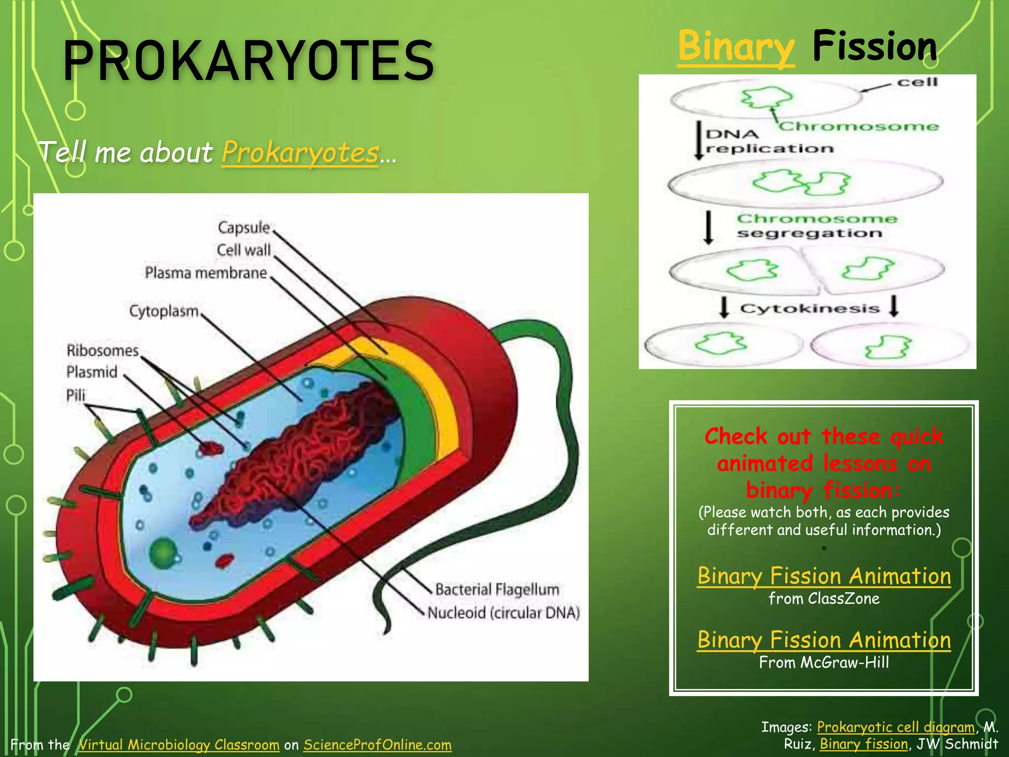
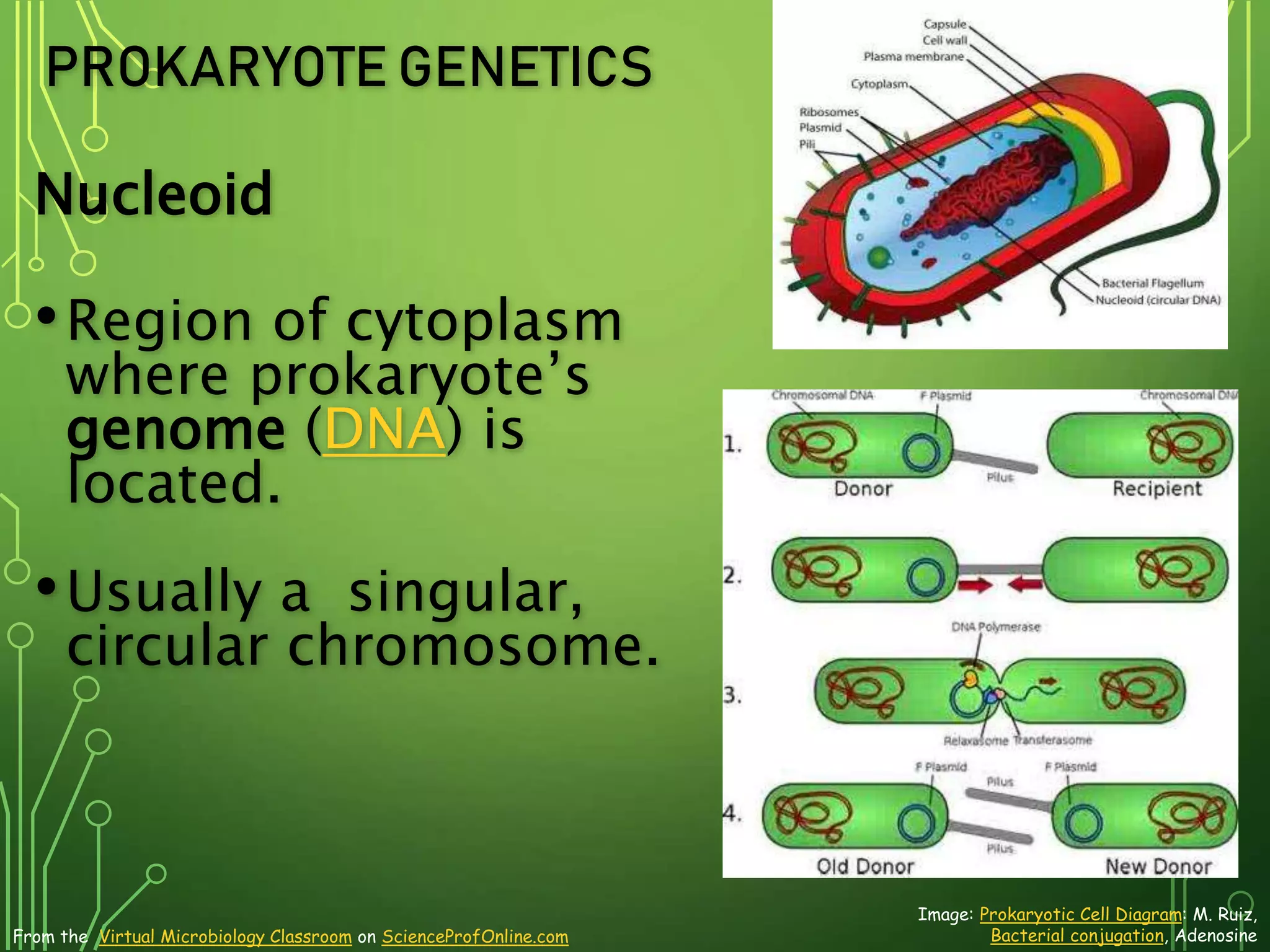
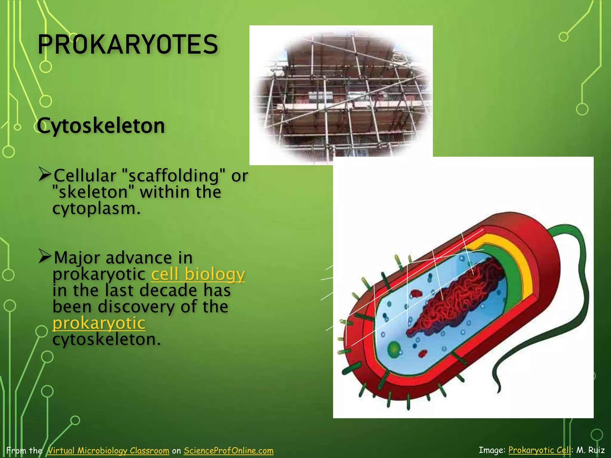
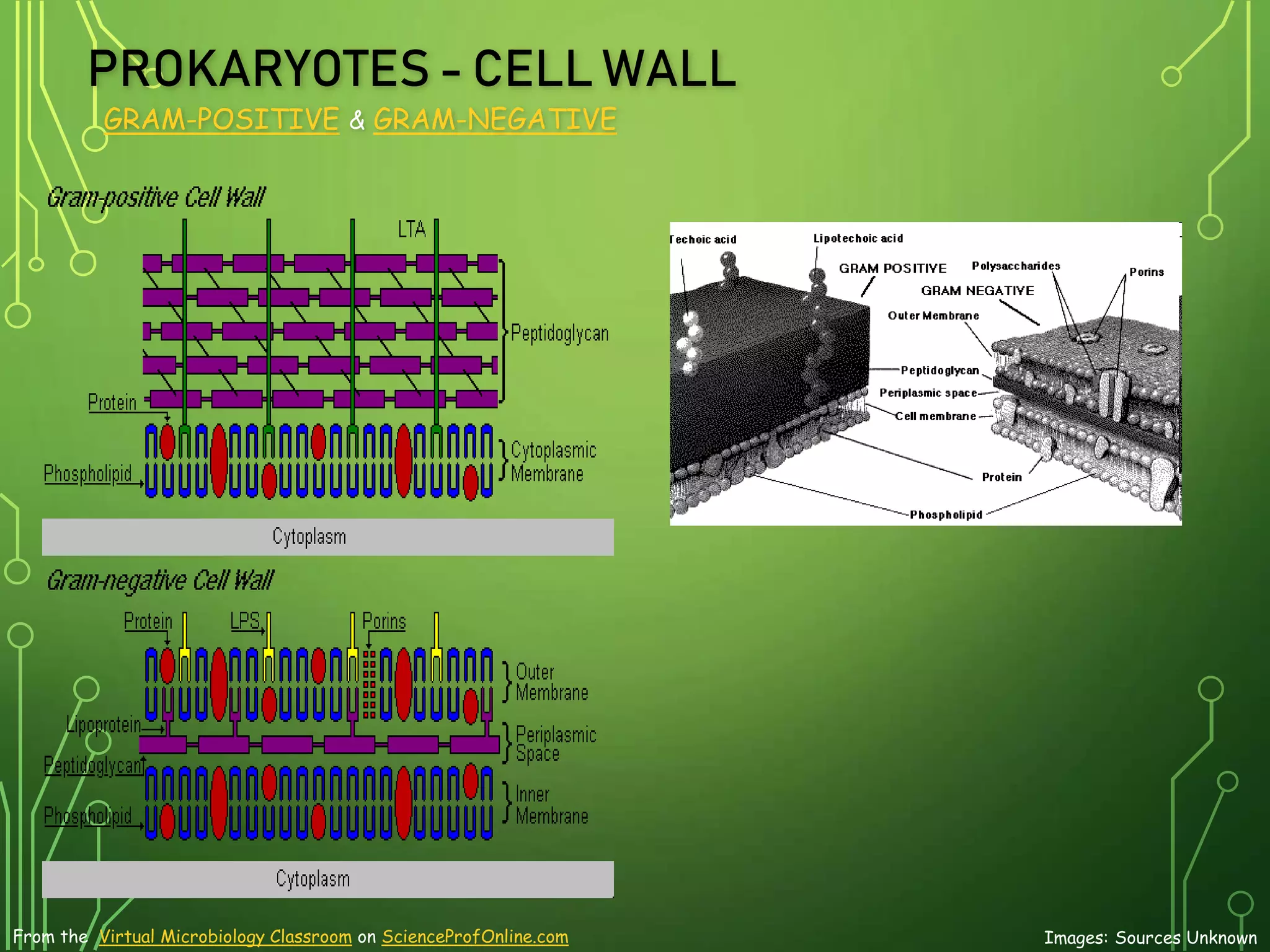
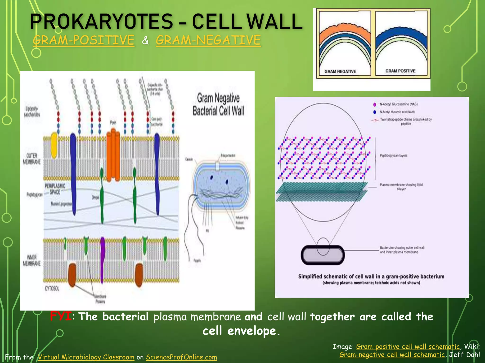
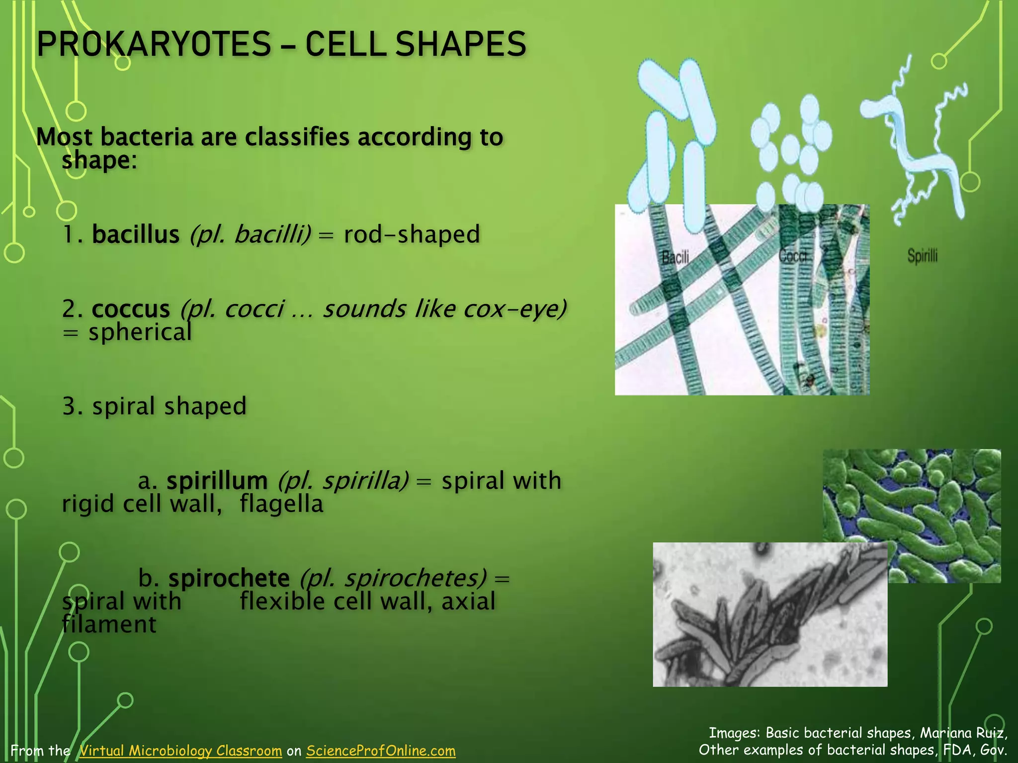
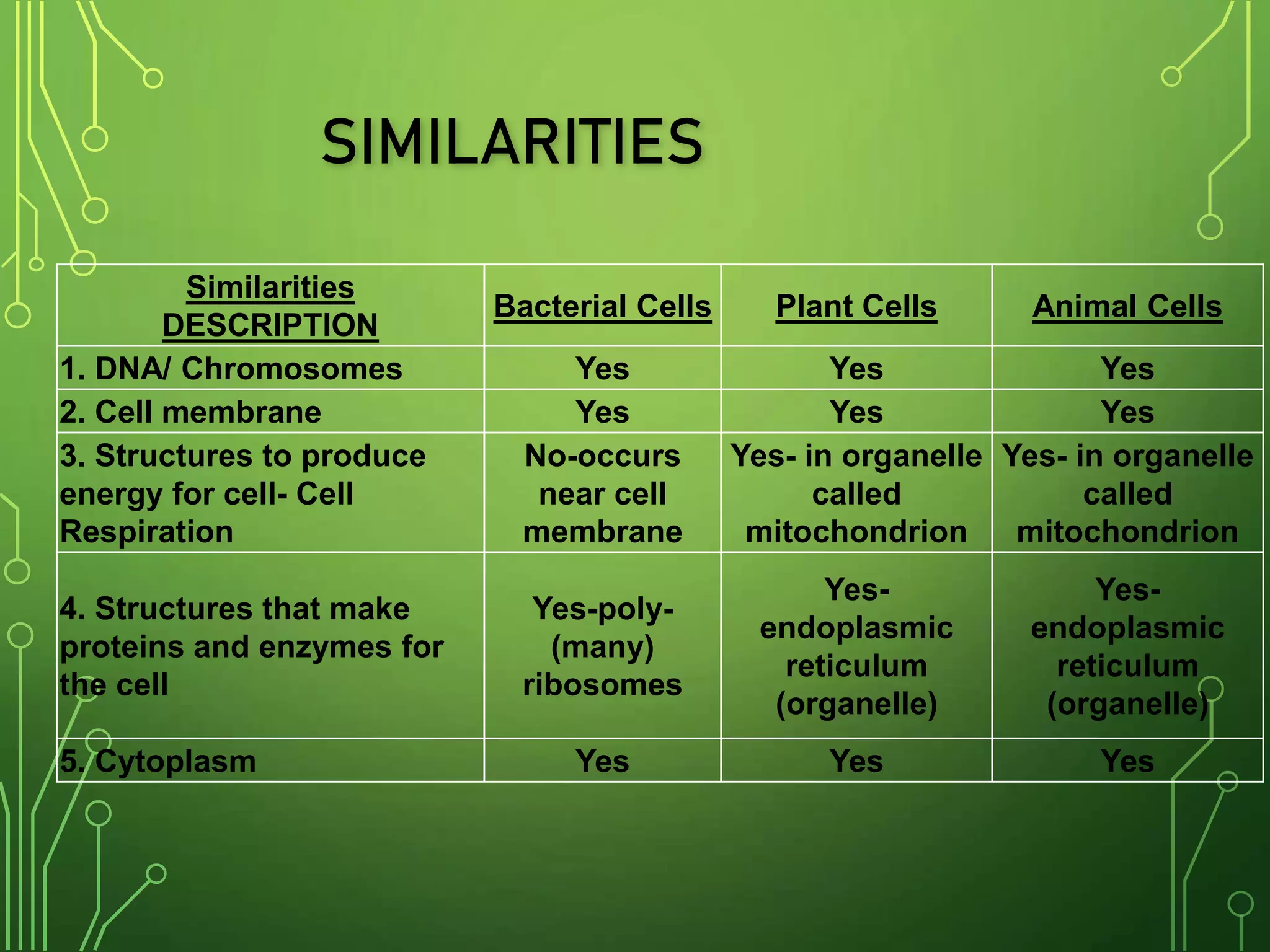
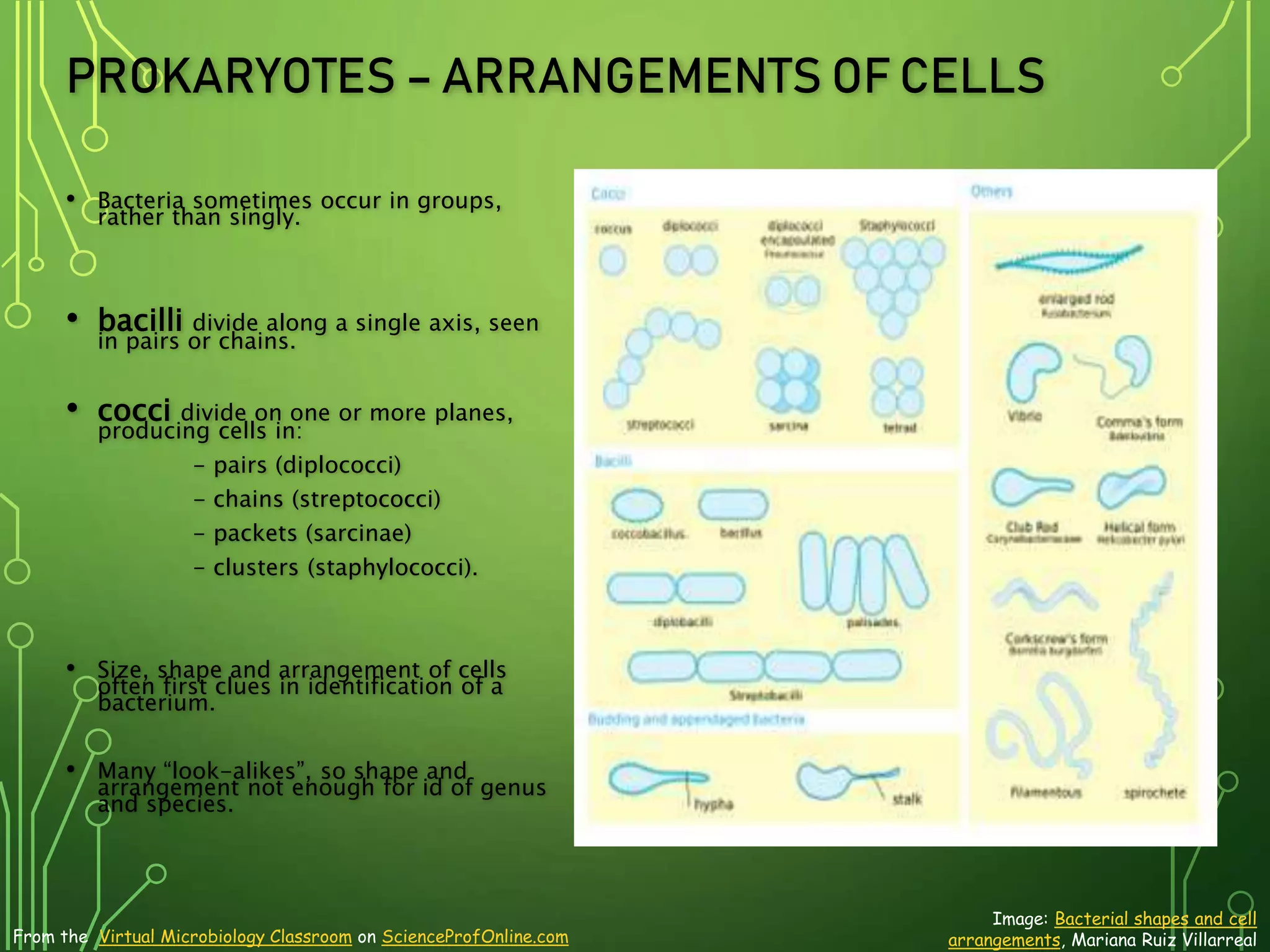
About Science Prof Online
PowerPoint Resources
• Science Prof Online (SPO) is a free science education website that provides fully-developed Virtual Science Classrooms,
science-related PowerPoints, articles and images. The site is designed to be a helpful resource for students, educators, and
anyone interested in learning about science.
• The SPO Virtual Classrooms offer many educational resources, including practice test questions, review questions, lecture
PowerPoints, video tutorials, sample assignments and course syllabi. New materials are continually being developed, so check
back frequently, or follow us on Facebook (Science Prof Online) or Twitter (ScienceProfSPO) for updates.
• Many SPO PowerPoints are available in a variety of formats, such as fully editable PowerPoint files (.ppt), as well as
uneditable versions in smaller file sizes, such as PowerPoint Shows (.pps) and Portable Document Format (.pdf), for ease of
printing. The font “Jokerman” is used frequently in titles. It has a microbiology feel to it. If you do not have this font, some
titles may appear odd, oversized and off-center. Find free downloads of Jokerman by Googling “download jokerman font
microsoft”.
• Images used on this resource, and on the SPO website are, wherever possible, credited and linked to their source. Any
words underlined and appearing in blue are links that can be clicked on for more information. PPT files must be viewed in
slide show mode to use the hyperlinks directly.
• Several helpful links to fun and interactive learning tools are included throughout the PPT and on the Smart Links slide,
near the end of each presentation. You must be in slide show mode to utilize hyperlinks and animations.
•This digital resource is licensed under Creative Commons Attribution-ShareAlike 3.0:
http://creativecommons.org/licenses/by-sa/3.0/
Alicia Cepaitis, MS
Chief Creative Nerd
Science Prof Online
Online Education Resources, LLC
alicia@scienceprofonline.com
From the Virtual Microbiology Classroom on ScienceProfOnline.com Image: Compound microscope objectives, T. Port
Tami Port, MS
Creator of Science Prof Online
Chief Executive Nerd
Science Prof Online
Online Education Resources, LLC
info@scienceprofonline.com
WHAT’S THE DIFFERENCE?
• Living cells are divided into two types:
• Eukaryotic (Plant & Animal cells)
• Prokaryotic (Bacteria)
PROKARYOTIC CELL
STRUCTURE & FUNCTION
Image: Prokaryotic cell diagram: M. Ruiz
From the Virtual Microbiology Classroom on ScienceProfOnline.com
TWO BASIC TYPES OF CELLS
Eukaryotic Cell
Prokaryotic Cell
Images: Prokaryotic cell diagram &
Eukaryotic cell diagram, M. Ruiz
From the Virtual Microbiology Classroom on ScienceProfOnline.com
SIZE OF LIVING THINGS
1 m = 100 cm = 1,000mm = 1,000,000 µm = 1,000,000,000nm
1mm = 1000 µm = 1000000nm
1 µm = 1000nm
Click link for an interactive Size of
Microscopic Things” animation on
Cells Alive.
From the Virtual Microbiology Classroom on ScienceProfOnline.com
HOW ARE PROKARYOTES DIFFERENT
FROM EUKARYOTES?
• The way their DNA is packaged
•No nucleus
•Not wrapped around histones
•The makeup of their cell wall
•Bacteria- peptidoglycan
•Archae- tough and made of other
chemicals, distinct to them
•Their internal structures
•No complex, membrane-bound
organelles
4.1 PROKARYOTIC FORM AND
FUNCTION
PROKARYOTES
Tell me about Prokaryotes…
Binary Fission
Images: Prokaryotic cell diagram, M.
Ruiz, Binary fission, JW Schmidt
From the Virtual Microbiology Classroom on ScienceProfOnline.com
Check out these quick
animated lessons on
binary fission:
(Please watch both, as each provides
different and useful information.)
Binary Fission Animation
from ClassZone
Binary Fission Animation
From McGraw-Hill
PROKARYOTE GENETICS
Nucleoid
•Region of cytoplasm
where prokaryote’s
genome (DNA) is
located.
•Usually a singular,
circular chromosome.
Image: Prokaryotic Cell Diagram: M. Ruiz,
Bacterial conjugation, Adenosine
From the Virtual Microbiology Classroom on ScienceProfOnline.com
Plasmid
• Small extra piece of chromosome/genetic
material.
• 5 - 100 genes
• Not critical to everyday functions.
• Can provide genetic information to promote:
- Antibiotic resistance
- Virulence factors
(molecules produced by pathogen that
specifically influence host's function to allow
the pathogen to thrive)
- Promote conjugation
(transfer of genetic material
between bacteria through cell- to-
cell contact)
PROKARYOTES
Cytoplasm
• Also known as proto-plasm.
• Gel-like matrix of water,
enzymes, nutrients, wastes,
and gases and
contains cell structures.
• this is the gel-like fluid that
the cell is filled with, inside
the plasma membrane;
liquid with all of the cellular
organelles suspended
within.
•
Image: Prokaryotic cell diagram: M.
Ruiz, Granules, Source Unknown From the Virtual Microbiology Classroom on ScienceProfOnline.com
•Location of growth, metabolism,
and replication.
Granules
•Bacteria’s way of storing nutrients.
•Staining of some granules aids in
identification
PROKARYOTES
Cytoskeleton
Cellular "scaffolding" or
"skeleton" within the
cytoplasm.
Major advance in
prokaryotic cell biology
in the last decade has
been discovery of the
prokaryotic
cytoskeleton.
Image: Prokaryotic Cell: M. Ruiz
From the Virtual Microbiology Classroom on ScienceProfOnline.com
•Up until recently, thought to be a
feature only of eukaryotic cells.
PROKARYOTES
Ribosomes
Found within cytoplasm or
attached to plasma
membrane.
Made of protein & rRNA.
Composed of two subunits.
Cell may contain thousands
.
Animation: Ribosome translating
protein,Xvazquez; Ribosome Structure, Vossman
From the Virtual Microbiology Classroom on ScienceProfOnline.com
Click here for
animation of
ribosome building a
protein.
q: what do ribosomes do?
q: what’s the relationship
between the job that the
ribosomes do and the genetic
instructions (nucleic acids) of
the cell?
PROKARYOTES
PLASMA MEMBRANE
Separates cell
from its
environment.
Phospholipid
molecules
oriented so that
hydrophilic water-
loving heads
directed outward
and hydrophobic
water-hating tails
directed inward.
Image: Cell Membrane diagram, Dhatfield
From the Virtual Microbiology Classroom on ScienceProfOnline.com
PROTEINS EMBEDDED IN TWO
LAYERS OF LIPIDS (LIPID
BILAYER).
MEMBRANE IS SEMI-PERMEABLE.
Q: WHAT DOES THAT MEAN?
From the peptidoglycan inwards all bacteria are very similar.
Going further out, the bacterial world divides into two major
classes (plus a couple of odd types). These are:
Gram-positive Gram-negative
Images: Staph, Gram Stain, SPO Microbiology
Images, T. Port; E coli, Y tambe
From the Virtual Microbiology Classroom on ScienceProfOnline.com
Prokaryotes - Cell Wall
Peptidoglycan is a huge polymer of
interlocking chains of alternating
monomers.
Provides rigid support while freely
permeable to solutes.
.
Bacterial Cell Wall
Image: Bonding structure peptidoglycan,
Mouagip; Other Image Source Unknown
From the Virtual Microbiology Classroom on ScienceProfOnline.com
Backbone of peptidoglycan molecule
composed of two amino sugar
derivatives of glucose. The “glycan”
part of peptidoglycan:
•- N-acetylglucosamine (NAG)
•- N-acetlymuramic acid (NAM)
NAG / NAM strands are
• connected by interlocking
• peptide bridges.
• The “peptid” part
PROKARYOTES - CELL WALL
GRAM-POSITIVE & GRAM-NEGATIVE
Images: Sources Unknown
From the Virtual Microbiology Classroom on ScienceProfOnline.com
PROKARYOTES - CELL WALL
GRAM-POSITIVE & GRAM-NEGATIVE
Image: Gram-positive cell wall schematic, Wiki;
Gram-negative cell wall schematic, Jeff Dahl
From the Virtual Microbiology Classroom on ScienceProfOnline.com
FYI: The bacterial plasma membrane and cell wall together are called the
cell envelope.
Q: WHY ARE THESE DIFFERENCES IN BACTERIAL CELL
WALL STRUCTURE SO IMPORTANT?
From the Virtual Microbiology Classroom on ScienceProfOnline.com
Image: Lipopolysaccharide, Wiki; Gram+ and Gram- cell wall diagram,
source unknown; ; Gram-negative cell wall schematic, Jeff Dahl
PROKARYOTES -
GLYCOCALYX
Some bacteria have an additional layer outside
of the cell wall called the glycocalyx.
This additional layer can come in
one of two forms:
1. Slime Layer
- Glycoproteins loosely associated with the cell
wall.
- Slime layer causes bacteria to adhere to solid
surfaces and helps prevent the cell from drying
out.
- Streptococcus
The slime layer of Gram+ Streptococcus mutans
allows it to accumulate on tooth enamel (yuck mouth
and one of the causes of cavities).
Other bacteria in the mouth become trapped in the
slime and form a biofilm & eventually a buildup of
plaque.
Images: Slime layer, Encyclopedia Britannica; Biofilm, PHIL # 11706; Dirty
toilet, T. Port Sweat on face; Staphylococcus & Mannitol Salt agar, T. Port
From the Virtual Microbiology Classroom on ScienceProfOnline.com
Mannitol
Salt
PROKARYOTES - GLYCOCALYX
2. Capsule
• Polysaccharides firmly attached to the
cell wall.
• Capsules adhere to solid surfaces and
to nutrients in the environment.
• Adhesive power of capsules is a major
factor in the initiation of some bacterial
diseases.
• Capsule also protect bacteria from
being phagocytized by cells of the
hosts immune system.
Image: Prokaryotic Cell Diagram: M. Ruiz, Other Images Unknown Source
From the Virtual Microbiology Classroom on ScienceProfOnline.com
PROKARYOTES - ENDOSPORES
Dormant, tough, non-reproductive structure
produced by small number of bacteria.
Resistant to radiation, desiccation,
lysozyme, temperature, starvation, and
chemical disinfectants.
Endospores are commonly found in soil and
water, where they may survive for very long
periods of time.
Q: How and why do endospores form? Watch
the animated lesson “Bacterial Spore
Formation” to find out. Link also provides quiz
questions to test your understanding of the
material.
Q: What are the two endospore producing
bacterial genera that were introduced in our
History of Microbiology lecture?
Image: Bacillus subtilis, SPO Science Image Library, Endospore
stain from Dr. Ronald E. Hurlbert, Microbiology 101 lab manual
From the Virtual Microbiology Classroom on ScienceProfOnline.com
An endospore stained bacterial smear of
Bacillus subtilis showing endospores as
green and vegetative cells as red.
GRAM-POSITIVE
Obligate or facultative anaerobes,
endospore producers
bacillus-shaped
Q: Which two groups of bacteria produce
endospores?
Common in soil. Only a few species cause disease
in humans.
Extremely diverse group of bacteria, includes:
- causative agent of anthrax (Bacillus
anthracis)
- species that synthesize important
antibiotics, and enzymes for detergents.
Due to extreme tolerance to both heat and
disinfectants, used to test heat sterilization
techniques and chemical disinfectants.
Bacterial Genus: __________
Images: Bacullus subtilis, T. Port: B. anthracis,
Gram stained, CDC; Endospore stain, Tami Port
This is our lab
friend Bacillus
subtilis.
Gram Stain
Endospore
Stain
From the Virtual Microbiology Classroom on ScienceProfOnline.com
PROKARYOTES – SURFACE APPENDAGES
Some prokaryotes have distinct
appendages that allow them to
move about or adhere to solid
surfaces.
Consist of delicate stands of
proteins.
flagella: Long, thin extensions
that allow some bacteria to move
about freely in aqueous
environments. (singular:
flagellum)
axial filament (endoflagella):
Wind around bacteria, causing
movement in waves.
Images: Helicobacter pylori ; Axial filament, Source unknown
From the Virtual Microbiology Classroom on ScienceProfOnline.com
PROKARYOTES – SURFACE APPENDAGES
fimbriae: Most Gram-negative
bacteria have these short, fine
appendages surrounding the cell.
Gram+ bacteria don’t have.
No role in motility. Help bacteria
adhere to solid surfaces. Major
factor in virulence.
(singular: fimbria)
Images: E. coli fimbriae, Manu Forero; Bacterial conjugation, Adenosine
From the Virtual Microbiology Classroom on ScienceProfOnline.com
Pili: Tubes that are longer than
fimbriae, usually shorter than
flagella.
Use for movement, like
grappling
hooks, and also use conjugation
pili
to transfer plasmids. (singular =
pilus)
MEET THE MICROBE! NEISSERIA AND ITS FIMBIRIAE
Image: Neisseria photo, Textbook of Bacteriology, Gram
stain of Neisseria gonorrhoeae, Souce PHIL #3798
From the Virtual Microbiology Classroom on ScienceProfOnline.com
• Gram- diplococci, resemble coffee beans when
viewed microscopically.
• Neisseria gonorrhoeae causes sexually
transmitted disease gonorrhoeae.
• Antibiotics applied to the eyes of neonates as a
preventive measure against gonorrhoea.
• One of the most communicable disease in the U.S.
• 125 cases per 100,000. Teens 15-19 yo 634 cases per
100,000. Young adults 20-25 460 per 100,000.
• N. meningitidis most common causes of bacterial meningitis
in young adults.
Q: What makes Neisseria so tough?
• Lipopolysaccharide (LPS) of the cell wall of Neisseria acts as an
endotoxin.
• Polysaccharide capsule prevents host phagocytosis and aids in
evasion of the host immune response.
• Use fimbriae to attach onto host cells; avirulent without.
Fimbriae have adhesion proteins (adhesins) on their tips that
match, lock and key, with proteins on host epithelial cell
surface.
Figure 4.22
PROKARYOTES – CELL SHAPES
Most bacteria are classifies according to
shape:
1. bacillus (pl. bacilli) = rod-shaped
2. coccus (pl. cocci … sounds like cox-eye)
= spherical
3. spiral shaped
a. spirillum (pl. spirilla) = spiral with
rigid cell wall, flagella
b. spirochete (pl. spirochetes) =
spiral with flexible cell wall, axial
filament
Images: Basic bacterial shapes, Mariana Ruiz,
Other examples of bacterial shapes, FDA, Gov.
From the Virtual Microbiology Classroom on ScienceProfOnline.com
There are many more shapes beyond
these basic ones. A few examples:
• Coccobacilli = elongated coccal form
• Filamentous = bacilli that occur in
long threads
• Vibrios = short, slightly curved rods
• Fusiform = bacilli with tapered ends
PROKARYOTES – ARRANGEMENTS OF CELLS
• Bacteria sometimes occur in groups,
rather than singly.
• bacilli divide along a single axis, seen
in pairs or chains.
• cocci divide on one or more planes,
producing cells in:
- pairs (diplococci)
- chains (streptococci)
- packets (sarcinae)
- clusters (staphylococci).
• Size, shape and arrangement of cells
often first clues in identification of a
bacterium.
• Many “look-alikes”, so shape and
arrangement not enough for id of genus
and species.
Image: Bacterial shapes and cell
arrangements, Mariana Ruiz Villarreal
From the Virtual Microbiology Classroom on ScienceProfOnline.com
Similarities
DESCRIPTION
Bacterial Cells Plant Cells Animal Cells
1. DNA/ Chromosomes Yes Yes Yes
2. Cell membrane Yes Yes Yes
3. Structures to produce
energy for cell- Cell
Respiration
No-occurs
near cell
membrane
Yes- in organelle
called
mitochondrion
Yes- in organelle
called
mitochondrion
4. Structures that make
proteins and enzymes for
the cell
Yes-poly-
(many)
ribosomes
Yes-
endoplasmic
reticulum
(organelle)
Yes-
endoplasmic
reticulum
(organelle)
5. Cytoplasm Yes Yes Yes
SIMILARITIES
PROKARYOTES – ARRANGEMENTS OF CELLS
• Bacteria sometimes occur in groups,
rather than singly.
• bacilli divide along a single axis, seen
in pairs or chains.
• cocci divide on one or more planes,
producing cells in:
- pairs (diplococci)
- chains (streptococci)
- packets (sarcinae)
- clusters (staphylococci).
• Size, shape and arrangement of cells
often first clues in identification of a
bacterium.
• Many “look-alikes”, so shape and
arrangement not enough for id of genus
and species.
Image: Bacterial shapes and cell
arrangements, Mariana Ruiz Villarreal
From the Virtual Microbiology Classroom on ScienceProfOnline.com
Differences
DESCRIPTION
Bacterial Cells
Plant
Cells
Animal
Cells
1. Cell Wall Yes Yes No
2. Nucleus-nuclear membrane No Yes Yes
3. Fimbria-DNA transfer Some No No
4. Vacuoles No Yes Yes
5. Chloroplasts (organelles) (for
photosynthesis)
Yes-blue-green
bacteria has a
green pigment
that makes its
own food.
Yes No
6. Flagella-mobility Yes No No
7. Capsule Some No No
DIFFERENCES
Identify Each Type of
Cell Shape
&
Arrangement
Images: A. Staph; B. E. coli, T. Port; C. Bacillus anthracis,
PHIL #2105; D. Streptococcus bacteria, PHIL #2110.
A B
C D
From the Virtual Microbiology Classroom on ScienceProfOnline.com
REVIEW!
Here’s an excellent interactive lesson on
Prokaryote Cell Structure
From the Virtual Microbiology Classroom on ScienceProfOnline.com Images: Prokaryotic cell diagram, M. Ruiz
Confused?
Here are links to fun resources that further explain aerobic
respiration:
• Cell Structure: Prokaryotes Main Page on the Virtual Micrboiology
Classroom of Science Prof Online.
• Prokaryotic Cell: Structures, Functions & Diagrams, an article from SPO.
• Prokaryotic & Eukaryotic: Two Types of Biological Cells, an article from SPO.
• “Got the Time” music video by Anthrax.
• Prokaryotic Cell interactive diagram from Cells Alive website.
• “How big is a…” interactive diagram from Cells Alive website.
• Cell Structure tutorials and quizzes from Interactive Concepts in Biochemistry.
• How Osmosis Works, animation from McGraw-Hill.
• “Germs”. Music by Weird Al Yankovic. Video by RevLucio.
• Bacterial Pathogen Pronunciation Station, a webpage with links to audio
files containing the pronunciation of the bacterial names, created by Neal R.
Chamberlain, Ph.D.
• Biology4Kids – Cell Biology Main Page by Raders.
(You must be in PPT slideshow view to click on links.)
From the Virtual Microbiology Classroom on ScienceProfOnline.com
ARE MICROBES INTIMIDATING YOU?
DO YOURSELF A FAVOR. USE THE…
VIRTUAL
MICROBIOLOGY
CLASSROOM (VMC) !
THE VMC IS FULL OF RESOURCES TO HELP YOU
SUCCEED, INCLUDING:
• PRACTICE TEST QUESTIONS
• REVIEW QUESTIONS
• STUDY GUIDES AND LEARNING OBJECTIVES
You can access the VMC by going to the Science Prof Online website
www.ScienceProfOnline.com
Images: Clostridium difficile, Giant Microbes; Prokaryotic cell, Mariana Ruiz

Prokaryotic-Cell-Structure-Function-Biology-Lecture-PowerPoint-VMCct.pptx

  • 1.
    About Science ProfOnline PowerPoint Resources • Science Prof Online (SPO) is a free science education website that provides fully-developed Virtual Science Classrooms, science-related PowerPoints, articles and images. The site is designed to be a helpful resource for students, educators, and anyone interested in learning about science. • The SPO Virtual Classrooms offer many educational resources, including practice test questions, review questions, lecture PowerPoints, video tutorials, sample assignments and course syllabi. New materials are continually being developed, so check back frequently, or follow us on Facebook (Science Prof Online) or Twitter (ScienceProfSPO) for updates. • Many SPO PowerPoints are available in a variety of formats, such as fully editable PowerPoint files (.ppt), as well as uneditable versions in smaller file sizes, such as PowerPoint Shows (.pps) and Portable Document Format (.pdf), for ease of printing. The font “Jokerman” is used frequently in titles. It has a microbiology feel to it. If you do not have this font, some titles may appear odd, oversized and off-center. Find free downloads of Jokerman by Googling “download jokerman font microsoft”. • Images used on this resource, and on the SPO website are, wherever possible, credited and linked to their source. Any words underlined and appearing in blue are links that can be clicked on for more information. PPT files must be viewed in slide show mode to use the hyperlinks directly. • Several helpful links to fun and interactive learning tools are included throughout the PPT and on the Smart Links slide, near the end of each presentation. You must be in slide show mode to utilize hyperlinks and animations. •This digital resource is licensed under Creative Commons Attribution-ShareAlike 3.0: http://creativecommons.org/licenses/by-sa/3.0/ Alicia Cepaitis, MS Chief Creative Nerd Science Prof Online Online Education Resources, LLC alicia@scienceprofonline.com From the Virtual Microbiology Classroom on ScienceProfOnline.com Image: Compound microscope objectives, T. Port Tami Port, MS Creator of Science Prof Online Chief Executive Nerd Science Prof Online Online Education Resources, LLC info@scienceprofonline.com
  • 2.
    WHAT’S THE DIFFERENCE? •Living cells are divided into two types: • Eukaryotic (Plant & Animal cells) • Prokaryotic (Bacteria)
  • 3.
    PROKARYOTIC CELL STRUCTURE &FUNCTION Image: Prokaryotic cell diagram: M. Ruiz From the Virtual Microbiology Classroom on ScienceProfOnline.com
  • 4.
    TWO BASIC TYPESOF CELLS Eukaryotic Cell Prokaryotic Cell Images: Prokaryotic cell diagram & Eukaryotic cell diagram, M. Ruiz From the Virtual Microbiology Classroom on ScienceProfOnline.com
  • 5.
    SIZE OF LIVINGTHINGS 1 m = 100 cm = 1,000mm = 1,000,000 µm = 1,000,000,000nm 1mm = 1000 µm = 1000000nm 1 µm = 1000nm Click link for an interactive Size of Microscopic Things” animation on Cells Alive. From the Virtual Microbiology Classroom on ScienceProfOnline.com
  • 6.
    HOW ARE PROKARYOTESDIFFERENT FROM EUKARYOTES? • The way their DNA is packaged •No nucleus •Not wrapped around histones •The makeup of their cell wall •Bacteria- peptidoglycan •Archae- tough and made of other chemicals, distinct to them •Their internal structures •No complex, membrane-bound organelles
  • 7.
  • 8.
    PROKARYOTES Tell me aboutProkaryotes… Binary Fission Images: Prokaryotic cell diagram, M. Ruiz, Binary fission, JW Schmidt From the Virtual Microbiology Classroom on ScienceProfOnline.com Check out these quick animated lessons on binary fission: (Please watch both, as each provides different and useful information.) Binary Fission Animation from ClassZone Binary Fission Animation From McGraw-Hill
  • 9.
    PROKARYOTE GENETICS Nucleoid •Region ofcytoplasm where prokaryote’s genome (DNA) is located. •Usually a singular, circular chromosome. Image: Prokaryotic Cell Diagram: M. Ruiz, Bacterial conjugation, Adenosine From the Virtual Microbiology Classroom on ScienceProfOnline.com
  • 10.
    Plasmid • Small extrapiece of chromosome/genetic material. • 5 - 100 genes • Not critical to everyday functions. • Can provide genetic information to promote: - Antibiotic resistance - Virulence factors (molecules produced by pathogen that specifically influence host's function to allow the pathogen to thrive) - Promote conjugation (transfer of genetic material between bacteria through cell- to- cell contact)
  • 11.
    PROKARYOTES Cytoplasm • Also knownas proto-plasm. • Gel-like matrix of water, enzymes, nutrients, wastes, and gases and contains cell structures. • this is the gel-like fluid that the cell is filled with, inside the plasma membrane; liquid with all of the cellular organelles suspended within. • Image: Prokaryotic cell diagram: M. Ruiz, Granules, Source Unknown From the Virtual Microbiology Classroom on ScienceProfOnline.com
  • 12.
    •Location of growth,metabolism, and replication. Granules •Bacteria’s way of storing nutrients. •Staining of some granules aids in identification
  • 13.
    PROKARYOTES Cytoskeleton Cellular "scaffolding" or "skeleton"within the cytoplasm. Major advance in prokaryotic cell biology in the last decade has been discovery of the prokaryotic cytoskeleton. Image: Prokaryotic Cell: M. Ruiz From the Virtual Microbiology Classroom on ScienceProfOnline.com
  • 14.
    •Up until recently,thought to be a feature only of eukaryotic cells.
  • 15.
    PROKARYOTES Ribosomes Found within cytoplasmor attached to plasma membrane. Made of protein & rRNA. Composed of two subunits. Cell may contain thousands . Animation: Ribosome translating protein,Xvazquez; Ribosome Structure, Vossman From the Virtual Microbiology Classroom on ScienceProfOnline.com Click here for animation of ribosome building a protein.
  • 16.
    q: what doribosomes do? q: what’s the relationship between the job that the ribosomes do and the genetic instructions (nucleic acids) of the cell?
  • 17.
    PROKARYOTES PLASMA MEMBRANE Separates cell fromits environment. Phospholipid molecules oriented so that hydrophilic water- loving heads directed outward and hydrophobic water-hating tails directed inward. Image: Cell Membrane diagram, Dhatfield From the Virtual Microbiology Classroom on ScienceProfOnline.com
  • 18.
    PROTEINS EMBEDDED INTWO LAYERS OF LIPIDS (LIPID BILAYER). MEMBRANE IS SEMI-PERMEABLE. Q: WHAT DOES THAT MEAN?
  • 19.
    From the peptidoglycaninwards all bacteria are very similar. Going further out, the bacterial world divides into two major classes (plus a couple of odd types). These are: Gram-positive Gram-negative Images: Staph, Gram Stain, SPO Microbiology Images, T. Port; E coli, Y tambe From the Virtual Microbiology Classroom on ScienceProfOnline.com Prokaryotes - Cell Wall
  • 20.
    Peptidoglycan is ahuge polymer of interlocking chains of alternating monomers. Provides rigid support while freely permeable to solutes. . Bacterial Cell Wall Image: Bonding structure peptidoglycan, Mouagip; Other Image Source Unknown From the Virtual Microbiology Classroom on ScienceProfOnline.com
  • 21.
    Backbone of peptidoglycanmolecule composed of two amino sugar derivatives of glucose. The “glycan” part of peptidoglycan: •- N-acetylglucosamine (NAG) •- N-acetlymuramic acid (NAM) NAG / NAM strands are • connected by interlocking • peptide bridges. • The “peptid” part
  • 22.
    PROKARYOTES - CELLWALL GRAM-POSITIVE & GRAM-NEGATIVE Images: Sources Unknown From the Virtual Microbiology Classroom on ScienceProfOnline.com
  • 23.
    PROKARYOTES - CELLWALL GRAM-POSITIVE & GRAM-NEGATIVE Image: Gram-positive cell wall schematic, Wiki; Gram-negative cell wall schematic, Jeff Dahl From the Virtual Microbiology Classroom on ScienceProfOnline.com FYI: The bacterial plasma membrane and cell wall together are called the cell envelope.
  • 24.
    Q: WHY ARETHESE DIFFERENCES IN BACTERIAL CELL WALL STRUCTURE SO IMPORTANT? From the Virtual Microbiology Classroom on ScienceProfOnline.com Image: Lipopolysaccharide, Wiki; Gram+ and Gram- cell wall diagram, source unknown; ; Gram-negative cell wall schematic, Jeff Dahl
  • 25.
    PROKARYOTES - GLYCOCALYX Some bacteriahave an additional layer outside of the cell wall called the glycocalyx. This additional layer can come in one of two forms: 1. Slime Layer - Glycoproteins loosely associated with the cell wall. - Slime layer causes bacteria to adhere to solid surfaces and helps prevent the cell from drying out. - Streptococcus The slime layer of Gram+ Streptococcus mutans allows it to accumulate on tooth enamel (yuck mouth and one of the causes of cavities). Other bacteria in the mouth become trapped in the slime and form a biofilm & eventually a buildup of plaque. Images: Slime layer, Encyclopedia Britannica; Biofilm, PHIL # 11706; Dirty toilet, T. Port Sweat on face; Staphylococcus & Mannitol Salt agar, T. Port From the Virtual Microbiology Classroom on ScienceProfOnline.com Mannitol Salt
  • 26.
    PROKARYOTES - GLYCOCALYX 2.Capsule • Polysaccharides firmly attached to the cell wall. • Capsules adhere to solid surfaces and to nutrients in the environment. • Adhesive power of capsules is a major factor in the initiation of some bacterial diseases. • Capsule also protect bacteria from being phagocytized by cells of the hosts immune system. Image: Prokaryotic Cell Diagram: M. Ruiz, Other Images Unknown Source From the Virtual Microbiology Classroom on ScienceProfOnline.com From the Virtual Microbiology Classroom on ScienceProfOnline.com
  • 27.
    PROKARYOTES - ENDOSPORES Dormant,tough, non-reproductive structure produced by small number of bacteria. Resistant to radiation, desiccation, lysozyme, temperature, starvation, and chemical disinfectants. Endospores are commonly found in soil and water, where they may survive for very long periods of time. Q: How and why do endospores form? Watch the animated lesson “Bacterial Spore Formation” to find out. Link also provides quiz questions to test your understanding of the material. Q: What are the two endospore producing bacterial genera that were introduced in our History of Microbiology lecture? Image: Bacillus subtilis, SPO Science Image Library, Endospore stain from Dr. Ronald E. Hurlbert, Microbiology 101 lab manual From the Virtual Microbiology Classroom on ScienceProfOnline.com An endospore stained bacterial smear of Bacillus subtilis showing endospores as green and vegetative cells as red.
  • 28.
    GRAM-POSITIVE Obligate or facultativeanaerobes, endospore producers bacillus-shaped Q: Which two groups of bacteria produce endospores? Common in soil. Only a few species cause disease in humans. Extremely diverse group of bacteria, includes: - causative agent of anthrax (Bacillus anthracis) - species that synthesize important antibiotics, and enzymes for detergents. Due to extreme tolerance to both heat and disinfectants, used to test heat sterilization techniques and chemical disinfectants. Bacterial Genus: __________ Images: Bacullus subtilis, T. Port: B. anthracis, Gram stained, CDC; Endospore stain, Tami Port This is our lab friend Bacillus subtilis. Gram Stain Endospore Stain From the Virtual Microbiology Classroom on ScienceProfOnline.com
  • 29.
    PROKARYOTES – SURFACEAPPENDAGES Some prokaryotes have distinct appendages that allow them to move about or adhere to solid surfaces. Consist of delicate stands of proteins. flagella: Long, thin extensions that allow some bacteria to move about freely in aqueous environments. (singular: flagellum) axial filament (endoflagella): Wind around bacteria, causing movement in waves. Images: Helicobacter pylori ; Axial filament, Source unknown From the Virtual Microbiology Classroom on ScienceProfOnline.com
  • 30.
    PROKARYOTES – SURFACEAPPENDAGES fimbriae: Most Gram-negative bacteria have these short, fine appendages surrounding the cell. Gram+ bacteria don’t have. No role in motility. Help bacteria adhere to solid surfaces. Major factor in virulence. (singular: fimbria) Images: E. coli fimbriae, Manu Forero; Bacterial conjugation, Adenosine From the Virtual Microbiology Classroom on ScienceProfOnline.com
  • 31.
    Pili: Tubes thatare longer than fimbriae, usually shorter than flagella. Use for movement, like grappling hooks, and also use conjugation pili to transfer plasmids. (singular = pilus)
  • 32.
    MEET THE MICROBE!NEISSERIA AND ITS FIMBIRIAE Image: Neisseria photo, Textbook of Bacteriology, Gram stain of Neisseria gonorrhoeae, Souce PHIL #3798 From the Virtual Microbiology Classroom on ScienceProfOnline.com • Gram- diplococci, resemble coffee beans when viewed microscopically. • Neisseria gonorrhoeae causes sexually transmitted disease gonorrhoeae. • Antibiotics applied to the eyes of neonates as a preventive measure against gonorrhoea.
  • 33.
    • One ofthe most communicable disease in the U.S. • 125 cases per 100,000. Teens 15-19 yo 634 cases per 100,000. Young adults 20-25 460 per 100,000. • N. meningitidis most common causes of bacterial meningitis in young adults. Q: What makes Neisseria so tough? • Lipopolysaccharide (LPS) of the cell wall of Neisseria acts as an endotoxin. • Polysaccharide capsule prevents host phagocytosis and aids in evasion of the host immune response. • Use fimbriae to attach onto host cells; avirulent without. Fimbriae have adhesion proteins (adhesins) on their tips that match, lock and key, with proteins on host epithelial cell surface.
  • 34.
  • 35.
    PROKARYOTES – CELLSHAPES Most bacteria are classifies according to shape: 1. bacillus (pl. bacilli) = rod-shaped 2. coccus (pl. cocci … sounds like cox-eye) = spherical 3. spiral shaped a. spirillum (pl. spirilla) = spiral with rigid cell wall, flagella b. spirochete (pl. spirochetes) = spiral with flexible cell wall, axial filament Images: Basic bacterial shapes, Mariana Ruiz, Other examples of bacterial shapes, FDA, Gov. From the Virtual Microbiology Classroom on ScienceProfOnline.com
  • 36.
    There are manymore shapes beyond these basic ones. A few examples: • Coccobacilli = elongated coccal form • Filamentous = bacilli that occur in long threads • Vibrios = short, slightly curved rods • Fusiform = bacilli with tapered ends
  • 37.
    PROKARYOTES – ARRANGEMENTSOF CELLS • Bacteria sometimes occur in groups, rather than singly. • bacilli divide along a single axis, seen in pairs or chains. • cocci divide on one or more planes, producing cells in: - pairs (diplococci) - chains (streptococci) - packets (sarcinae) - clusters (staphylococci). • Size, shape and arrangement of cells often first clues in identification of a bacterium. • Many “look-alikes”, so shape and arrangement not enough for id of genus and species. Image: Bacterial shapes and cell arrangements, Mariana Ruiz Villarreal From the Virtual Microbiology Classroom on ScienceProfOnline.com
  • 38.
    Similarities DESCRIPTION Bacterial Cells PlantCells Animal Cells 1. DNA/ Chromosomes Yes Yes Yes 2. Cell membrane Yes Yes Yes 3. Structures to produce energy for cell- Cell Respiration No-occurs near cell membrane Yes- in organelle called mitochondrion Yes- in organelle called mitochondrion 4. Structures that make proteins and enzymes for the cell Yes-poly- (many) ribosomes Yes- endoplasmic reticulum (organelle) Yes- endoplasmic reticulum (organelle) 5. Cytoplasm Yes Yes Yes SIMILARITIES
  • 39.
    PROKARYOTES – ARRANGEMENTSOF CELLS • Bacteria sometimes occur in groups, rather than singly. • bacilli divide along a single axis, seen in pairs or chains. • cocci divide on one or more planes, producing cells in: - pairs (diplococci) - chains (streptococci) - packets (sarcinae) - clusters (staphylococci). • Size, shape and arrangement of cells often first clues in identification of a bacterium. • Many “look-alikes”, so shape and arrangement not enough for id of genus and species. Image: Bacterial shapes and cell arrangements, Mariana Ruiz Villarreal From the Virtual Microbiology Classroom on ScienceProfOnline.com
  • 40.
    Differences DESCRIPTION Bacterial Cells Plant Cells Animal Cells 1. CellWall Yes Yes No 2. Nucleus-nuclear membrane No Yes Yes 3. Fimbria-DNA transfer Some No No 4. Vacuoles No Yes Yes 5. Chloroplasts (organelles) (for photosynthesis) Yes-blue-green bacteria has a green pigment that makes its own food. Yes No 6. Flagella-mobility Yes No No 7. Capsule Some No No DIFFERENCES
  • 41.
    Identify Each Typeof Cell Shape & Arrangement Images: A. Staph; B. E. coli, T. Port; C. Bacillus anthracis, PHIL #2105; D. Streptococcus bacteria, PHIL #2110. A B C D From the Virtual Microbiology Classroom on ScienceProfOnline.com
  • 42.
    REVIEW! Here’s an excellentinteractive lesson on Prokaryote Cell Structure From the Virtual Microbiology Classroom on ScienceProfOnline.com Images: Prokaryotic cell diagram, M. Ruiz
  • 43.
    Confused? Here are linksto fun resources that further explain aerobic respiration: • Cell Structure: Prokaryotes Main Page on the Virtual Micrboiology Classroom of Science Prof Online. • Prokaryotic Cell: Structures, Functions & Diagrams, an article from SPO. • Prokaryotic & Eukaryotic: Two Types of Biological Cells, an article from SPO. • “Got the Time” music video by Anthrax. • Prokaryotic Cell interactive diagram from Cells Alive website. • “How big is a…” interactive diagram from Cells Alive website. • Cell Structure tutorials and quizzes from Interactive Concepts in Biochemistry. • How Osmosis Works, animation from McGraw-Hill. • “Germs”. Music by Weird Al Yankovic. Video by RevLucio. • Bacterial Pathogen Pronunciation Station, a webpage with links to audio files containing the pronunciation of the bacterial names, created by Neal R. Chamberlain, Ph.D. • Biology4Kids – Cell Biology Main Page by Raders. (You must be in PPT slideshow view to click on links.) From the Virtual Microbiology Classroom on ScienceProfOnline.com
  • 44.
    ARE MICROBES INTIMIDATINGYOU? DO YOURSELF A FAVOR. USE THE… VIRTUAL MICROBIOLOGY CLASSROOM (VMC) ! THE VMC IS FULL OF RESOURCES TO HELP YOU SUCCEED, INCLUDING: • PRACTICE TEST QUESTIONS • REVIEW QUESTIONS • STUDY GUIDES AND LEARNING OBJECTIVES You can access the VMC by going to the Science Prof Online website www.ScienceProfOnline.com Images: Clostridium difficile, Giant Microbes; Prokaryotic cell, Mariana Ruiz