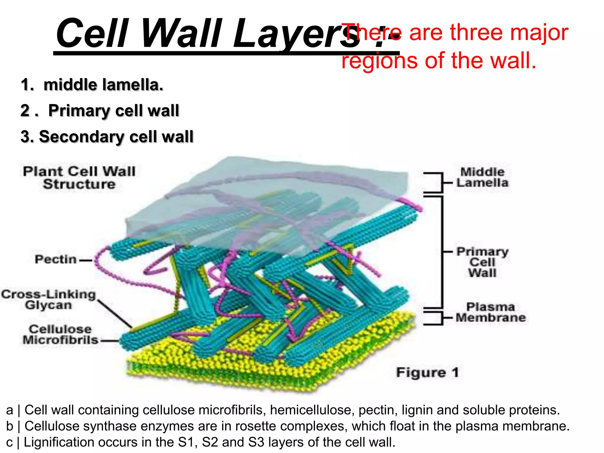
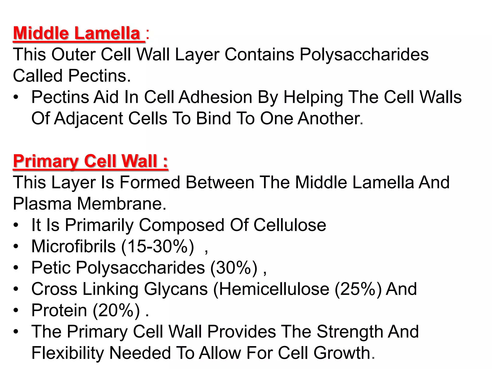
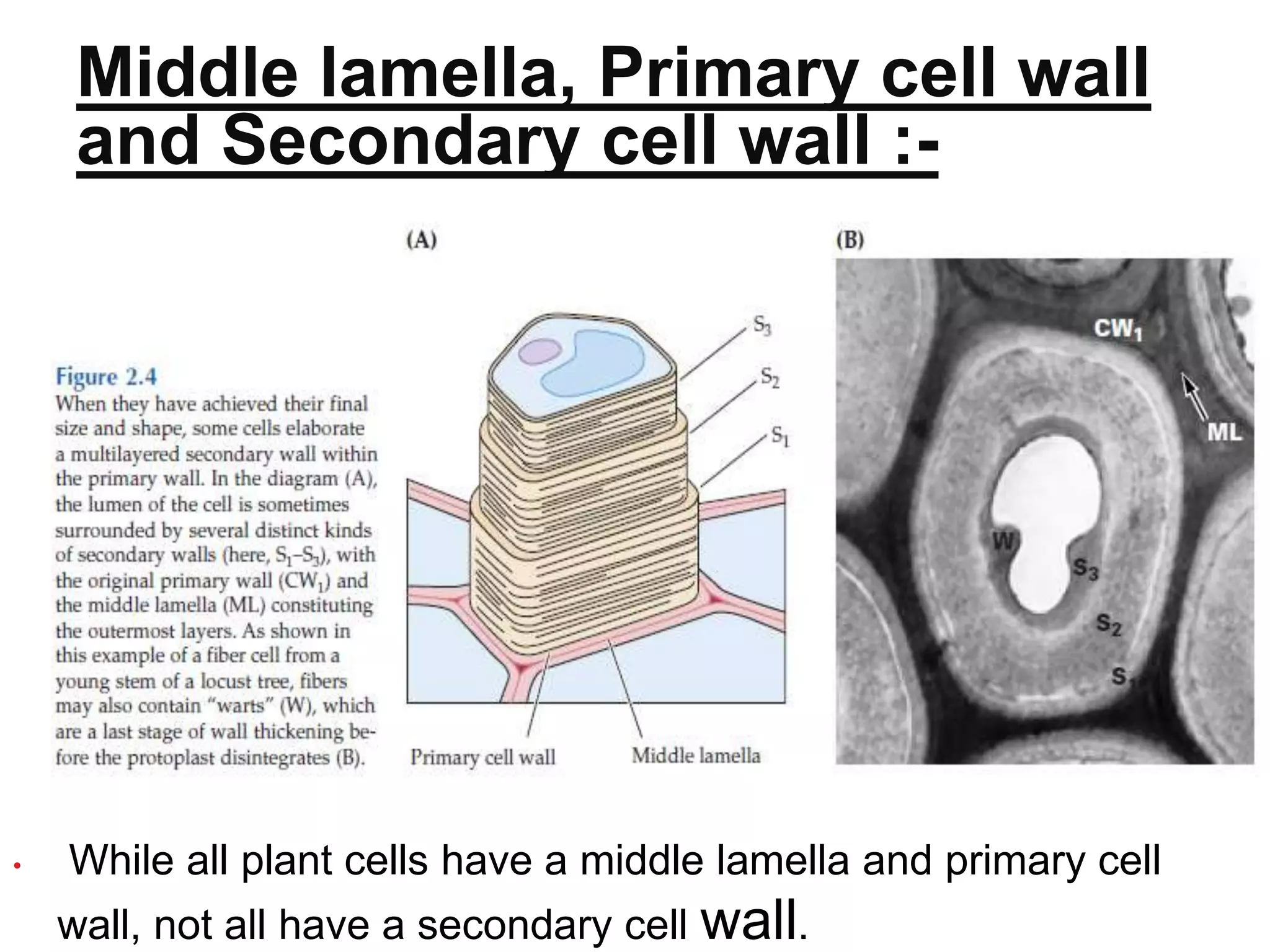
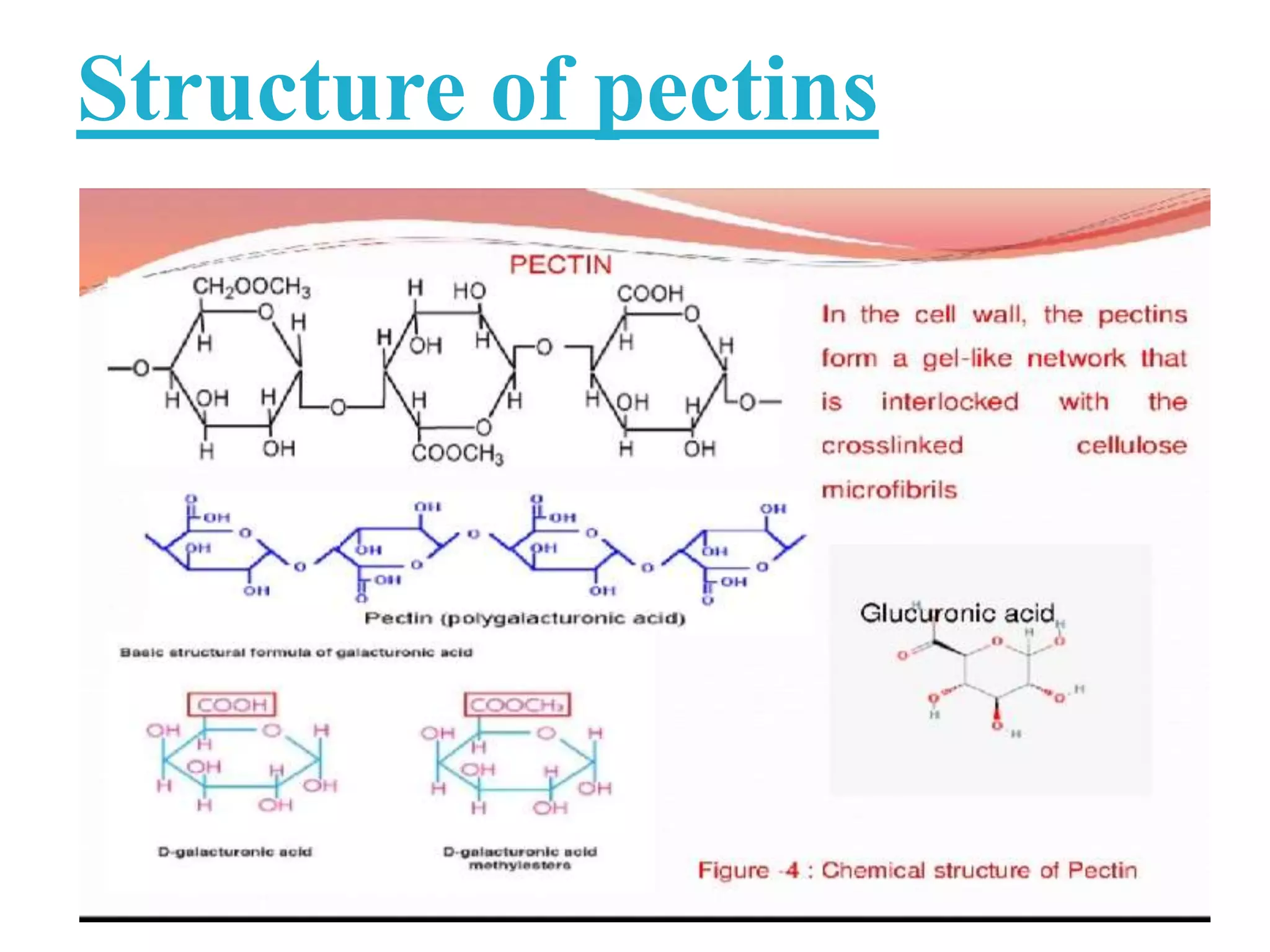
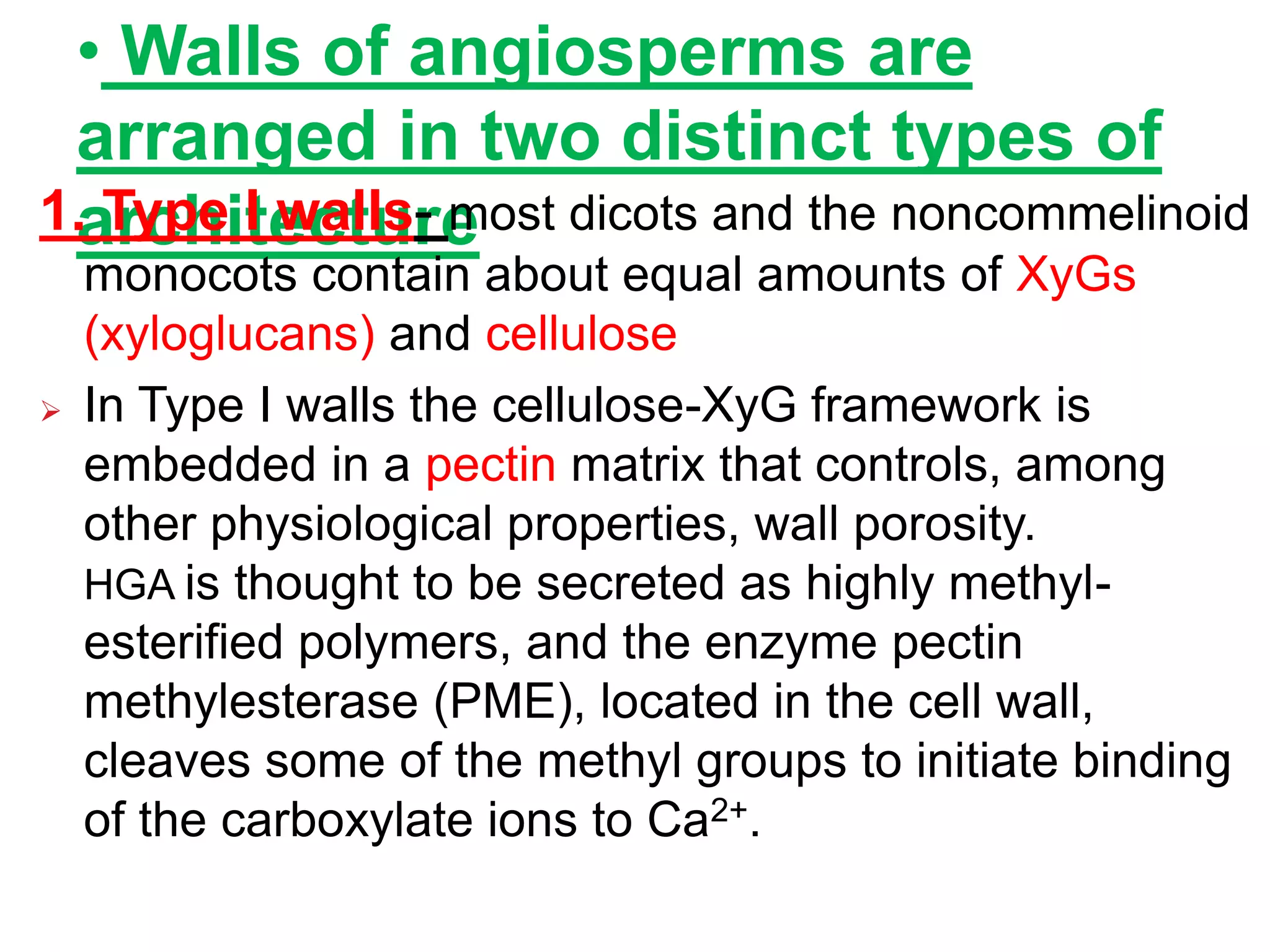
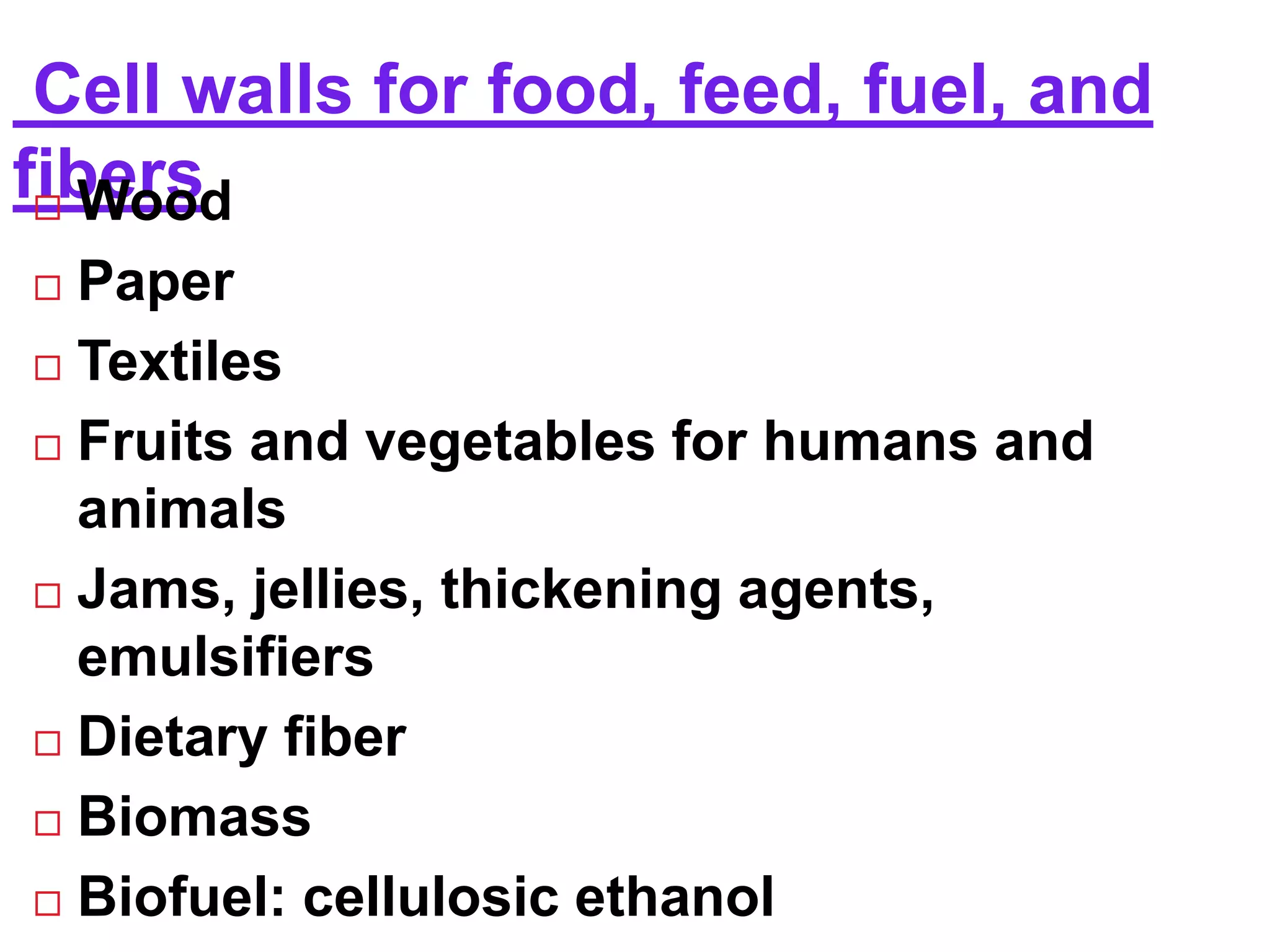
Cell wall in plants- introduction, cell wall layers, functions, sugars the building blocks of cell wall, macromolecules of cell wall, cell wall architecture, biosynthesis and assembly
This power point presentation consists of 64 slides including information about plant and other type of cell wall. Chemical composition, structure, function and properties of cell wall have been explained. Ultra structure of plant cell wall has also been high lighted. Algal,Fungal,Bacterial and Archaeal cell walls have also been explained.
Derived from the word latex meaning juice in latin. sometimes called lactiferous cells or vessels from the latin word for milk, lac
According to origin simple laticifer derived from a single cell or union of cells.
Laticifers can be defined as a specialized cell or a row of such cells that secrete the milky fluid termed latex. The word laticifer is used as a general term to denote the various latex-secreting structures latex cell, latex vessel, latex duct, latex tube and laticiferous duct. The laticiferous duct is a cavity into which latex is secreted.
A laticifer is a type of elongated secretory cell found in the leaves and/or stems of plants that produce latex and rubber as secondary metabolites.
Thin walled
Greately elongated
Much branched structure
Which contain milky juice
Complex composition called latex
These are following two types:-
Articulated laticifers
Non-articulated laticifers
Vascular Cambium & Seasonal activity & its Role in Stem & RootFatima Ramay
Vascular Cambium & Seasonal activity & its Role in Stem & Root:
The vascular cambium (pl. cambia or cambiums) is a lateral meristem in the vascular tissue of plants.
The vascular cambium is a cylindrical layer of cambium that runs through the stem of a plant that undergoes secondary growth.
In Dicots:
The vascular cambium is in dicot stems and roots, located between the xylem and the phloem in the stem and root of a vascular plant, and is the source of both the secondary xylem growth (inwards, towards the pith) and the secondary phloem growth (outwards).
In Monocots:
Monocot stems, such as corn, palms and bamboos, do not have a vascular cambium and do not exhibit secondary growth by the production of concentric annual rings. They cannot increase in girth by adding lateral layers of cells as in conifers and woody dicots.
Cambium of some plants remains active for the entire period of their life, i.e., cambial cells divide and resulting cells mature to form xylem and phloem elements.
This type of seasonal activity usually found in the plants present in the tropical regions, and not all plants show cambial activity.
Percentage of ringless trees in the rain forests of;India : 75%Amazon : 43%Malaysia : 15%
In regions with definite seasonal climate; seasonal activity of cambium ceased with onset of unfavorable conditions; In Autumn, it enters the dormant state and lasts for the end of summer; In Spring, cambium again becomes active.
Duration of cambial activity is also affected by day-length, e.g., In Robinia pseudoacacia, cambium is dormant under short-day condition.
The cambium cells formed in circular in cross section from the beginning onwards.
The cambial ring is partially primary (fascicular cambium) and partially secondary (interfascicular cambium).
Periderm originates from the cortical cells (extra stelar in origin).
In Dicot stem, for mechanical support xylem is with comparatively smaller vessels, greater fibers and less parenchyma.
More amount of cork is produces for protection.
Lenticels on periderm are very prominent.
The cambial ring formed is wavy in the beginning and later becomes circular.
The cambium ring is completely secondary in origin.
Periderm originates from the pericycle (intra stelar in origin).
In Dicot root, xylem is with big thin walled vessels with few fibers and more parenchyma.
Less amount of cork is produced as root is underground.
Lenticels on periderm are not very prominent.
Structure, Development & Function of PeridermFatima Ramay
A group of secondary tissues forming a protective layer which replaces the epidermis of many plant stems, roots, and other parts.
Although periderm may develop in leaves and fruits, its main function is to protects stems and roots.
The periderm consists of three different layers:
Phelloderm
Phellogen (cork cambium)
Phellem (cork)
Its main function is to protect the underlying tissues from:
Desiccation
Freezing
Heat injury
Mechanical destruction
Disease
Loss of epidermis.
Bounding tissue restricting the pathogen & insects.
Allowing gaseous exchange through lenticels.
It explains about what is plant tissue & both the types i.e meristem & permanent tissue. It also explains about the general characteristic, and how it has been classified based on origin, position, function and plane. It also furnish further information regarding the above
The Shoot apex is also known as the terminal bud of plants that grows from 0.1-1.0 mm and consists of the apical meristem, developing leaves and the immediate surrounding leaf primordial. The shoot apex is present in both dicot and monocot plants.
Living material is organized in unit and microorganism were living form of microscopical size and usually unicellular in structure originally classification is unsatisfied.
Plant systems: Extracellular matrix components of plants-cell wall, cellulose and hemicelluloses, extensins, WAKs, secondary wall structure, pits-primary and secondary pits and their development, plasmodesmota-structure and functions, pectins, cutins, lignins, turnover of cell wall components
(May 29th, 2024) Advancements in Intravital Microscopy- Insights for Preclini...Scintica Instrumentation
Intravital microscopy (IVM) is a powerful tool utilized to study cellular behavior over time and space in vivo. Much of our understanding of cell biology has been accomplished using various in vitro and ex vivo methods; however, these studies do not necessarily reflect the natural dynamics of biological processes. Unlike traditional cell culture or fixed tissue imaging, IVM allows for the ultra-fast high-resolution imaging of cellular processes over time and space and were studied in its natural environment. Real-time visualization of biological processes in the context of an intact organism helps maintain physiological relevance and provide insights into the progression of disease, response to treatments or developmental processes.
In this webinar we give an overview of advanced applications of the IVM system in preclinical research. IVIM technology is a provider of all-in-one intravital microscopy systems and solutions optimized for in vivo imaging of live animal models at sub-micron resolution. The system’s unique features and user-friendly software enables researchers to probe fast dynamic biological processes such as immune cell tracking, cell-cell interaction as well as vascularization and tumor metastasis with exceptional detail. This webinar will also give an overview of IVM being utilized in drug development, offering a view into the intricate interaction between drugs/nanoparticles and tissues in vivo and allows for the evaluation of therapeutic intervention in a variety of tissues and organs. This interdisciplinary collaboration continues to drive the advancements of novel therapeutic strategies.
This pdf is about the Schizophrenia.
For more details visit on YouTube; @SELF-EXPLANATORY;
https://www.youtube.com/channel/UCAiarMZDNhe1A3Rnpr_WkzA/videos
Thanks...!
A brief information about the SCOP protein database used in bioinformatics.
The Structural Classification of Proteins (SCOP) database is a comprehensive and authoritative resource for the structural and evolutionary relationships of proteins. It provides a detailed and curated classification of protein structures, grouping them into families, superfamilies, and folds based on their structural and sequence similarities.
Richard's entangled aventures in wonderlandRichard Gill
Since the loophole-free Bell experiments of 2020 and the Nobel prizes in physics of 2022, critics of Bell's work have retreated to the fortress of super-determinism. Now, super-determinism is a derogatory word - it just means "determinism". Palmer, Hance and Hossenfelder argue that quantum mechanics and determinism are not incompatible, using a sophisticated mathematical construction based on a subtle thinning of allowed states and measurements in quantum mechanics, such that what is left appears to make Bell's argument fail, without altering the empirical predictions of quantum mechanics. I think however that it is a smoke screen, and the slogan "lost in math" comes to my mind. I will discuss some other recent disproofs of Bell's theorem using the language of causality based on causal graphs. Causal thinking is also central to law and justice. I will mention surprising connections to my work on serial killer nurse cases, in particular the Dutch case of Lucia de Berk and the current UK case of Lucy Letby.
Observation of Io’s Resurfacing via Plume Deposition Using Ground-based Adapt...Sérgio Sacani
Since volcanic activity was first discovered on Io from Voyager images in 1979, changes
on Io’s surface have been monitored from both spacecraft and ground-based telescopes.
Here, we present the highest spatial resolution images of Io ever obtained from a groundbased telescope. These images, acquired by the SHARK-VIS instrument on the Large
Binocular Telescope, show evidence of a major resurfacing event on Io’s trailing hemisphere. When compared to the most recent spacecraft images, the SHARK-VIS images
show that a plume deposit from a powerful eruption at Pillan Patera has covered part
of the long-lived Pele plume deposit. Although this type of resurfacing event may be common on Io, few have been detected due to the rarity of spacecraft visits and the previously low spatial resolution available from Earth-based telescopes. The SHARK-VIS instrument ushers in a new era of high resolution imaging of Io’s surface using adaptive
optics at visible wavelengths.
Mammalian Pineal Body Structure and Also Functions
Cell wall in plants
1. JAI NARAIN VYAS UNIVERSITY, JODHPUR
Department of botany
Paper- 101
Seminar topic : - Cell
wall
Submitted
to :
Dr . VINOD
KATARIA
Submitted by :
Anmol Mertiya
M.sc Previous
1
stsem
2. CONTENS
1. Introduction
2. Cell Wall Layers
3. Functions Of The Plant Cell Wall
4. Sugars :- Building Blocks Of The Cell
Wall
5. Macromolecules Of The Cell Wall
6. Cell wall architecture
7. Cell wall biosynthesis and assembly
8. References
3. INTRODUTCTION
:-
Discovery of cell :- Cell wall was
first observed and name by
Robert Hooke in 1665.
In 1804, Karl Rudolphi and
J.H.F.Link proved that cells had
indepent cell wall .
The outermost structure of most
plant cells is a dyanamic and
rigid layer called cell wall (
4. Cell Wall Layers :-
There are three major
regions of the wall.
1. middle lamella.
2 . Primary cell wall
3. Secondary cell wall
a | Cell wall containing cellulose microfibrils, hemicellulose, pectin, lignin and soluble proteins.
b | Cellulose synthase enzymes are in rosette complexes, which float in the plasma membrane.
c | Lignification occurs in the S1, S2 and S3 layers of the cell wall.
5. Middle Lamella :
This Outer Cell Wall Layer Contains Polysaccharides
Called Pectins.
• Pectins Aid In Cell Adhesion By Helping The Cell Walls
Of Adjacent Cells To Bind To One Another.
Primary Cell Wall :
This Layer Is Formed Between The Middle Lamella And
Plasma Membrane.
• It Is Primarily Composed Of Cellulose
• Microfibrils (15-30%) ,
• Petic Polysaccharides (30%) ,
• Cross Linking Glycans (Hemicellulose (25%) And
• Protein (20%) .
• The Primary Cell Wall Provides The Strength And
Flexibility Needed To Allow For Cell Growth.
6. This layer is formed between the primary
cell wall and in some plant cells.
This rigid layer strengthens and supports
the cell. In addition to cellulose and
hemicellulose, some secondary cell walls
contain lignin.
• Lignin strengthens the cell wall and aids in
water conductivity in plant vascular tissue
cells.
Secondary wall :
7. Middle lamella, Primary cell wall
and Secondary cell wall :-
• While all plant cells have a middle lamella and primary cell
wall, not all have a secondary cell wall.
8. Functions of the plant cell wall :
• maintaining/determining cell shape .
• support and mechanical strength .
• prevents the cell membrane from bursting in a hypotonic medium.
• controls the rate and direction of cell growth and regulates cell
volume
• ultimately responsible for the plant architectural design and
controlling plant morphogenesis .
• metabolic role.
• physical barrier to: (a) pathogens
• (b) water in suberized cells.
• However, remember that the wall is very porous and allows the free
passage of small molecules, including proteins up to 60,000.
• economic products - cell walls are important for products such as
paper, wood, fiber, energy, shelter, and even roughage in our diet.
9. Sugars : Building
Blocks Of The Cell Wall
:-
Polysaccharides are long chains of sugar
molecules covalently linked at various
positions, some being decorated with side
chain of various lengths.
Sugars represent a vast spectrum of
polyhydroxyl “aldehydes” ( aldoses ) and
“ketones” ( ketoses ) that can be grouped
according to their chemical formula,
configuration , and stereochemical
conformation.
10. Many sugars have the empirical
formula ( CH2O )n, from which the
term carbohydrate is derived.
Haworth
project
1. Pyranose 2.
Furanose
(5 membered ring
)
( 6 membered ring
)
Chair Puckere
11. Sugars: building blocks of the cell wall
The monosaccharides in cell wall polymers are derived from glucose.
12. Macromolecules of the cell
wall
1. Cellulose
2. Callose
3. Cross-linking glycans
4. Pectin
5. Protein
6. Aromatic substance
13. o Cellulose is the principal scaffolding component
of all plant cell walls.
o Cellulose is the most adundant plant
polysaccharide for 15-30% of the dry mass of
all primary cell and a much larger percentace of
secondary wall
o Made of (1→4)β-D-glucan chains hydrogen bonded
to one another along their length
o Groups of 30 to 40 of these chains laterally
hydrogen bond to form crystalline or para-
crystalline microfibrils
1. Cellulose
14.
15. 2. Callose
Callose differs from cellulose in consisting of
(1→3)β-D-glucan chains, which can form
helical duplexes and triplexes
Callose is made by a few cell types at specific
stages of wall development, such as in
growing pollen tubes and in the cell plates of
dividing cells
Callose is also made in response to wounding
. Callose synthesis may also occue at
the wall in response to abiotic or biotic stress.
16.
17. 3. Cross-linking glycans
The most common hemicelluloses in cell walls are
xyloglucans (XyGs) and Glucuronoarabinoxylans ( GAXs
).
The XyG having β(1,4)-linked glucose residues that have
α(1,6)-linked xylosyl side chains.
The GAXs having β(1,4)-linked xylose residues. For ex
. glucuronic acid to produce glucuroxylan .
Both XyGs & GAXs may also be modified by acetylation,
which affects their capacity to cross-link to other cell wall
components.
XyG is main hemicellulose in dicots primary walls &
functions to cross-link cellulose microfibrils .
Although GAXs are major hemicelluloses in secondary
cell walls, they are also prominent in primary walls of
19. 4. Pectins
Pectins a mixture of heterogeneous, branched, and
highly hydrated polysaccharides rich in D-
galacturonic acid—have been defined classically as
material extracted from the cell wall by Ca2+-
chelators such as ammonium oxalate, EDTA,
EGTA, or cyclohexane diamine tetraacetate.
Pectins perform many functions:
• determining wall porosity
• providing charged surfaces that modulate wall pH
and ion balance.
• regulating cell–cell adhesion at the middle lamella.
• serving as recognition molecules that alert plant cells
to the presence of symbiotic organisms, pathogens,
20. Two types of pectine
1. HGA 2. RGⅠ
( Homogolacturonan) (Rhamnogalacturonan-Ⅰ
Homopolymers of (1→4)α-D Gal A
2 parts
1.xylogalacturonamn 2. RG Ⅱ
(Rhamnogalacturonan -Ⅱ)
Rod- like
heteropolymer of
peating (1 → 2) α -1
Rha ( 1→ 4) α -
D Gal A disaccaride
unite
22. 5. Structural proteins
Structural proteins of the cell wall are encoded by
large multigene families.
Although the structural framework of the cell wall
is largely carbohydrate , structural proteins may
also form networks in the wall.
There are four major classes are structural
proteins :
1. hydroxy proline - rich glycoproteins ( HRGPs)
2. proline – rich proteins ( PRPs )
3. glycine – rich proteins ( GRPs )
4. Arabinogalactan proteins ( AGPs )
24. 6 .Aromatic substances :
Aromatic substances are present in the
nonlignified walls of commelinoid species.
The primary walls of the commelinoid orders
of monocots and the Chenopodiaceae
contain significant amounts of aromatic
substances in their nonlignified cell walls-a
feature that makes them fluorescent under
ultraviolet (UV) light.
Hydroxycinnamic acid are also reduced in the
plant to hydroxycinnamoyl alcohols, which
form the common precursors for lignin and
25. Cell wall architecture
The primary wall consists of
three structural networks:
o Cellulose and cross-linking
glycans
o Matrix pectic polysaccharides
o Structural proteins or a
phenylpropanoid network
26. • Walls of angiosperms are
arranged in two distinct types of
architecture
1. Type I walls- most dicots and the noncommelinoid
monocots contain about equal amounts of XyGs
(xyloglucans) and cellulose
In Type I walls the cellulose-XyG framework is
embedded in a pectin matrix that controls, among
other physiological properties, wall porosity.
HGA is thought to be secreted as highly methyl-
esterified polymers, and the enzyme pectin
methylesterase (PME), located in the cell wall,
cleaves some of the methyl groups to initiate binding
of the carboxylate ions to Ca2+.
27. 2. Type II walls- commelinoid monocots
contain cellulose microfibrils similar to those of
the Type I wall; instead of XyG, however, the
principal polymers that interlock the microfibrils
are GAXs (glucuronoarbinoxylans).
In general, Type II walls are pectin-poor, but an
additional contribution to the charge density of
the wall is provided by the α-L-GlcA units on
GAX. These walls have very little structural
protein compared with dicots and other
monocots but they can accumulate extensive
interconnecting networks of phenylpropanoids,
particularly as the cells stop expanding.
28.
29. Cell wall biosynthesis and assembly :
Origin of cell wall takes place from cell
plate during cytokinesis.
Many cell wall vesicles provided by GB
and ER combine to form a cell plate.
After some physical and chemical
changes, the cell plate ( rich in pectin )
grows on both sides to form a middle
lamella, which glues neighbouring plant
cells.
After which primary wall and secondary
walls are laid down on the middle lamella
30. After a wall forms, it can grow
and mature through a process:-
1. Synthesis
2. Secretion
3. Assembly
4. Expansion ( in growing cells )
5. Cross-linking and secondary wall
fomation
31.
32. Cell walls for food, feed, fuel, and
fibers
Wood
Paper
Textiles
Fruits and vegetables for humans and
animals
Jams, jellies, thickening agents,
emulsifiers
Dietary fiber
Biomass
Biofuel: cellulosic ethanol
33. REFERENCES
Alberts , B., Bray, D., Lewis, J., Raff, m.,
Roberts, K. And Watson, J. D. 1999. Molecular
Biology Of Cell . Garland Publishing , Inc. , New
York.
Buchanan, B. B. ,Gruissem, W. And Jones , R. L.
2000. Biochemistry And Molecular Biology Of
Plants. American Society Of Plant Physiologists,
Maryland, USA
https://jcs.biologist.org