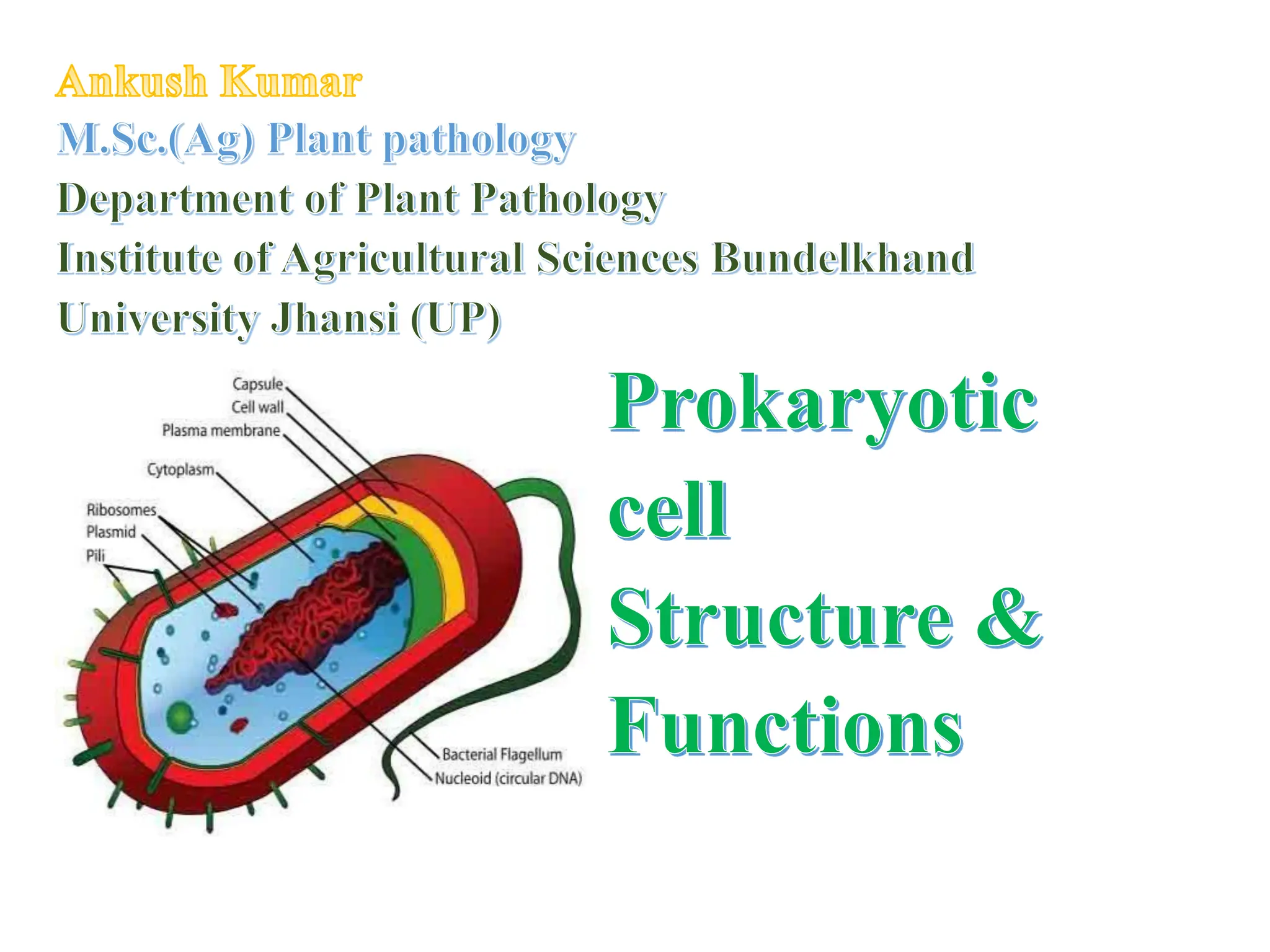
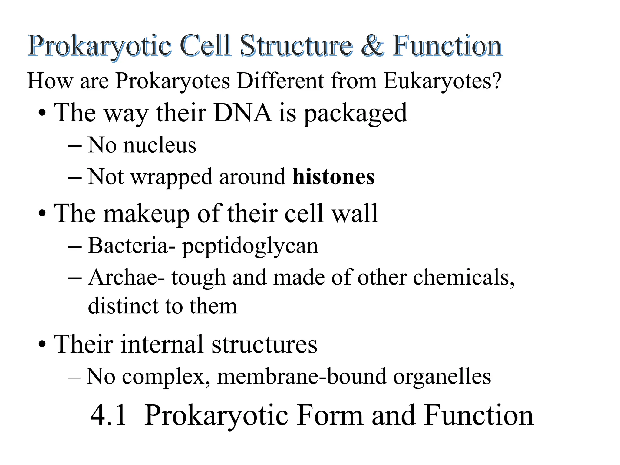
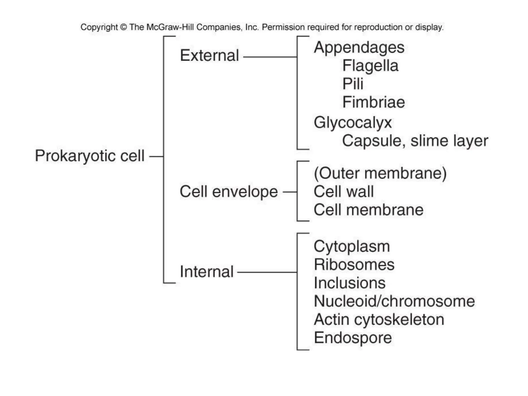
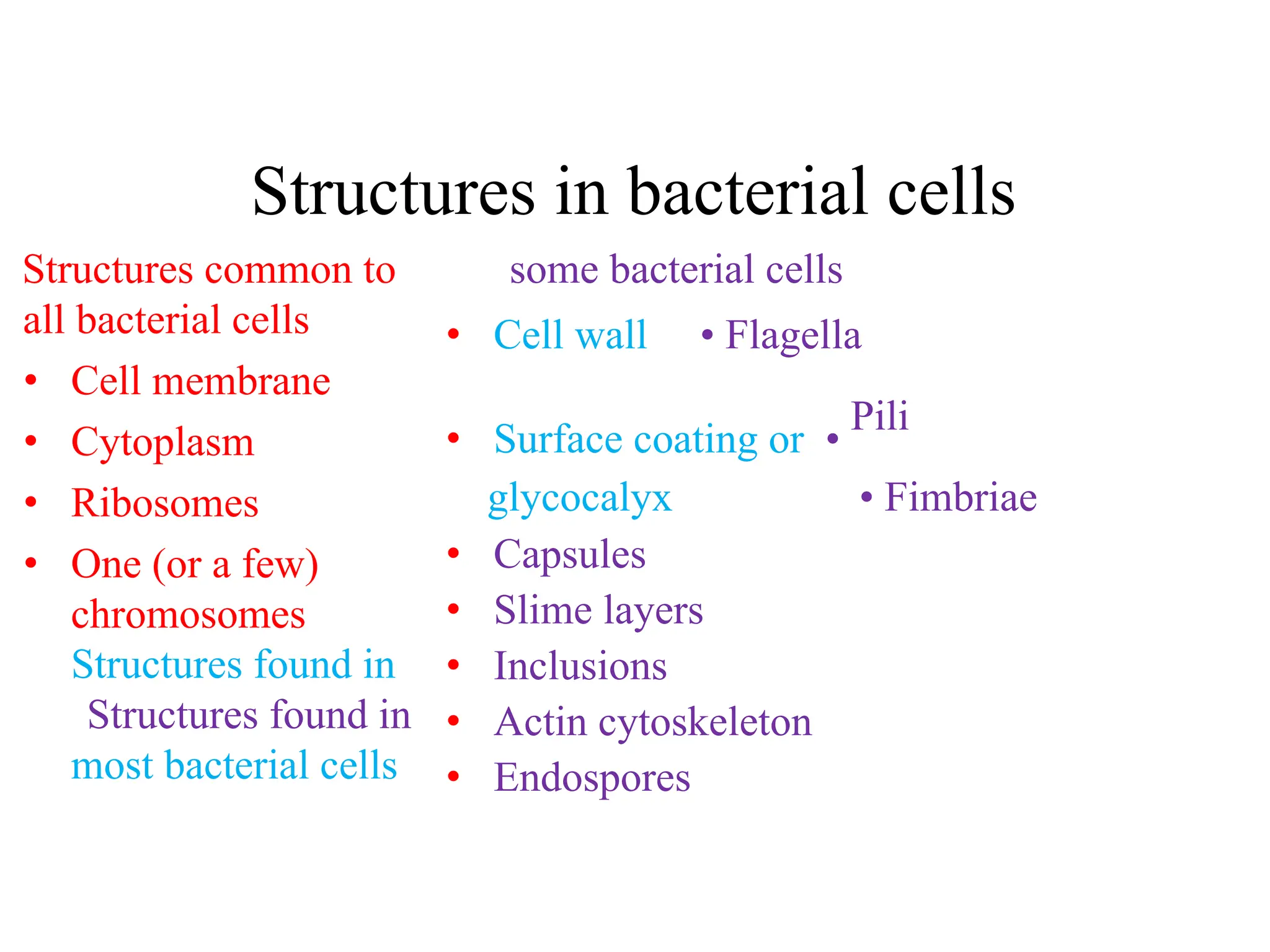
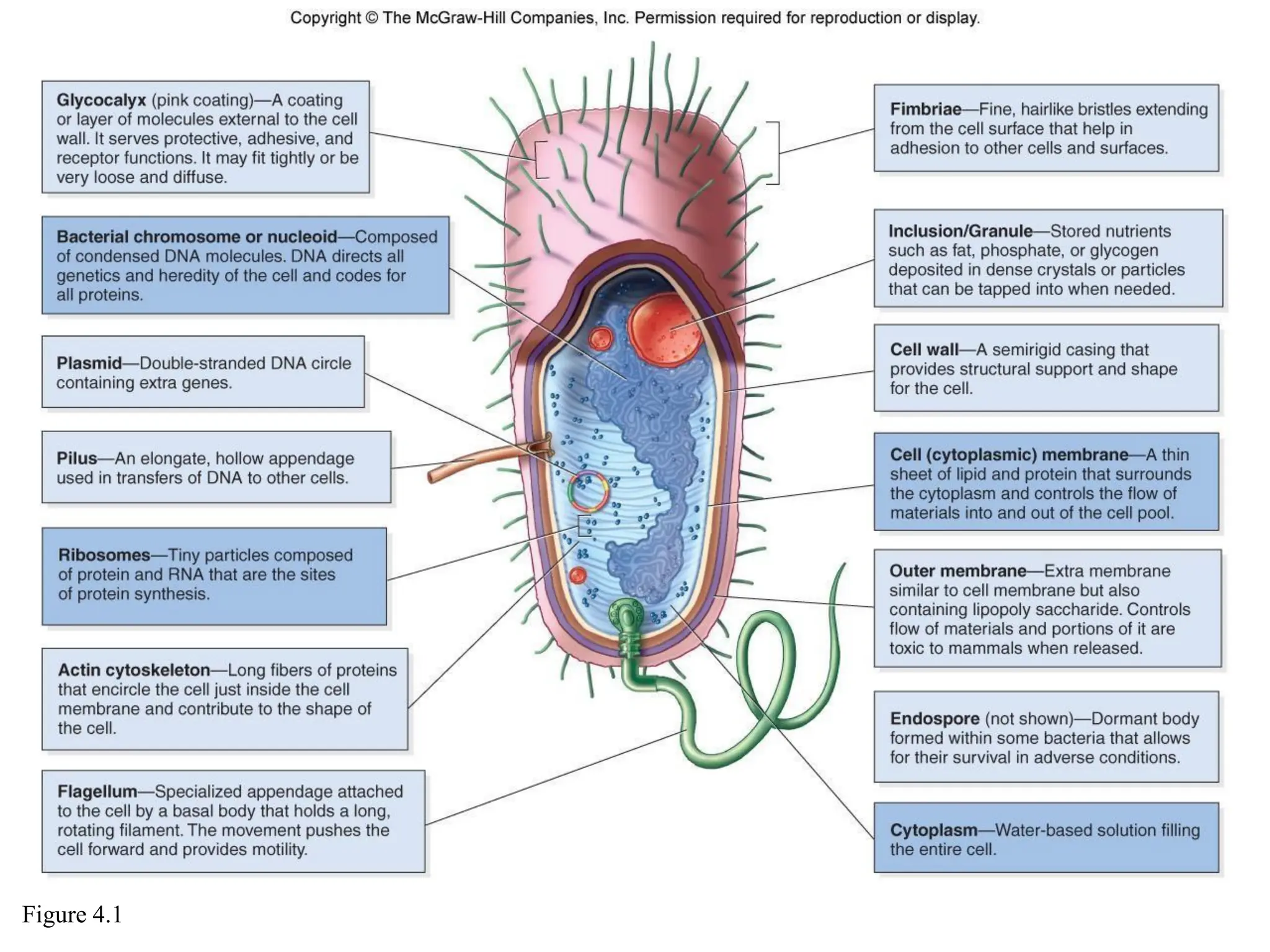
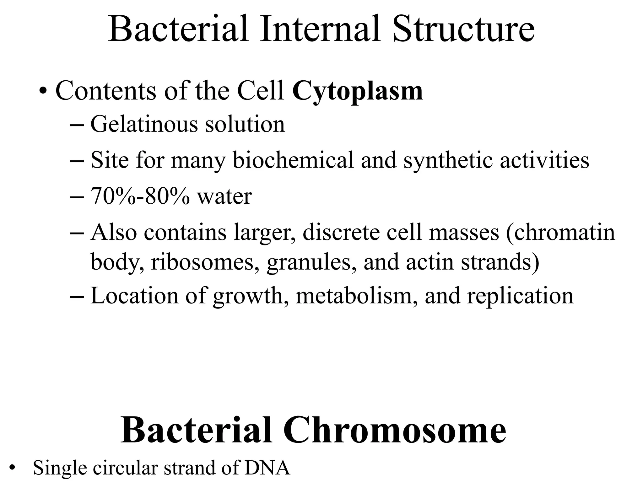
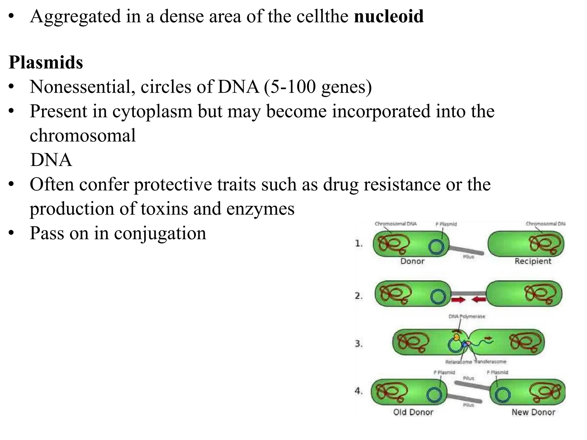
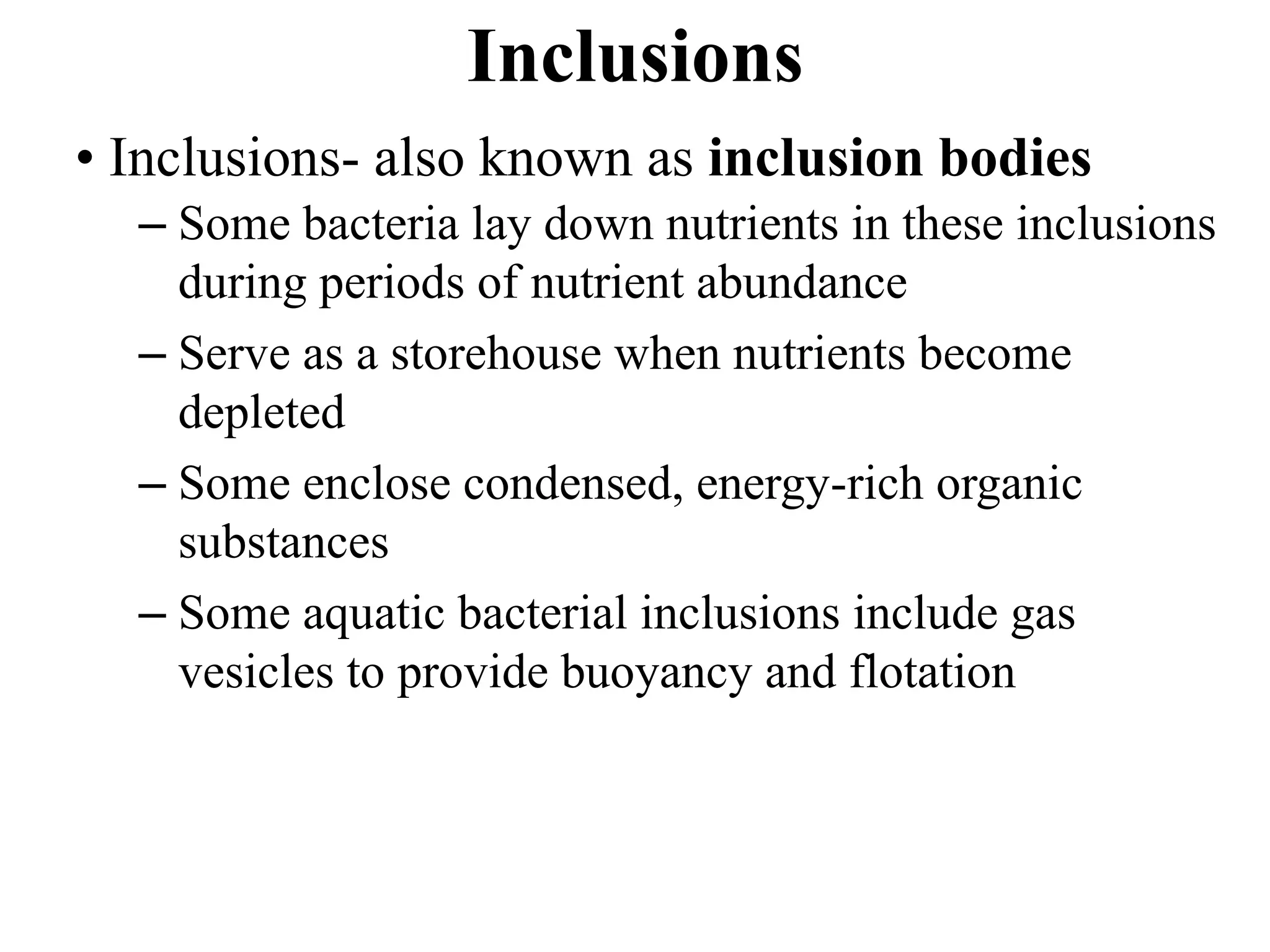
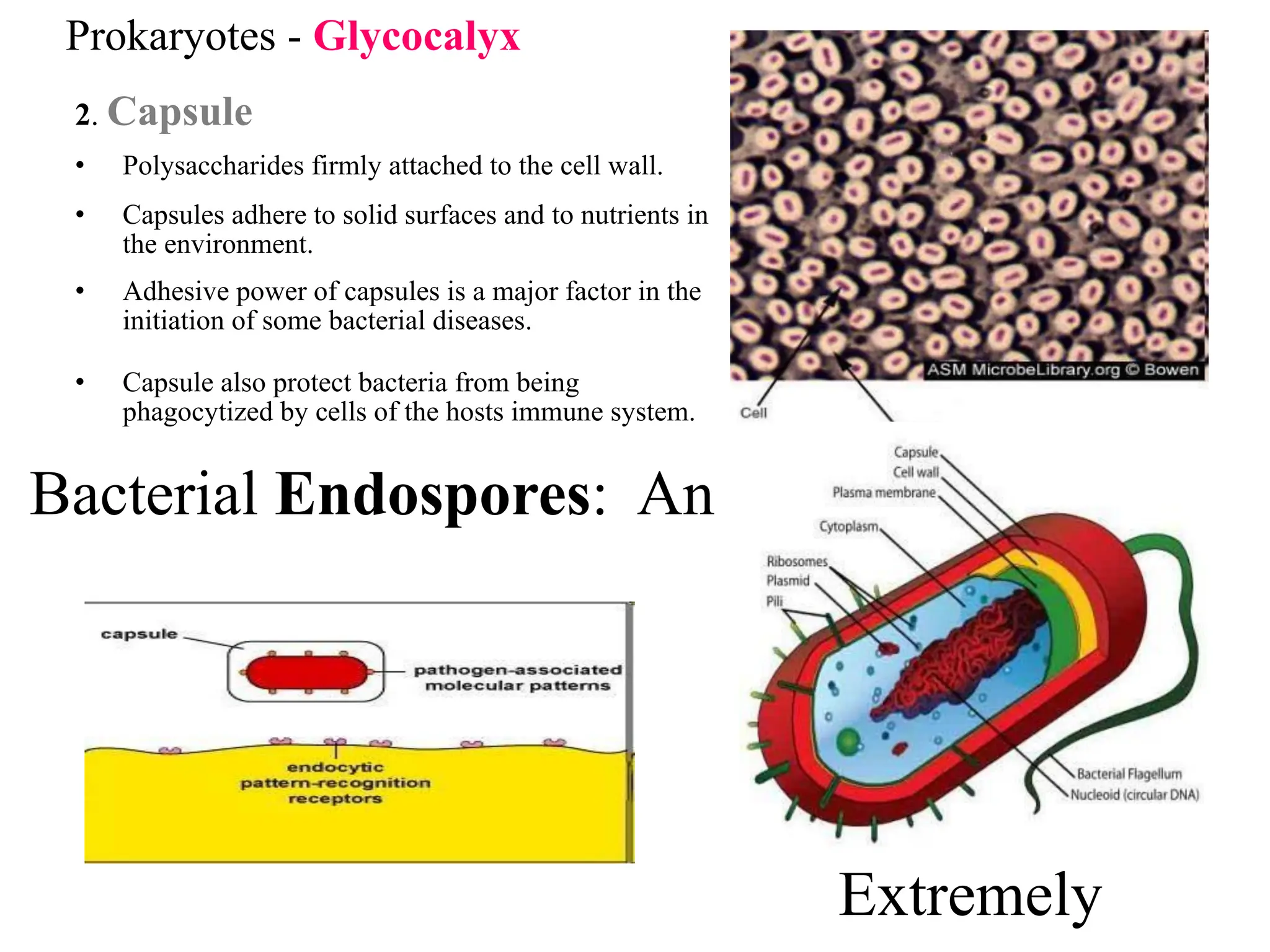
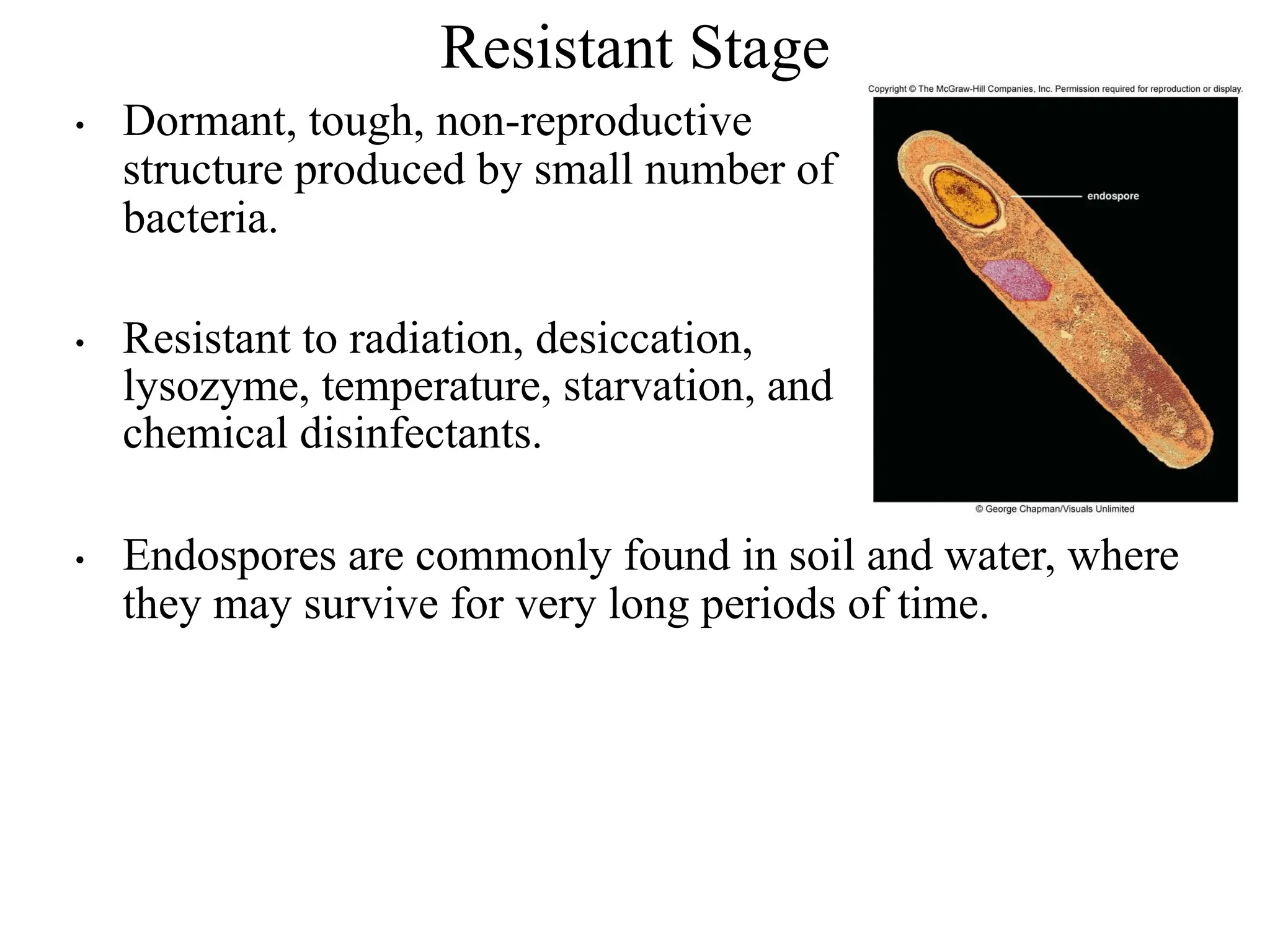
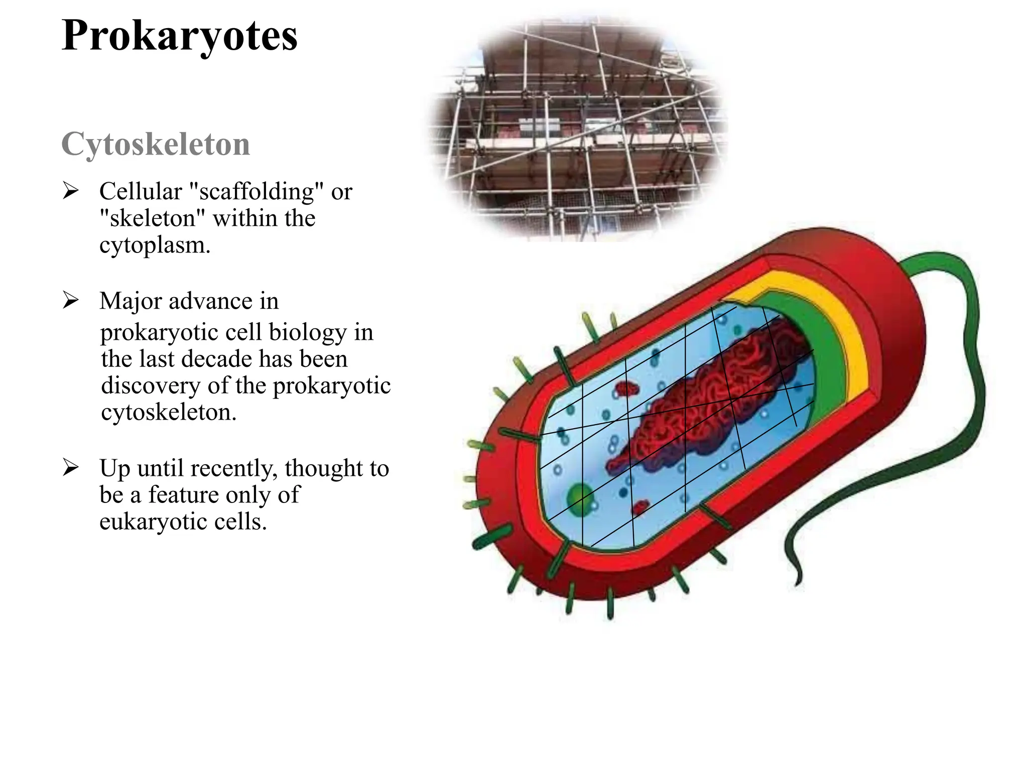
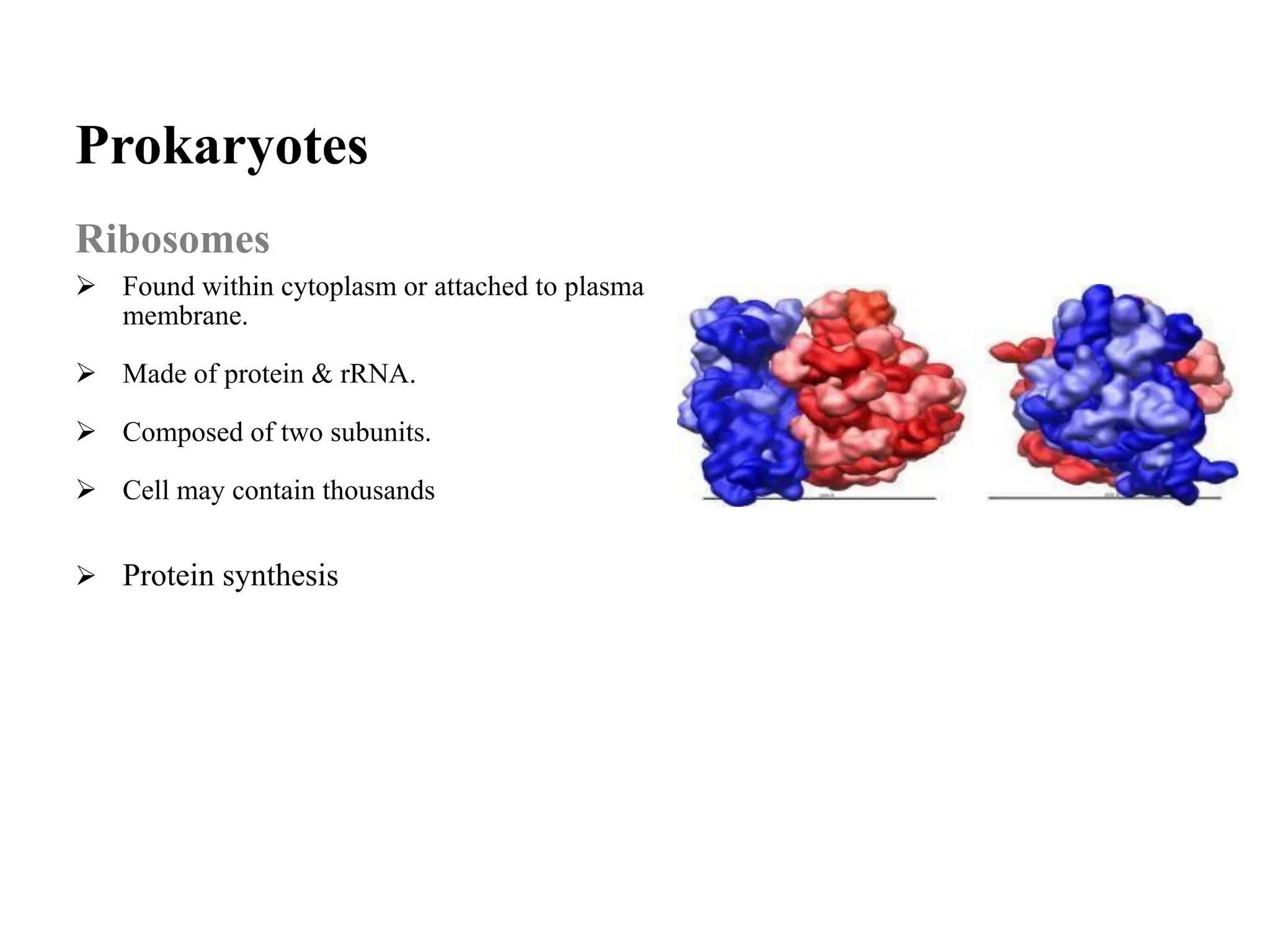
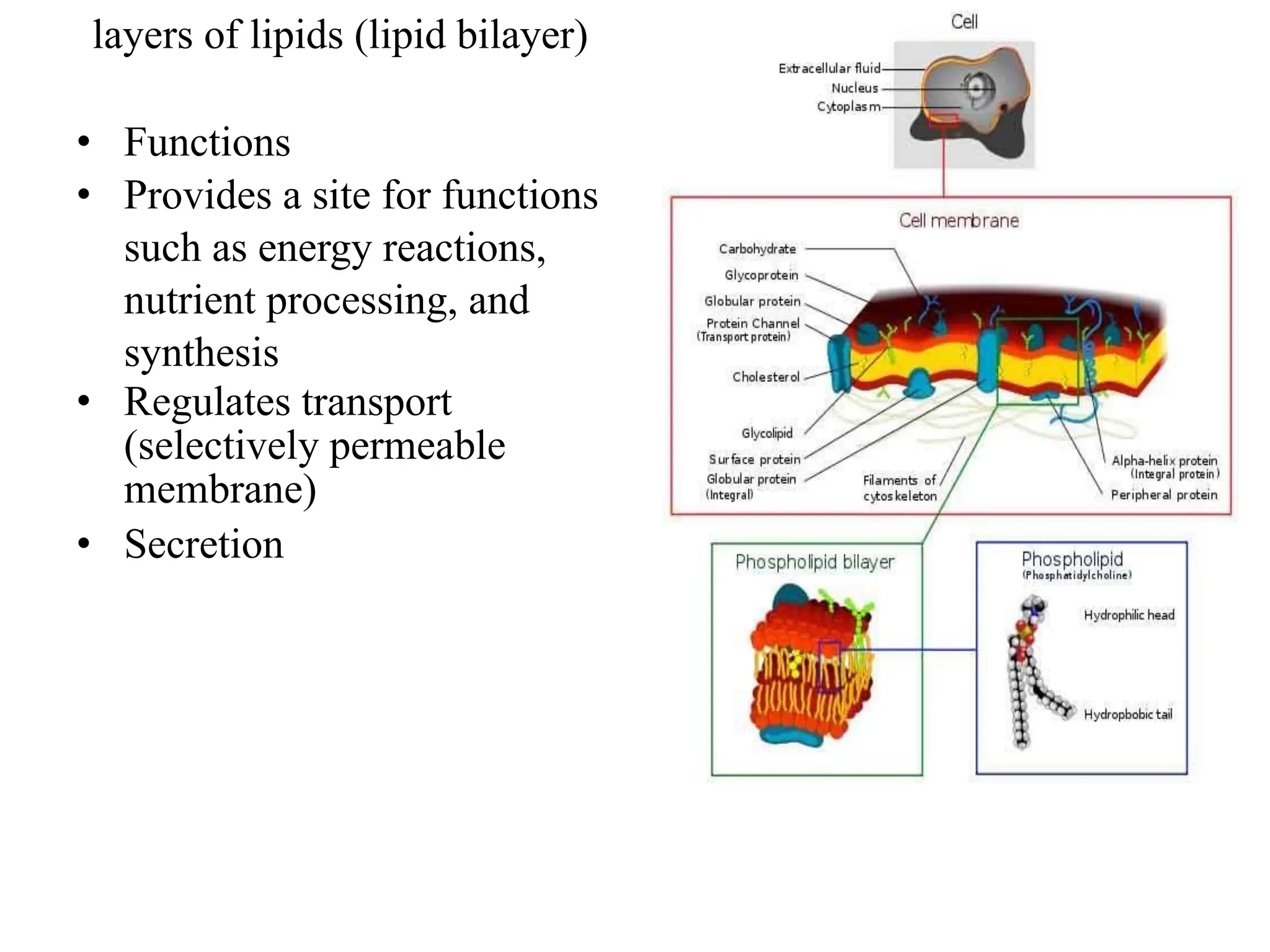
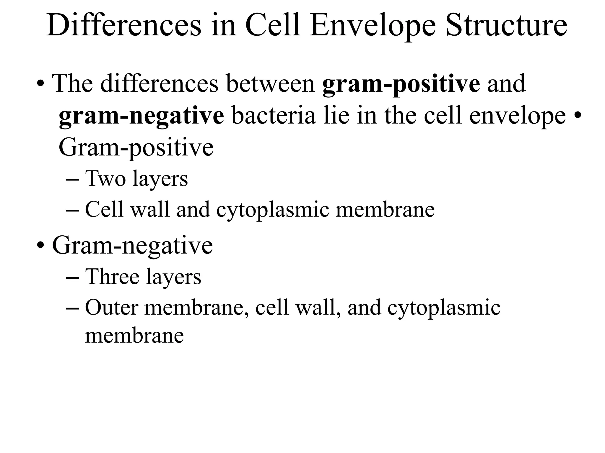
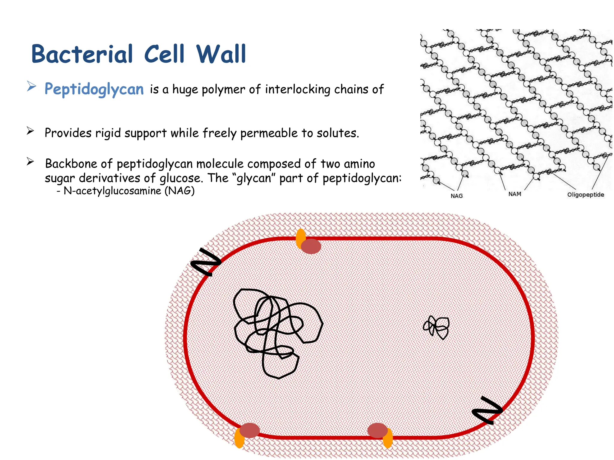
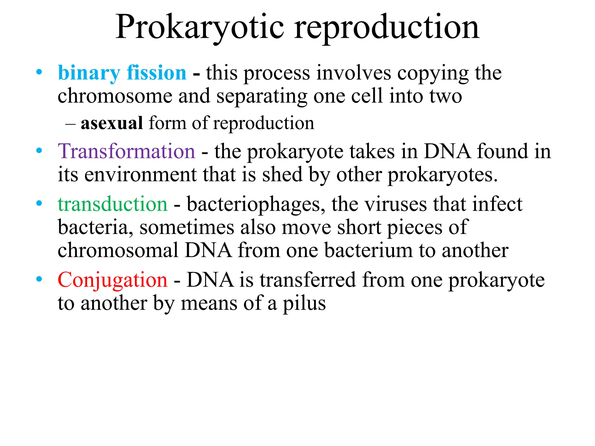
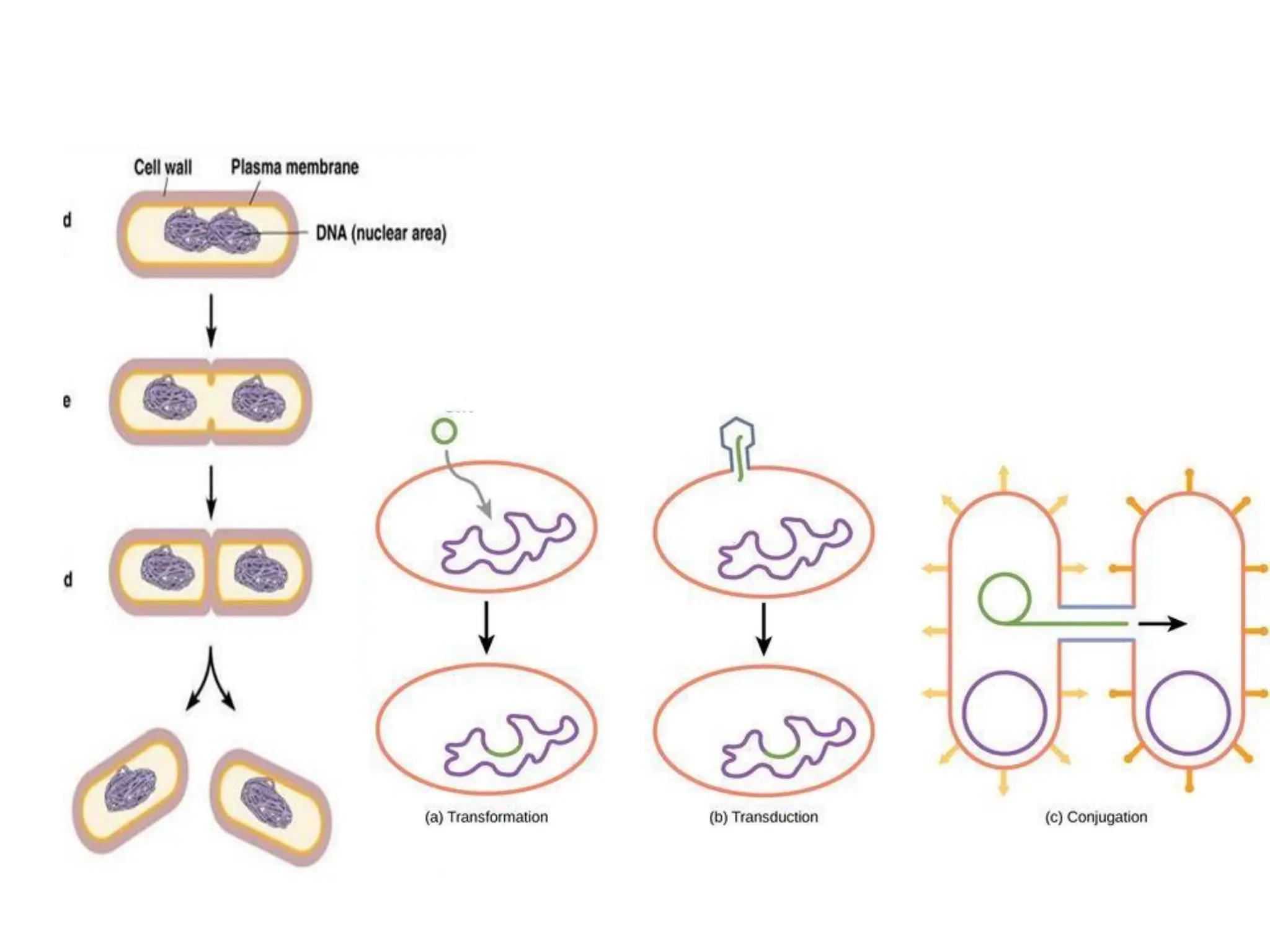
Prokaryotic
Structure &
Functions
How are Prokaryotes Different from Eukaryotes?
• The way their DNA is packaged
– No nucleus
– Not wrapped around histones
• The makeup of their cell wall
– Bacteria- peptidoglycan
– Archae- tough and made of other chemicals,
distinct to them
• Their internal structures
– No complex, membrane-bound organelles
4.1 Prokaryotic Form and Function
Structures in bacterial cells
Structures common to
all bacterial cells
• Cell membrane
• Cytoplasm
• Ribosomes
• One (or a few)
chromosomes
Structures found in
Structures found in
most bacterial cells
some bacterial cells
• Cell wall • Flagella
• Surface coating or •
Pili
glycocalyx • Fimbriae
• Capsules
• Slime layers
• Inclusions
• Actin cytoskeleton
• Endospores
Figure 4.1
Bacterial Internal Structure
• Contents of the Cell Cytoplasm
– Gelatinous solution
– Site for many biochemical and synthetic activities
– 70%-80% water
– Also contains larger, discrete cell masses (chromatin
body, ribosomes, granules, and actin strands)
– Location of growth, metabolism, and replication
Bacterial Chromosome
• Single circular strand of DNA
• Aggregated in a dense area of the cellthe nucleoid
Plasmids
• Nonessential, circles of DNA (5-100 genes)
• Present in cytoplasm but may become incorporated into the
chromosomal
DNA
• Often confer protective traits such as drug resistance or the
production of toxins and enzymes
• Pass on in conjugation
Inclusions
• Inclusions- also known as inclusion bodies
– Some bacteria lay down nutrients in these inclusions
during periods of nutrient abundance
– Serve as a storehouse when nutrients become
depleted
– Some enclose condensed, energy-rich organic
substances
– Some aquatic bacterial inclusions include gas
vesicles to provide buoyancy and flotation
Granules
• A type of inclusion body
• Contain crystals of inorganic compounds
• Are not enclosed by membranes
• Staining of some granules aids in identification.
Figure 4.19
The Glycocalyx
• a coating of repeating polysaccharide, protein, or
both
• Protects the cell
• Can help the cell adhere to the environment
• Slime layer- a loose shield that protects some
bacteria from loss of water and nutrients
• Capsule- when the glycocalyx is bound more
tightly to the cell and is denser and thicker
Functions of the Glycocalyx
Many pathogenic bacteria have glycocalyces
• Protect the bacteria against phagocytes
• Important in formation of biofilms
• Streptococcus
– form a biofilm & eventually a buildup of plaque.
– The slime layer of Gram+ Streptococcus mutans allows
it to accumulate on tooth enamel (yuck mouth and one
of the causes of cavities).
– Other bacteria in the mouth become trapped in the
slime
Prokaryotes - Glycocalyx
2. Capsule
• Polysaccharides firmly attached to the cell wall.
• Capsules adhere to solid surfaces and to nutrients in
the environment.
• Adhesive power of capsules is a major factor in the
initiation of some bacterial diseases.
• Capsule also protect bacteria from being
phagocytized by cells of the hosts immune system.
Bacterial Endospores: An
Extremely
Resistant Stage
• Dormant, tough, non-reproductive
structure produced by small number of
bacteria.
• Resistant to radiation, desiccation,
lysozyme, temperature, starvation, and
chemical disinfectants.
• Endospores are commonly found in soil and water, where
they may survive for very long periods of time.
Prokaryotes
Cytoskeleton
➢ Cellular "scaffolding" or
"skeleton" within the
cytoplasm.
➢ Major advance in
prokaryotic cell biology in
the last decade has been
discovery of the prokaryotic
cytoskeleton.
➢ Up until recently, thought to
be a feature only of
eukaryotic cells.
Prokaryotes
Ribosomes
➢ Found within cytoplasm or attached to plasma
membrane.
➢ Made of protein & rRNA.
➢ Composed of two subunits.
➢ Cell may contain thousands
➢ Protein synthesis
The Cell Envelope: The Boundary layer of
Bacteria
• Majority of bacteria have a cell envelope • Lies
outside of the cytoplasm • Composed of two or
three basic layers
– Cell membrane
– Cell wall
– In some bacteria, the outer membrane
Plasma Membrane
• Separates the cell from its environment
• Phospholipid bilayer with proteins embedded in two
layers of lipids (lipid bilayer)
• Functions
• Provides a site for functions
such as energy reactions,
nutrient processing, and
synthesis
• Regulates transport
(selectively permeable
membrane)
• Secretion
Differences in Cell Envelope Structure
• The differences between gram-positive and
gram-negative bacteria lie in the cell envelope •
Gram-positive
– Two layers
– Cell wall and cytoplasmic membrane
• Gram-negative
– Three layers
– Outer membrane, cell wall, and cytoplasmic
membrane
➢ Peptidoglycan is a huge polymer of interlocking chains of
➢ Provides rigid support while freely permeable to solutes.
➢ Backbone of peptidoglycan molecule composed of two amino
sugar derivatives of glucose. The “glycan” part of peptidoglycan:
- N-acetylglucosamine (NAG)
Bacterial Cell Wall
Structure of the Cell Wall
• Provides shape and strong structural support
• Most are rigid because of peptidoglycan content
• Target of many antibiotics- disrupt the cell wall, and cells
have little protection from lysis
• Gram-positive cell (2 layers)
– A thick (20 to 80 nm) petidoglycan cell wall and membrane
• Gram-Negative Cell (3 layers)
– Outer membrane
– Single, thin (1 to 3 nm) sheet of peptidoglycan (Periplasmic
space surrounds the peptidoglycan)
– Cell membrane
Figure 4.12
Figure 4.14
The Gram-Negative Outer Membrane
• Similar to the cell membrane, except it contains
specialized polysaccharides and proteins
• Outermost layer- contains lipopolysaccharide
(LPS)
• Innermost layer- phospholipid layer anchored by
lipoproteins to the peptidoglycan layer below
• Outer membrane serves as a partial chemical sieve
– Only relatively small molecules can penetrate
– Access provided by special membrane channels formed
by porin proteins
Practical Considerations of Differences in
Cell Envelope Structure
• Outer membrane- an extra barrier in gramnegative
bacteria
– Makes them impervious to some antrimicrobial
chemicals
– Generally more difficult to inhibit or kill than
grampositive bacteria
• Cell envelope can interact with human tissues and
cause disease
– Corynebacterium diphtheriae
– Streptococcus pyogenes
Prokaryotes - Cell Wall
From the peptidoglycan inwards all bacteria are very similar. Going further out, the
bacterial world divides into two major classes (plus a couple of odd types).
These are:
Gram-positive Gram-negative
Prokaryotes - Cell Wall
Gram-Positive & Gram-Negative
Q: Why are these differences in bacterial cell wall
structure so important?
Nontypical Cell Walls
• Some aren’t characterized as either grampositive
or gram-negative • For example, Mycobacterium
and Nocardiaunique types of lipids (acid-fast) •
Archaea – no peptidoglycan • Mycoplasmas-
lack cell wall entirely
External Structures
• Appendages: Cell extensions
– Common but not present on all species
– Can provide motility (flagella and axial filaments)
– Can be used for attachment and mating (pili and
fimbriae)
Prokaryotes – Surface Appendages
➢ fimbriae: Most Gram-negative bacteria have these short, fine
appendages surrounding the cell. Gram+ bacteria don’t have.
No role in motility. Help bacteria
adhere to solid surfaces. Major factor in
virulence.
(singular: fimbria)
➢ pili:Tubes that are longer than fimbriae,
usually shorter than flagella.
Use for movement, like grappling hooks,
and also use conjugation pili to transfer
plasmids. (singular = pilus)
Prokaryotes – Cell Shapes
Most bacteria are classifies according to shape:
1. bacillus (pl. bacilli) = rod-shaped
2. coccus (pl. cocci … sounds like cox-eye) = spherical
3. spiral shaped
a. spirillum (pl. spirilla) = spiral with rigid cell wall, flagella
b. spirochete (pl. spirochetes) = spiral with flexible cell wall, axial filament
Pleomorphism- when cells of a single species vary to some extent in shape and size
There are many more shapes beyond these basic ones. A few examples:
– Coccobacilli = elongated coccal form
– Filamentous = bacilli that occur in long threads
– Vibrios = short, slightly curved rods
– Fusiform = bacilli with tapered ends
Figure 4.22
Arrangement, or Grouping
• Cocci- greatest variety in arrangement
– Single
– Pairs (diplococci)
– Tetrads
– Irregular clusters (staphylococci and micrococci)
– Chains (streptococci)
– Cubical packet (sarcina)
• Bacilli- less varied
– Single
– Pairs (diplobacilli)
– Chain (streptobacilli)
– Row of cells oriented side by side (palisades)
• Spirilla
– Occasionally found in short chains
Prokaryotes – Arrangements of Cells
• Bacteria sometimes occur in groups, rather
than singly.
• bacilli divide along a single axis, seen in
pairs or chains.
• cocci divide on one or more planes,
producing cells in:
- pairs (diplococci)
- chains (streptococci) - packets
(sarcinae) - clusters
(staphylococci).
• Size, shape and arrangement of cells often
first clues in identification of a bacterium.
• Many “look-alikes”, so shape and
arrangement not enough for id of genus and
species.
Prokaryotic reproduction
• binary fission - this process involves copying the
chromosome and separating one cell into two
– asexual form of reproduction
• Transformation - the prokaryote takes in DNA found in
its environment that is shed by other prokaryotes.
• transduction - bacteriophages, the viruses that infect
bacteria, sometimes also move short pieces of
chromosomal DNA from one bacterium to another
• Conjugation - DNA is transferred from one prokaryote
to another by means of a pilus
prokaryotic cell structure

prokaryotic cell structure

  • 1.
  • 2.
    How are ProkaryotesDifferent from Eukaryotes? • The way their DNA is packaged – No nucleus – Not wrapped around histones • The makeup of their cell wall – Bacteria- peptidoglycan – Archae- tough and made of other chemicals, distinct to them • Their internal structures – No complex, membrane-bound organelles 4.1 Prokaryotic Form and Function
  • 4.
    Structures in bacterialcells Structures common to all bacterial cells • Cell membrane • Cytoplasm • Ribosomes • One (or a few) chromosomes Structures found in Structures found in most bacterial cells some bacterial cells • Cell wall • Flagella • Surface coating or • Pili glycocalyx • Fimbriae • Capsules • Slime layers • Inclusions • Actin cytoskeleton • Endospores
  • 5.
  • 6.
    Bacterial Internal Structure •Contents of the Cell Cytoplasm – Gelatinous solution – Site for many biochemical and synthetic activities – 70%-80% water – Also contains larger, discrete cell masses (chromatin body, ribosomes, granules, and actin strands) – Location of growth, metabolism, and replication Bacterial Chromosome • Single circular strand of DNA
  • 7.
    • Aggregated ina dense area of the cellthe nucleoid Plasmids • Nonessential, circles of DNA (5-100 genes) • Present in cytoplasm but may become incorporated into the chromosomal DNA • Often confer protective traits such as drug resistance or the production of toxins and enzymes • Pass on in conjugation
  • 8.
    Inclusions • Inclusions- alsoknown as inclusion bodies – Some bacteria lay down nutrients in these inclusions during periods of nutrient abundance – Serve as a storehouse when nutrients become depleted – Some enclose condensed, energy-rich organic substances – Some aquatic bacterial inclusions include gas vesicles to provide buoyancy and flotation
  • 9.
    Granules • A typeof inclusion body • Contain crystals of inorganic compounds • Are not enclosed by membranes • Staining of some granules aids in identification. Figure 4.19
  • 10.
    The Glycocalyx • acoating of repeating polysaccharide, protein, or both • Protects the cell • Can help the cell adhere to the environment • Slime layer- a loose shield that protects some bacteria from loss of water and nutrients • Capsule- when the glycocalyx is bound more tightly to the cell and is denser and thicker
  • 11.
    Functions of theGlycocalyx Many pathogenic bacteria have glycocalyces • Protect the bacteria against phagocytes • Important in formation of biofilms • Streptococcus – form a biofilm & eventually a buildup of plaque. – The slime layer of Gram+ Streptococcus mutans allows it to accumulate on tooth enamel (yuck mouth and one of the causes of cavities).
  • 12.
    – Other bacteriain the mouth become trapped in the slime
  • 13.
    Prokaryotes - Glycocalyx 2.Capsule • Polysaccharides firmly attached to the cell wall. • Capsules adhere to solid surfaces and to nutrients in the environment. • Adhesive power of capsules is a major factor in the initiation of some bacterial diseases. • Capsule also protect bacteria from being phagocytized by cells of the hosts immune system. Bacterial Endospores: An Extremely
  • 14.
    Resistant Stage • Dormant,tough, non-reproductive structure produced by small number of bacteria. • Resistant to radiation, desiccation, lysozyme, temperature, starvation, and chemical disinfectants. • Endospores are commonly found in soil and water, where they may survive for very long periods of time.
  • 15.
    Prokaryotes Cytoskeleton ➢ Cellular "scaffolding"or "skeleton" within the cytoplasm. ➢ Major advance in prokaryotic cell biology in the last decade has been discovery of the prokaryotic cytoskeleton. ➢ Up until recently, thought to be a feature only of eukaryotic cells.
  • 16.
    Prokaryotes Ribosomes ➢ Found withincytoplasm or attached to plasma membrane. ➢ Made of protein & rRNA. ➢ Composed of two subunits. ➢ Cell may contain thousands ➢ Protein synthesis
  • 17.
    The Cell Envelope:The Boundary layer of Bacteria • Majority of bacteria have a cell envelope • Lies outside of the cytoplasm • Composed of two or three basic layers – Cell membrane – Cell wall – In some bacteria, the outer membrane Plasma Membrane • Separates the cell from its environment • Phospholipid bilayer with proteins embedded in two
  • 18.
    layers of lipids(lipid bilayer) • Functions • Provides a site for functions such as energy reactions, nutrient processing, and synthesis • Regulates transport (selectively permeable membrane) • Secretion
  • 19.
    Differences in CellEnvelope Structure • The differences between gram-positive and gram-negative bacteria lie in the cell envelope • Gram-positive – Two layers – Cell wall and cytoplasmic membrane • Gram-negative – Three layers – Outer membrane, cell wall, and cytoplasmic membrane
  • 20.
    ➢ Peptidoglycan isa huge polymer of interlocking chains of ➢ Provides rigid support while freely permeable to solutes. ➢ Backbone of peptidoglycan molecule composed of two amino sugar derivatives of glucose. The “glycan” part of peptidoglycan: - N-acetylglucosamine (NAG) Bacterial Cell Wall
  • 21.
    Structure of theCell Wall • Provides shape and strong structural support • Most are rigid because of peptidoglycan content • Target of many antibiotics- disrupt the cell wall, and cells have little protection from lysis • Gram-positive cell (2 layers) – A thick (20 to 80 nm) petidoglycan cell wall and membrane • Gram-Negative Cell (3 layers) – Outer membrane – Single, thin (1 to 3 nm) sheet of peptidoglycan (Periplasmic space surrounds the peptidoglycan) – Cell membrane
  • 22.
  • 23.
  • 24.
    The Gram-Negative OuterMembrane • Similar to the cell membrane, except it contains specialized polysaccharides and proteins • Outermost layer- contains lipopolysaccharide (LPS) • Innermost layer- phospholipid layer anchored by lipoproteins to the peptidoglycan layer below • Outer membrane serves as a partial chemical sieve – Only relatively small molecules can penetrate – Access provided by special membrane channels formed by porin proteins Practical Considerations of Differences in Cell Envelope Structure
  • 25.
    • Outer membrane-an extra barrier in gramnegative bacteria – Makes them impervious to some antrimicrobial chemicals – Generally more difficult to inhibit or kill than grampositive bacteria • Cell envelope can interact with human tissues and cause disease – Corynebacterium diphtheriae – Streptococcus pyogenes Prokaryotes - Cell Wall From the peptidoglycan inwards all bacteria are very similar. Going further out, the bacterial world divides into two major classes (plus a couple of odd types). These are:
  • 26.
    Gram-positive Gram-negative Prokaryotes -Cell Wall Gram-Positive & Gram-Negative
  • 28.
    Q: Why arethese differences in bacterial cell wall structure so important?
  • 29.
    Nontypical Cell Walls •Some aren’t characterized as either grampositive or gram-negative • For example, Mycobacterium and Nocardiaunique types of lipids (acid-fast) • Archaea – no peptidoglycan • Mycoplasmas- lack cell wall entirely External Structures • Appendages: Cell extensions – Common but not present on all species – Can provide motility (flagella and axial filaments) – Can be used for attachment and mating (pili and fimbriae)
  • 31.
    Prokaryotes – SurfaceAppendages ➢ fimbriae: Most Gram-negative bacteria have these short, fine appendages surrounding the cell. Gram+ bacteria don’t have. No role in motility. Help bacteria adhere to solid surfaces. Major factor in virulence. (singular: fimbria) ➢ pili:Tubes that are longer than fimbriae, usually shorter than flagella. Use for movement, like grappling hooks, and also use conjugation pili to transfer plasmids. (singular = pilus) Prokaryotes – Cell Shapes Most bacteria are classifies according to shape:
  • 32.
    1. bacillus (pl.bacilli) = rod-shaped 2. coccus (pl. cocci … sounds like cox-eye) = spherical 3. spiral shaped a. spirillum (pl. spirilla) = spiral with rigid cell wall, flagella b. spirochete (pl. spirochetes) = spiral with flexible cell wall, axial filament Pleomorphism- when cells of a single species vary to some extent in shape and size There are many more shapes beyond these basic ones. A few examples: – Coccobacilli = elongated coccal form – Filamentous = bacilli that occur in long threads – Vibrios = short, slightly curved rods – Fusiform = bacilli with tapered ends
  • 33.
  • 34.
    Arrangement, or Grouping •Cocci- greatest variety in arrangement – Single – Pairs (diplococci) – Tetrads – Irregular clusters (staphylococci and micrococci) – Chains (streptococci) – Cubical packet (sarcina) • Bacilli- less varied – Single – Pairs (diplobacilli) – Chain (streptobacilli) – Row of cells oriented side by side (palisades) • Spirilla – Occasionally found in short chains
  • 35.
    Prokaryotes – Arrangementsof Cells • Bacteria sometimes occur in groups, rather than singly. • bacilli divide along a single axis, seen in pairs or chains. • cocci divide on one or more planes, producing cells in: - pairs (diplococci) - chains (streptococci) - packets (sarcinae) - clusters (staphylococci). • Size, shape and arrangement of cells often first clues in identification of a bacterium. • Many “look-alikes”, so shape and arrangement not enough for id of genus and species.
  • 36.
    Prokaryotic reproduction • binaryfission - this process involves copying the chromosome and separating one cell into two – asexual form of reproduction • Transformation - the prokaryote takes in DNA found in its environment that is shed by other prokaryotes. • transduction - bacteriophages, the viruses that infect bacteria, sometimes also move short pieces of chromosomal DNA from one bacterium to another • Conjugation - DNA is transferred from one prokaryote to another by means of a pilus