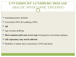
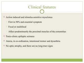
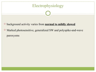
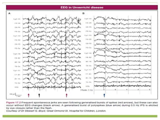
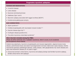
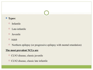
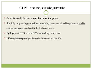
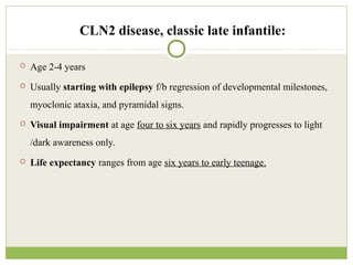
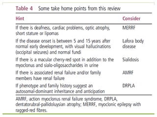
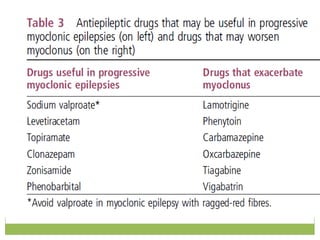
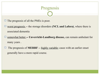
This document discusses progressive myoclonic epilepsy (PME), a group of rare genetic neurological disorders characterized by myoclonus and epileptic seizures with progressive neurological decline. It describes several specific forms of PME, including neuronal ceroid lipofuscinoses (NCLs), Lafora body disease, Unverricht-Lundborg disease, and myoclonic epilepsy with ragged-red fibers. For each, it covers clinical features, genetics, investigations such as EEG and MRI findings, pathology, treatment approaches, and prognosis. The document provides a detailed review and comparison of these progressive myoclonic epilepsy syndromes.
This presentation looks at generalised periodic epileptiform discharges and the various disorders like Creutzfeldt Jacob disease (CJD), SSPE and metabolic encephalopathies in which it is seen. SIRPID is also discussed. Triphasic waves are described. Radermacker complexes in SSPE are described.
Not epileptic
•Wrong seizure type (semiology)
•Wrong epileptic syndrome
•Wrong interpretation of EEG and imaging
When to start a drug?
•Which drug and in what dose?
•When to change the drug?
•When (and how) to add a second drug (and which one)?
•When to stop the drug(s)?
•When to consider alternative therapies, including surgery?
This presentation consist information about unspoken and less well known variants of GBS as well as CIDP. Also it includes information about diagnosis and management.
Dr. john millichap kcnq2 Cure summit professional track learn more at kcnq2cu...scottyandjim
Dr. John Millichap speaking at 2014 Denver KCNQ2 Cure summit professionals track at Children's Hospital of Colorado. More information at www.kcnq2cure.org
Prix Galien International 2024 Forum ProgramLevi Shapiro
June 20, 2024, Prix Galien International and Jerusalem Ethics Forum in ROME. Detailed agenda including panels:
- ADVANCES IN CARDIOLOGY: A NEW PARADIGM IS COMING
- WOMEN’S HEALTH: FERTILITY PRESERVATION
- WHAT’S NEW IN THE TREATMENT OF INFECTIOUS,
ONCOLOGICAL AND INFLAMMATORY SKIN DISEASES?
- ARTIFICIAL INTELLIGENCE AND ETHICS
- GENE THERAPY
- BEYOND BORDERS: GLOBAL INITIATIVES FOR DEMOCRATIZING LIFE SCIENCE TECHNOLOGIES AND PROMOTING ACCESS TO HEALTHCARE
- ETHICAL CHALLENGES IN LIFE SCIENCES
- Prix Galien International Awards Ceremony
New Directions in Targeted Therapeutic Approaches for Older Adults With Mantl...i3 Health
i3 Health is pleased to make the speaker slides from this activity available for use as a non-accredited self-study or teaching resource.
This slide deck presented by Dr. Kami Maddocks, Professor-Clinical in the Division of Hematology and
Associate Division Director for Ambulatory Operations
The Ohio State University Comprehensive Cancer Center, will provide insight into new directions in targeted therapeutic approaches for older adults with mantle cell lymphoma.
STATEMENT OF NEED
Mantle cell lymphoma (MCL) is a rare, aggressive B-cell non-Hodgkin lymphoma (NHL) accounting for 5% to 7% of all lymphomas. Its prognosis ranges from indolent disease that does not require treatment for years to very aggressive disease, which is associated with poor survival (Silkenstedt et al, 2021). Typically, MCL is diagnosed at advanced stage and in older patients who cannot tolerate intensive therapy (NCCN, 2022). Although recent advances have slightly increased remission rates, recurrence and relapse remain very common, leading to a median overall survival between 3 and 6 years (LLS, 2021). Though there are several effective options, progress is still needed towards establishing an accepted frontline approach for MCL (Castellino et al, 2022). Treatment selection and management of MCL are complicated by the heterogeneity of prognosis, advanced age and comorbidities of patients, and lack of an established standard approach for treatment, making it vital that clinicians be familiar with the latest research and advances in this area. In this activity chaired by Michael Wang, MD, Professor in the Department of Lymphoma & Myeloma at MD Anderson Cancer Center, expert faculty will discuss prognostic factors informing treatment, the promising results of recent trials in new therapeutic approaches, and the implications of treatment resistance in therapeutic selection for MCL.
Target Audience
Hematology/oncology fellows, attending faculty, and other health care professionals involved in the treatment of patients with mantle cell lymphoma (MCL).
Learning Objectives
1.) Identify clinical and biological prognostic factors that can guide treatment decision making for older adults with MCL
2.) Evaluate emerging data on targeted therapeutic approaches for treatment-naive and relapsed/refractory MCL and their applicability to older adults
3.) Assess mechanisms of resistance to targeted therapies for MCL and their implications for treatment selection
- Video recording of this lecture in English language: https://youtu.be/lK81BzxMqdo
- Video recording of this lecture in Arabic language: https://youtu.be/Ve4P0COk9OI
- Link to download the book free: https://nephrotube.blogspot.com/p/nephrotube-nephrology-books.html
- Link to NephroTube website: www.NephroTube.com
- Link to NephroTube social media accounts: https://nephrotube.blogspot.com/p/join-nephrotube-on-social-media.html
TEST BANK for Operations Management, 14th Edition by William J. Stevenson, Ve...kevinkariuki227
TEST BANK for Operations Management, 14th Edition by William J. Stevenson, Verified Chapters 1 - 19, Complete Newest Version.pdf
Factory Supply Best Quality Pmk Oil CAS 28578–16–7 PMK Powder in Stockrebeccabio
Factory Supply Best Quality Pmk Oil CAS 28578–16–7 PMK Powder in Stock
Telegram: bmksupplier
signal: +85264872720
threema: TUD4A6YC
You can contact me on Telegram or Threema
Communicate promptly and reply
Free of customs clearance, Double Clearance 100% pass delivery to USA, Canada, Spain, Germany, Netherland, Poland, Italy, Sweden, UK, Czech Republic, Australia, Mexico, Russia, Ukraine, Kazakhstan.Door to door service
Hot Selling Organic intermediates
HOT NEW PRODUCT! BIG SALES FAST SHIPPING NOW FROM CHINA!! EU KU DB BK substit...GL Anaacs
Contact us if you are interested:
Email / Skype : kefaya1771@gmail.com
Threema: PXHY5PDH
New BATCH Ku !!! MUCH IN DEMAND FAST SALE EVERY BATCH HAPPY GOOD EFFECT BIG BATCH !
Contact me on Threema or skype to start big business!!
Hot-sale products:
NEW HOT EUTYLONE WHITE CRYSTAL!!
5cl-adba precursor (semi finished )
5cl-adba raw materials
ADBB precursor (semi finished )
ADBB raw materials
APVP powder
5fadb/4f-adb
Jwh018 / Jwh210
Eutylone crystal
Protonitazene (hydrochloride) CAS: 119276-01-6
Flubrotizolam CAS: 57801-95-3
Metonitazene CAS: 14680-51-4
Payment terms: Western Union,MoneyGram,Bitcoin or USDT.
Deliver Time: Usually 7-15days
Shipping method: FedEx, TNT, DHL,UPS etc.Our deliveries are 100% safe, fast, reliable and discreet.
Samples will be sent for your evaluation!If you are interested in, please contact me, let's talk details.
We specializes in exporting high quality Research chemical, medical intermediate, Pharmaceutical chemicals and so on. Products are exported to USA, Canada, France, Korea, Japan,Russia, Southeast Asia and other countries.
Flu Vaccine Alert in Bangalore Karnatakaaddon Scans
As flu season approaches, health officials in Bangalore, Karnataka, are urging residents to get their flu vaccinations. The seasonal flu, while common, can lead to severe health complications, particularly for vulnerable populations such as young children, the elderly, and those with underlying health conditions.
Dr. Vidisha Kumari, a leading epidemiologist in Bangalore, emphasizes the importance of getting vaccinated. "The flu vaccine is our best defense against the influenza virus. It not only protects individuals but also helps prevent the spread of the virus in our communities," he says.
This year, the flu season is expected to coincide with a potential increase in other respiratory illnesses. The Karnataka Health Department has launched an awareness campaign highlighting the significance of flu vaccinations. They have set up multiple vaccination centers across Bangalore, making it convenient for residents to receive their shots.
To encourage widespread vaccination, the government is also collaborating with local schools, workplaces, and community centers to facilitate vaccination drives. Special attention is being given to ensuring that the vaccine is accessible to all, including marginalized communities who may have limited access to healthcare.
Residents are reminded that the flu vaccine is safe and effective. Common side effects are mild and may include soreness at the injection site, mild fever, or muscle aches. These side effects are generally short-lived and far less severe than the flu itself.
Healthcare providers are also stressing the importance of continuing COVID-19 precautions. Wearing masks, practicing good hand hygiene, and maintaining social distancing are still crucial, especially in crowded places.
Protect yourself and your loved ones by getting vaccinated. Together, we can help keep Bangalore healthy and safe this flu season. For more information on vaccination centers and schedules, residents can visit the Karnataka Health Department’s official website or follow their social media pages.
Stay informed, stay safe, and get your flu shot today!
Couples presenting to the infertility clinic- Do they really have infertility...Sujoy Dasgupta
Dr Sujoy Dasgupta presented the study on "Couples presenting to the infertility clinic- Do they really have infertility? – The unexplored stories of non-consummation" in the 13th Congress of the Asia Pacific Initiative on Reproduction (ASPIRE 2024) at Manila on 24 May, 2024.
2. PROGRESSIVE MYOCLONIC EPILEPSY
“group of familial neurodegenerative disorders c/b myoclonus with
epileptic seizures and progressive neurologic decline”
3.
4. Neurodegenerative, Lysosomal storage disorders
AR
Characterized by progressive intellectual and motor deterioration, seizures, and
early death
Visual loss is a feature of most forms
NEURONAL CEROID-LIPOFUSCINOSES (NCLs)
5. Types:
Infantile
Late-infantile
Juvenile
Adult
Northern epilepsy (or progressive epilepsy with mental retardation)
The most prevalent NCLs are
CLN3 disease, classic juvenile
CLN2 disease, classic late infantile
6. CLN3 disease, classic juvenile
Onset is usually between ages four and ten years.
Rapidly progressing visual loss resulting in severe visual impairment within
one to two years is often the first clinical sign.
Epilepsy – GTCS and/or CPS- around age ten years.
Life expectancy ranges from the late teens to the 30s.
7. CLN2 disease, classic late infantile:
Age 2-4 years
Usually starting with epilepsy f/b regression of developmental milestones,
myoclonic ataxia, and pyramidal signs.
Visual impairment at age four to six years and rapidly progresses to light
/dark awareness only.
Life expectancy ranges from age six years to early teenage.
8. Adult NCL (ANCL)
Onset: around age 30 years,
Death occurs about ten years later.
Ophthalmologic studies are normal.
9. Histopathology and Ultrastructural Studies
Light microscopy:
PAS and Luxol Fast Blue positive, auto fluorescent intracellular ceroid material, neurons and astrocytes
in the grey matter
Electron Microscopy (skin)
(1) Infantile NCL—granular bodies/GRODs
(2) Late infantile NCL—curvilinear bodies (CV)
(3) Juvenile NCL—finger print bodies (FP)
(4) Adult onset NCL -- varied forms and combination of inclusions
Electron microscopy (Brain)
Curvilinear , lamellar and electron dense inclusions in neurons, astrocytes and vascular endothelial
cells
10. MRI findings
Presence of cerebellar/cerebral atrophy, leucoencphalopathy and thalamic
T2W-hypointensity
I-NCL: leucoencphalopathy and thalamic hypointensity (T2W)
LI-NCL: periventricular and parieto-occipital hyperintensities
J-NCL: cerebellar atrophy
11.
12. Treatment
• Lamotrigine (LTG), valproic acid (VPA), clonazepam (CZP)
• Lamotrigine may exacerbate Sz and myoclonus especially in CLN2 disease.
• Benzodiazepines -- benefit for seizures, anxiety, spasticity, and sleep disorders.
• Carbamazepine (CZP) and phenytoin -- may increase seizure activity and
myoclonus
13. LAFORA BODY DISEASE
• Autosomal recessive; stimulus-sensitive PME
• Two genes: Laforin (EPM2A) and Malin (NHLRC1)
• Onset in the late childhood or adolescence
• C/F
• Focal visual occipital seizure
• Myoclonus
• Visual deterioration
• Psychoses
• Rapid intellectual decline with the development of dementia
• Imaging
• Diffuse cortical atrophy without any parenchymal changes
14. Electrophysiology
• EEG background slows, alpha-rhythm and sleep features are lost with
progression, and photosensitivity with fast frequency (>30 Hz)
stimulation
• replete with paroxysms of generalized irregular spike-wave
discharges with occipital predominance and focal, especially
occipital abnormalities
• Giant SSEP, VEP: Enhanced cortical excitability
15. Pathology
• Lafora body inclusions
• Oval to round shaped PAS positive, diastase resistant
• Positive for Lugol’s Iodine and ubiquitin immune-staining
• Inclusions (Lafora bodies) are seen in the cerebral and cerebellar cortex and
in brain stem nuclei
• Inclusions are also seen in other organs including liver, muscle, and skin
16. Treatment
Valproic acid : controls both GTCS and myoclonic jerks
Clonazepam - adjunctive
Zonisamide - both seizures and myoclonus
piracetam and levetiracetam - add-on treatment
17. UNVERRICHT LUNDBERG DISEASE
(BALTIC MYOCLONIC EPILEPSY)
neurodegenerative disorder
Unverricht (1891) & Lundborg (1903)
AR
Age of onset: 6-15 yrs
Most common and Least severe type of progressive myoclonus epilepsy
Life expectancy may not be affected
Disability is mainly due to myoclonus, GTCS and ataxia
18. Clinical features
Action induced and stimulus-sensitive myoclonus
First in 50% and essential symptom
Focal or multifocal
Affect predominantly the proximal muscles of the extremities
Tonic-clonic epileptic seizures
Ataxia, in co-ordination, intentional tremor and dysarthria
No optic atrophy, and there are no long-tract signs
21. Pathophysiology
Defective function of cystatin B, a cysteine protease inhibitor, as a
consequence of mutations in CSTB
The causative gene, EPM1, localized to chromosome 21q22.3
22. Treatment
• Symptomatic rehabilitative management are the mainstay
• Valproic acid: Drug of choice -- Diminishes myoclonus and freq of generalized
seizures
• Clonazepam: Only drug approved by FDA for myoclonic seizures --add-on
• Levetiracetam : -- effective for both myoclonus and generalized seizures
• Topiramate & zonisamide: Add-on
• High-dose piracetam -- useful in the treatment of myoclonus only
23. • Sodium channel blockers : should be avoided
• (carbamazepine, oxcarbazepine, phenytoin)
• GABA ergic drugs (tiagabine, vigabatrin)
• gabapentin and pregabalin
• May aggravate myoclonus and myoclonic seizures.
24. MYOCLONIC EPILEPSY WITH RAGGED-RED
FIBERS (MERRF)
Mitochondrial cytopathy
Mean age at onset 14.6 ± 5.8 years
Maternal inheritance
Mutations in the MT-TK gene are the most common cause of MERRF,
occurring in more than 80%
25. Clinical features
Myoclonus, myopathy and spasticity
Seizures, ataxia, peripheral neuropathy and dementia
Deafness and optic atrophy
Short stature and heart abnormalities, cardiomyopathy
Lipomas
26. Electrophysiology and Imaging
Electrophysiology
EEG:
Slowing of background activity
Generalized epileptiform discharges
ENMG:
Neuropathy and myopathy
SSEP
Giant potentials
Imaging
Diffuse atrophy of cerebrum, brainstem, and cerebellum
Basal ganglia calcification
27. Dentato rubral-pallidoluysian atrophy
AD
Triplet repeat expansion
Adoloscent or childhood onset
Ataxia, choreoathetosis, dementia
Neuronal loss and gliosis in dentatorubral and pallidoluysian systems
28. Differential diagnosis to PME
IGE syndrome patients treated with inappropriate AED
LGS/Symptomatic generalised epilepsy
Progressive encephalopathies with seizures
(Myoclonus is not the clinical core)
GM2 gangliosidosis
Non-ketotic hyperglycinemia
Niemen pick type C
Juvenile Huntington’s disease
Alzheimer's disease
29. Post anoxic myoclonus- No progression
Progressive myoclonic ataxia- No evidence of dementia
Overlaps with spinocerebellar ataxia, celiac disease, whipple disease
Benign myoclonic epilepsy of childhood and adult hood.
Benign familial myoclonus
30. PME – Neuro-ophthalmology
PME syndrome
Retinal degeneration/optic atrophy – MERRF
NCL
LBD
Cherry red spot – Neurosialidosis
NCL
With visual symptoms Without visual symptoms
ULD
37. Prognosis
The prognosis of all the PMEs is poor.
worst prognosis -- the storage disorders (NCL and Lafora), where there is
associated dementia
somewhat better -- Unverricht-Lundborg disease, can remain ambulant for
many years .
The prognosis of MERRF -- highly variable; cases with an earlier onset
generally have a more rapid course.
38. References
1. CP Panayiotopoulos. A Clinical Guide to Epileptic Syndrome and their
Treatment. Diseases frequently associated with epileptic seizures. Revised 2nd
edition. London. Springer Healthcare Ltd, 2010; 233-254.
2. Malek N, et al. The progressive myoclonic epilepsies. Pract Neurol
2015;15:164–171.
3. P. Satishchandra, S. Sinha. Progressive myoclonic epilepsy. Neurology India
2010.
4. Franceschetti S,Michelucci R,et al.Progressive myoclonic epilepsies: Definitive
and still undetermined causes. Neurology 2014;82;405-411
5. Shahwan A, Farrell M, Delanty N. Progressive myoclonic epilepsies: a review
of genetic and therapeutic aspects. Lancet Neurol 2005; 4: 239–48