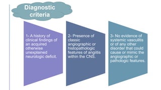
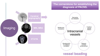
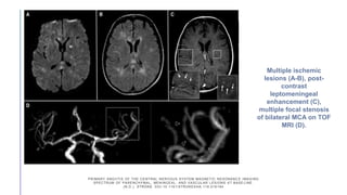
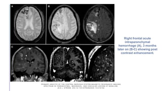
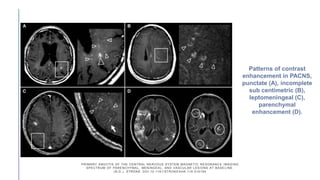
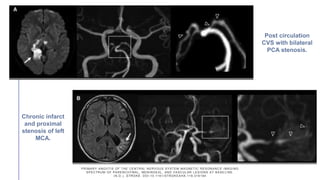
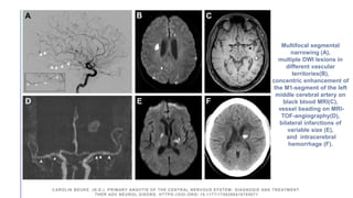
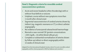
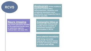
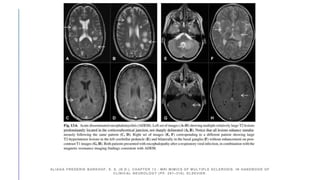
SlideShare a Scribd company logo
PRIMARY CNS
VASCULITIS
(PACNS)
By/ Lobna Ahmed
• Primary angiitis of the central nervous system (PACNS) is a rare inflammatory disorder of the blood
vessels affects the blood vessels supplying the brain parenchyma, spinal cord and leptomeninges,
and less frequently veins and venules, occurring in the absence of any evidence of systemic
vasculitis.
• Clinical manifestations are non-specific with various presenting symptoms.
• Headache is the most common presenting symptom in the majority of the PACNS cases.
• The exact etiology and pathogenesis of PACNS are unknown, but infectious agents have been
postulated as one of the triggers (VZV, HIV, HCV, Bacterial).
• There is also a notable association between PACNS and cerebral amyloid angiopathy. The amyloid
deposition has been identified as a possible trigger for PACNS in animal models.
The exact epidemiology of this rare
disorder have not been done (some data
shows an annual incidence rate of 2.4
cases per 1 million person-years).
Equal distribution among both sexes
and the median age of diagnosis of
about 50 years.
Epidemiology
PACNS mainly
affects younger
stroke patients
lacking CVS risk
factors.
Approximately 3-5%
of CVS in patients
<50 years are
caused by PACNS.
 Pathological findings can affect both
small and large vessels of the CNS.
 Specific activation of the immune system
(especially T-cells) by various cause
inflammation of blood vessels in the CNS.
 Immunohistochemical staining of biopsy
samples in PACNS showed an extensive
infiltration around the small cerebral arteries
by CD45R0+ T cells.
Infiltration of
immune cells
within vessels wall
destruction of the
vessels walls
Thickening of
vessels walls with
alternating
segments of
stenosis
Poor blood
circulation
Pathophysiology
On the other hand,
weak vessels walls can
rupture leading to
hemorrhage.
 Affection of parenchymal and leptomeningeal
arteries.
 Three different types of vasculitis patterns
have been described in PACNS
(granulomatous type, necrotizing type, and
lymphocytic type).
 The most common type, granulomatous,
shows numerous granulomas with
multinucleated cells.
 The necrotizing type presents with fibrinoid
necrosis, and lymphocytic type shows
extensive lymphocytic inflammation with
plasma cells.
Granulomatous
Necrotizing
Lymphocytic
Histopathology
The onset of PACNS is usually insidious, and the course
is slowly progressive. Acute presentations have also
been reported but are, however, less common.
Clinical
presentation
Non-specific
Multiple
symptoms
Headache is the most
common presenting
symptom
•Cognitive dysfunction
•Stroke
•Transient ischemic attack (TIA)
•Aphasia
•Visual symptoms including visual field
deficits, blurred vision, and double
vision
•Seizures
•Ataxia
•Papilledema
•Intracranial bleeding
•Amnestic syndrome
The presenting symptoms and signs from the
most common to the least common include:
Headache 60%
Cognitive
impairment 50%
Focal neurological
deficits
Marked constitutional symptoms
such as fever, weight loss and night
sweats are less frequent and can be
indicative of systemic vasculitides.
In about 5% of patients primary CNS
vasculitis can affect the spinal cord
and isolated vasculitic myelopathies
have also been reported.
A combination of symptoms is
usually present in most patients.
PACNS should always be
considered as a possibility in
cases of rapidly progressive
cognitive decline and personality
changes of unknown etiology.
Diagnostic
criteria
1- A history of
clinical findings of
an acquired
otherwise
unexplained
neurologic deficit.
2- Presence of
classic
angiographic or
histopathologic
features of angiitis
within the CNS.
3- No evidence of
systemic vasculitis
or of any other
disorder that could
cause or mimic the
angiographic or
pathologic features.
Definite • Confirmed by tissue biopsy.
Probable
• High probability finding on an angiogram with
abnormal findings on MRI and a CSF profile
consistent with PACNS (in the absence of tissue
confirmation).
Diagnostic
certainty
Subtypes of PACNS
Small vessels
Large/medium
vessels
According to the size
of affected vessel
In contrast, LV-PACNS is
more likely to present with
focal deficits and acute
ischemia on MRI.
Isolated SV-PACNS is associated with:
-A more severe encephalopathic
presentation with cognitive impairment
and seizures.
-A more abnormal CSF analysis and
more diffuse gad enhanced lesions on
MRI.
-More relapses.
CAROLIN BEUKE. (N.D.). PRIMARY ANGIITIS OF THE CENTRAL NERVOUS SYSTEM: DIAGNOSIS AND TREATMENT.
THER ADV NEUROL DISORD. HTTPS://DOI.ORG/ 10.1177/1756286418785071
Evaluation
Laboratory
• Detection of ANA, RF, ANCA, antiphospholipid Ab,
bacterial or viral Ag is usually suggestive of systemic
vasculitis or auto-immune disease.
• Elevated ESR and CRP neither sensitive nor specific.
• Serological tests are needed to exclude infection
including varicella, mycobacteria, syphilis, human HIV,
and fungi.
• No specific marker for PACNS.
CSF
The only useful laboratory investigation is CSF examination
which shows an abnormality in more than 90% of the cases.
Should be performed in all patients.
Inflammatory
findings:
lymphocytic
pleocytosis,
elevated
protein.
Normal CSF (cell count
<5 cells /μl or total
protein concentration
<45 mg/dl) >> search for
DD.
OCB and IgG are only
found occasionally.
Increased interleukin
(IL)17-producing CD41
cells in CSF can
differentiate PACNS
from ischemic stroke.
Values more than 250
cells/μl are indicative for
infection.
Malignant vasculitis can
be detected with CSF
cytology and flow
cytometry.
CSF
The cornerstone for establishing the
diagnosis of PACNS.
Imaging
Cerebral
angiography
MRI, MRA
CT,CTA
vessel beading
Intracranial
vessels
Narrowing
and dilatation
Fusiform
arterial
dilation
Delayed
contrast
enhancement
Development
of collateral
circulation
Multilocular
occlusions
Vessel beading is not
a specific feature for
PACNS
It is also seen in:
• Noninflammatory vasculopathies.
• Non-vasculitis conditions such as
atherosclerosis.
• After radiation.
• Neurofibromatosis.
• Atrial myxomas.
• Infections.
• Vasospasm.
DSA is the gold
standard imaging.
It’s not 100% specific.
Possibly due to the
waxing and waning
pattern of stenosis.
VALERIE L JEWELLS. (N.D.). CNS VASCULITIS; AN OVERVIEW OF THIS M ULTIPLE SCLEROSIS MIMIC; CLINICAL AND MRI
IMPLICATIONS. SEMINARS IN ULTRASOUND CT AND MRI. HTTPS://DOI.ORG /HTTPS://DOI.ORG/10.1053/J.SULT.2020.02.004
 MRI of the brain is abnormal in more than 90%
of patients, but the pattern of abnormal findings
is not specific.
 MRI should include T1, T2, FLAIR, DWI, ADC,
GRE, with and without gadolinium
administration.
Multifocal bilateral
T2, FLAIR lesions
in cortical,
subcortical, DWM
and gray matter.
New and old
coexisting ischemic
lesions.
Lesions can be
accompanied by
subarachnoid and
ICH.
Mass lesions that
can mimic a tumors
or abscess.
Gd enhancement of
parenchymal
lesions and the
leptomeninges.
Imaging
Cerebral
angiography
MRI, MRA
CT,CTA
MRI/MRA is the first
diagnostic test of choice.
VALERIE L JEWELLS. (N.D.). CNS VASCULITIS; AN OVERVIEW OF THIS M ULTIPLE SCLEROSIS MIMIC; CLINICAL AND MRI
IMPLICATIONS. SEMINARS IN ULTRASOUND CT AND MRI. HTTPS://DOI.ORG /HTTPS://DOI.ORG/10.1053/J.SULT.2020.02.004
MRA is a less invasive imaging modality and therefore can be used to monitor the
disease course, however, in comparison with DSA, MRA is less sensitive in the
detection of both lesions localized in the posterior circulation and in distal arteries.
Occipital
Temporal
Parietal
Frontal
WM lesions
distribution
2/3rd of CNSV cases
have WM involvement.
MCA>PCA>ACA
MRI
protocol
 Standard protocol is 3D T1, T2, FLAIR, and post contrast imaging
with MRA, perfusion and SWI (for depicting hemosiderin from
intraparenchymal hemorrhages and hemosiderosis).
 Recently, vessel wall imaging can be used as a part of the protocol
to detect vessel wall inflammation, preferentially performed at 3T
via black blood sequences using T2 TSE or T2* weighted-GRE
sequences with FLAIR and TOF MRI as well as fat sat T1 images.
VALERIE L JEWELLS. (N.D.). CNS VASCULITIS; AN OVERVIEW OF THIS M ULTIPLE SCLEROSIS MIMIC; CLINICAL AND MRI
IMPLICATIONS. SEMINARS IN ULTRASOUND CT AND MRI. HTTPS://DOI.ORG /HTTPS://DOI.ORG/10.1053/J.SULT.2020.02.004
 Vessel wall imaging uses different techniques to
suppress the signal of intraluminal blood (“black
blood imaging”), thus allowing evaluation of the
vessel wall and possibly the detection of
inflammatory changes within the vessel wall.
 Vessel wall contrast enhancement has been
reported as a potential sign of CNS vasculitis
 In particular, differentiating between inflammation,
intracranial atherosclerotic plaques, and other wall
abnormalities based on the typical enhancement
patterns was reported.
High resolution
contrast-enhanced
MRI
(HR-MRI)
“Black Blood MRI”
PATZIG, M., FORBRIG, R., KÜPPER, C. ET AL. DIAGNOSIS AND FOLLOW-UP EVALUATION OF CENTRAL NERVOUS SYSTEM
VASCULITIS: AN EVALUATION OF VESSEL-WALL MRI FINDINGS. J NEUROL 269, 982–996 (2022). HTTPS://DOI.ORG/10.1007/S00415-
021-10683-7
 CT /CTA may demonstrate occlusion, stenosis,
and wall thickening of large vessels.
 It’s limitations are insensitivity for WM
assessment, leptomeningeal enhancement or
depiction of small vessels.
Imaging
Cerebral
angiography
MRI, MRA
CT,CTA
PRIMARY ANGIITIS OF THE CENTRAL NERVOUS SYSTEM MAGNETIC RESONANC E IMAGING
SPECTRUM OF PARENCHYMAL, MENINGEAL, AND VASCULAR LESIONS AT BASE LINE.
(N.D.). STROKE. DOI:10.1161/STROKEAHA.116.016194
Multiple ischemic
lesions (A-B), post-
contrast
leptomeningeal
enhancement (C),
multiple focal stenosis
of bilateral MCA on TOF
MRI (D).
PRIMARY ANGIITIS OF THE CENTRAL NERVOUS SYSTEM MAGNETIC RESONANC E IMAGING
SPECTRUM OF PARENCHYMAL, MENINGEAL, AND VASCULAR LESIONS AT BASE LINE.
(N.D.). STROKE. DOI:10.1161/STROKEAHA.116.016194
Right frontal acute
intraparenchymal
hemorrhage (A), 3 months
later on (B-C) showing post
contrast enhancement.
PRIMARY ANGIITIS OF THE CENTRAL NERVOUS SYSTEM MAGNETIC RESONANC E IMAGING
SPECTRUM OF PARENCHYMAL, MENINGEAL, AND VASCULAR LESIONS AT BASE LINE.
(N.D.). STROKE. DOI:10.1161/STROKEAHA.116.016194
Patterns of contrast
enhancement in PACNS,
punctate (A), incomplete
sub centimetric (B),
leptomeningeal (C),
parenchymal
enhancement (D).
PRIMARY ANGIITIS OF THE CENTRAL NERVOUS SYSTEM MAGNETIC RESONANC E IMAGING
SPECTRUM OF PARENCHYMAL, MENINGEAL, AND VASCULAR LESIONS AT BASE LINE.
(N.D.). STROKE. DOI:10.1161/STROKEAHA.116.016194
Post circulation
CVS with bilateral
PCA stenosis.
Chronic infarct
and proximal
stenosis of left
MCA.
Multifocal segmental
narrowing (A),
multiple DWI lesions in
different vascular
territories(B),
concentric enhancement of
the M1-segment of the left
middle cerebral artery on
black blood MRI(C),
vessel beading on MRI-
TOF-angiography(D),
bilateral infarctions of
variable size (E),
and intracerebral
hemorrhage (F).
CAROLIN BEUKE. (N.D.). PRIMARY ANGIITIS OF THE CENTRAL NERVOUS SYSTEM: DIAGNOSIS AND TREATMENT.
THER ADV NEUROL DISORD. HTTPS://DOI.ORG/ 10.1177/1756286418785071
-Vessel wall contrast enhancement of
the right distal M1 segment is seen at
initial presentation on vessel wall
imaging (A), which remains unchanged
at two-months follow-up (B) despite
immunosuppressive therapy.
-Correlating TOF-MRA findings (C, D),
showing unchanged high-grade
stenosis of the affected segment.
PATZIG, M., FORBRIG, R., KÜPPER, C. ET AL. DIAGNOSIS AND FOLLOW-UP EVALUATION OF CENTRAL NERVOUS SYSTEM
VASCULITIS: AN EVALUATION OF VESSEL-WALL MRI FINDINGS. J NEUROL 269, 982–996 (2022). HTTPS://DOI.ORG/10.1007/S00415-
021-10683-7
-At initial presentation (A, C), there is marked
vessel wall contrast enhancement of the
posterior circulation, including the basilar
artery (arrow) and left PCOM (arrowhead).
-Follow-up vessel wall imaging after ten years
(B, D) shows complete resolution of vessel
wall contrast enhancement of the PCOM and
regressive but still persistent vessel wall
contrast enhancement of the basilar artery.
-Correlating TOF-MRA images (E, F)
demonstrate resolution of a high-grade
stenosis of the left PCOM.
PATZIG, M., FORBRIG, R., KÜPPER, C. ET AL. DIAGNOSIS AND FOLLOW-UP EVALUATION OF CENTRAL NERVOUS SYSTEM
VASCULITIS: AN EVALUATION OF VESSEL-WALL MRI FINDINGS. J NEUROL 269, 982–996 (2022). HTTPS://DOI.ORG/10.1007/S00415-
021-10683-7
Angio
Positive
• In large/ proximal
vessel affection.
Angio
Normal
• In small vessel (<500
μm in diameter)
affection.
In case of negative
angiographic findings >>
brain biopsy should be
considered as an important
step towards diagnosing
PACNS.
Biopsy
• Due to the focal and segmental distribution of the disease,
the sensitivity of brain biopsy is as a result of sampling
errors between 53–74%.
• The diagnostic yield can be increased by 80% by targeting
areas of imaging abnormalities.
• If affected lesions are not accessible for surgery, a biopsy
from the right (nondominant) frontal lobe with its overlying
leptomeninges tissue is recommended.
• Negative biopsy cannot rule out the diagnosis of cerebral
vasculitis, but histopathological examination often results
in alternative diagnoses (special attention is paid to PACNS
mimics like infections, neoplasms especially lymphoma,
and degenerative disease.
Biopsy
is safe! Brain biopsies are associated with rather low morbidity and
mortality rates; therefore, a biopsy should be obtained, if the
diagnosis is uncertain.
CAROLIN BEUKE. (N.D.). PRIMARY ANGIITIS OF THE CENTRAL NERVOUS SYSTEM: DIAGNOSIS AND TREATMENT.
THER ADV NEUROL DISORD. HTTPS://DOI.ORG/ 10.1177/1756286418785071
Differential
diagnosis
Non-
inflammatory
vascular
diseases.
Malignancy.
Systemic
inflammatory
diseases.
Infection.
RCVS
Formerly RCVS
was described as
a subtype of
PACNS, called
benign angiopathy
of the CNS.
Typical clinical features are sudden and
recurrent attacks of severe (usually
thunderclap) HEADACHE with or without
focal neurological deficits or seizures.
More in
Females.
Mean age of
onset 42
yrs.
The most
important non-
inflammatory
DD to PACNS.
Headache
RCVS is more often detected in patients
with exposure to vasoactive drugs,
migraine, hypertension, eclampsia or in
the postpartum period.
RCVS
Neuro imaging
may show infarctions (located
in the superficial border zone
or watershed regions),
cortical SAH, and lobar ICH.
Angiogram shows multifocal
segmental cerebral artery
vasoconstriction; dissection and
unruptured aneurysms occur
significantly more frequently than in
cerebral vasculitis.
Angiographic follow up
12 weeks after clinical onset
usually shows complete or
substantial resolution of
abnormalities in RCVS.
Intra-arterial nimodipine
use during convential
angiography showed reverse
of vessel narrowing in RCVS
in contrast with PACNS.
A-CT scan showing a small
cSAH,
B-MRI (FLAIR) showing a small
cSAH,
C- CT scan showing an
occipital intracerebral
haemorrhage,
D-MRI showing sequelae of
bilateral occipital infarcts and
left frontal-parietal infarct,
E-MRI (FLAIR) showing
hypersignals consistent with a
RPLS,
F-Control MRI in the same
patient after 28 days showing
resolution of the RPLS.
Diffuse intracerebral vasoconstriction on initial angiogram, after 2 wk of
headache then right hemiparesis and encephalopathy. B, Resolution of
vasoconstriction after 4 mo.
Systemic
vasculiti
s
CNS involvement
most often arises in
patients with:
 Bechet's disease.
 Wegener’s granulomatosis.
 Churg–Strauss syndrome.
Bechet’s
=
Variable vessel
size vasculitis
Large
vessel
vasculitis
 Involves >2mm size vessels, including the ICA,
M1 segment of the MCA, A1 segment of the
ACA, and P1 segment of the PCA.
 Takayasu’s arteritis (<50 yo), Giant cell arteritis
and temporal arteritis (>50 yo) are common
large vessel causes.
Medium
size vessel
vasculitis
 Most commonly Kawasaki’s
disease and polyarteritis
nodosa involve the M2, A2,
and P2 segments.
Small
vessel
vasculitis
 SLE, granulomatosis
with polyangiitis +/-
eosinophils, IgA
vasculitis, and
microscopic polyangiitis
Vasculitis
2nd to CT
diseases
SLE
Sjogren’s
syndrome
Dermatomyositis
Mixed CT
disease
RA
 Moyamoya angiopathy.
 Divry-van Bogaert syndrome.
 fibromuscular dysplasia.
 Fabry disease.
 Sneddon’s syndrome.
Non-
inflammatory
vascular
diseases
Malignancy
 In single cases, the occurrence of PACNS in
association with Hodgkin’s lymphoma, non- Hodgkin’s
lymphoma and angio-immuno-lympho-proliferative
lesions has been reported.
 Furthermore, intravascular lymphoma (IVL), a subtype
of extra-nodal diffuse large B-cell lymphoma, is an
important differential diagnosis of primary CNS
vasculitis characterized by intravascular proliferation
of lymphoma cells with a predilection for the CNS and
skin.
Infection
VZV vasculopathy:
 An infectious arteritis that causes ischemic infarction of the
brain and spinal cord, and cerebral hemorrhage, as well as
aneurysm and carotid dissection.
 Diagnosis of VZV infection can be made by detection of
anti-VZV IgG antibody in the CSF.
PACNS should be distinguished from other infectious agents
that can affect the CNS including:
Hepatitis C virus, HIV, Streptococcus pneumoniae, Neisseria
meningitidis, Bartonella spp ,Mycobacterium tuberculosis,
Borrelia burgdorferi, and Treponema pallidum.
Other
mimics
CADASIL
• An AD arteriopathy with
manifestation in early
adulthood.
• Clinical symptoms include:
migraine, recurrent
transient ischemic
attacks or strokes,
psychiatric disorders,
cognitive impairment,
and epilepsy.
• Brian MRI typically shows
large or small white matter
abnormalities.
• The most typical findings
are white matter
hyperintensities in the
anterior temporal lobe
(temporal pole) seen on T2
sequence.
Susac syndrome
• Autoimmune-mediated
endotheliopathy
characterized by the
clinical triad of:
Branch retinal artery
occlusions.
Encephalopathy.
Neurosensorial hearing
loss.
• It causes microangiopathic
occlusion of precapillary
arterioles in the brain,
retina, and inner ear.
Anti-phospholipid syndrome
• Present by diverse
neurologic manifestations
including: stroke, TIA,
seizures, movement
disorders,
neuropsychiatric
manifestations, and
cognitive decline.
• Angiography shows
intracranial abnormalities
suggestive of vasculitis.
ALIAGA FREDERIK BARKHOF, E. S. (N.D.). CHAPTER 13 - MRI MIMICS OF MULTIPLE SCLEROSIS. IN HANDBOOK OF
CLINICAL NEUROLOGY (PP. 291–316). ELSEVIER.
ADEM
Other
mimics
ALIAGA FREDERIK BARKHOF, E. S. (N.D.). CHAPTER 13 - MRI MIMICS OF MULTIPLE SCLEROSIS. IN HANDBOOK OF
CLINICAL NEUROLOGY (PP. 291–316). ELSEVIER.
ALIAGA FREDERIK BARKHOF, E. S. (N.D.). CHAPTER 13 - MRI MIMICS OF MULTIPLE SCLEROSIS. IN HANDBOOK OF
CLINICAL NEUROLOGY (PP. 291–316). ELSEVIER.
Treatment
Induction
therapy
Maintenance
therapy
Corticosteroids Immunosuppressants Biologics
Induction
therapy
Combination
therapy
Steroids Cyclophosphamide
High-dose of IV CST (1000 mg daily for
3–5 days),
or oral prednisone (1 mg/kg per day) are
currently the most frequently used
therapies.
IV is not more
effective than
oral!
Given either as an oral dose
(2 mg/kg/day) for 3–6 months,
or as IV pulse (750 mg/m2/
month) for 6 months.
Monitor:
 Leukocyte nadir and disease course.
 Side effects (infection, especially
bladder, cancer and infertility).
 It is contraindicated during pregnancy.
In patients who are intolerant to conventional immunosuppressive
therapeutic regimens ..
or in patients failing to respond to cyclophosphamide therapy ..
Biological
agents
Rituximab
TNF-α blockers
(infliximab and
etanercept)
Maintenance
therapy
Goal:
Limiting the
risk of relapses and
preventing long-term
disabilities.
Started 4–6 months
after initiation of the
induction therapy.
Corticosteroid-sparing low-
risk immunosuppressive
agents
Azathioprine Methotrexate
Mycophenolate
mofetil
1-2 mg/kg/day
1-2 g/day
20-25 mg/week
CAROLIN BEUKE. (N.D.). PRIMARY ANGIITIS OF THE CENTRAL NERVOUS SYSTEM: DIAGNOSIS AND TREATMENT.
THER ADV NEUROL DISORD. HTTPS://DOI.ORG/ 10.1177/1756286418785071
Duration of
treatment
• Validated data are lacking. In principle, induction
therapy should be administered for 6–12 months
based on individual response to treatment.
• Decisions on de-escalation of induction therapy
should depend on the achievement of clinical
stability.
Prognosis
• Older patients and those with infarctions on the MRI
scan had an increased mortality rate.
• Meningeal gadolinium enhancements on MRI and
seizures were associated with an increased risk of
relapse.
• Involvement of larger or rather proximal cerebral
vessels seems to be associated with a higher mortality
rate and poorer prognosis, requiring a more
aggressive treatment.
Disease monitoring
• Combination of repeated neurological examinations and periodic
neuroradiological imaging (e.g. MRI and MRA) during therapy and
afterwards is recommended for assessing disease activity.
• MRI should be performed 4–6 weeks after the initiation of therapy and
afterwards every 3–4 months in the first year of treatment.
• CSF examinations can additionally be helpful in the follow up to
document improvement in the inflammatory response.
• In patients with severe clinical course and worsening neurological
symptoms, serial conventional angiography might be necessary.
• Color duplex sonography might be useful for follow-up examinations
in patients with cerebral artery stenoses
Primary cns vasculitis

More Related Content

What's hot

Nmosd & mog
Nmosd & mogNmosd & mog
Nmosd & mog
NeurologyKota
 
Vasculitis undergrad: diagnosis & treatment.
Vasculitis undergrad: diagnosis & treatment.Vasculitis undergrad: diagnosis & treatment.
Vasculitis undergrad: diagnosis & treatment.
Faculty of Medicine, Ain Shams University
 
Inflammatory Myopathies
Inflammatory MyopathiesInflammatory Myopathies
Inflammatory Myopathies
Ipsita Panda
 
A Review on Neurosarcoidosis Dec 21, 2016
A Review on Neurosarcoidosis Dec 21, 2016A Review on Neurosarcoidosis Dec 21, 2016
A Review on Neurosarcoidosis Dec 21, 2016
Prisma Health Upstate
 
Approach to Leukodystrophy
Approach to Leukodystrophy Approach to Leukodystrophy
Approach to Leukodystrophy
NeurologyKota
 
Approach to peripheral neuropathy
Approach to peripheral neuropathyApproach to peripheral neuropathy
Approach to peripheral neuropathy
NeurologyKota
 
Autoimmune encephalitis current concepts
Autoimmune encephalitis current conceptsAutoimmune encephalitis current concepts
Autoimmune encephalitis current concepts
NeurologyKota
 
Approach to demyelinating diseases
Approach to demyelinating diseasesApproach to demyelinating diseases
Approach to demyelinating diseases
NeurologyKota
 
Stroke in children and young adult
Stroke in children and young adultStroke in children and young adult
Stroke in children and young adult
dahmed hamed
 
Stroke classification.pptx
Stroke classification.pptxStroke classification.pptx
Stroke classification.pptx
ArpanDutta51
 
Reversible cerebral vasoconstriction syndrome
Reversible cerebral vasoconstriction syndromeReversible cerebral vasoconstriction syndrome
Reversible cerebral vasoconstriction syndrome
Prisma Health Upstate
 
Neuroradiology in ms
Neuroradiology in msNeuroradiology in ms
Neuroradiology in ms
Amr Hassan
 
Autoimmune encephalitis ppt
Autoimmune encephalitis pptAutoimmune encephalitis ppt
Autoimmune encephalitis ppt
Sachin Adukia
 
Myelopathy 1
Myelopathy 1Myelopathy 1
Myelopathy 1
sunil bhatt
 
Autoimmune encephalitis
Autoimmune encephalitisAutoimmune encephalitis
Autoimmune encephalitis
NeurologyKota
 
Progressive multifocal leukoencephalopathy (PML)
Progressive multifocal leukoencephalopathy (PML)Progressive multifocal leukoencephalopathy (PML)
Progressive multifocal leukoencephalopathy (PML)
Amr Hassan
 
Iv thrombolysis in clinical practicefinal 11082021
Iv thrombolysis in clinical practicefinal 11082021Iv thrombolysis in clinical practicefinal 11082021
Iv thrombolysis in clinical practicefinal 11082021
Gillian Gordon Perue
 
Approach to a patient with vasculitis
Approach to a patient with vasculitisApproach to a patient with vasculitis
Approach to a patient with vasculitis
aminanurnova
 

What's hot (20)

Nmosd & mog
Nmosd & mogNmosd & mog
Nmosd & mog
 
Vasculitis undergrad: diagnosis & treatment.
Vasculitis undergrad: diagnosis & treatment.Vasculitis undergrad: diagnosis & treatment.
Vasculitis undergrad: diagnosis & treatment.
 
Inflammatory Myopathies
Inflammatory MyopathiesInflammatory Myopathies
Inflammatory Myopathies
 
A Review on Neurosarcoidosis Dec 21, 2016
A Review on Neurosarcoidosis Dec 21, 2016A Review on Neurosarcoidosis Dec 21, 2016
A Review on Neurosarcoidosis Dec 21, 2016
 
Approach to Leukodystrophy
Approach to Leukodystrophy Approach to Leukodystrophy
Approach to Leukodystrophy
 
Approach to peripheral neuropathy
Approach to peripheral neuropathyApproach to peripheral neuropathy
Approach to peripheral neuropathy
 
Autoimmune encephalitis current concepts
Autoimmune encephalitis current conceptsAutoimmune encephalitis current concepts
Autoimmune encephalitis current concepts
 
Approach to demyelinating diseases
Approach to demyelinating diseasesApproach to demyelinating diseases
Approach to demyelinating diseases
 
Stroke in children and young adult
Stroke in children and young adultStroke in children and young adult
Stroke in children and young adult
 
Stroke classification.pptx
Stroke classification.pptxStroke classification.pptx
Stroke classification.pptx
 
Reversible cerebral vasoconstriction syndrome
Reversible cerebral vasoconstriction syndromeReversible cerebral vasoconstriction syndrome
Reversible cerebral vasoconstriction syndrome
 
Neuroradiology in ms
Neuroradiology in msNeuroradiology in ms
Neuroradiology in ms
 
Autoimmune encephalitis ppt
Autoimmune encephalitis pptAutoimmune encephalitis ppt
Autoimmune encephalitis ppt
 
Myelopathy 1
Myelopathy 1Myelopathy 1
Myelopathy 1
 
Ring enhancing lesions
Ring enhancing lesionsRing enhancing lesions
Ring enhancing lesions
 
Normal pressure hydrocephalus
Normal pressure hydrocephalusNormal pressure hydrocephalus
Normal pressure hydrocephalus
 
Autoimmune encephalitis
Autoimmune encephalitisAutoimmune encephalitis
Autoimmune encephalitis
 
Progressive multifocal leukoencephalopathy (PML)
Progressive multifocal leukoencephalopathy (PML)Progressive multifocal leukoencephalopathy (PML)
Progressive multifocal leukoencephalopathy (PML)
 
Iv thrombolysis in clinical practicefinal 11082021
Iv thrombolysis in clinical practicefinal 11082021Iv thrombolysis in clinical practicefinal 11082021
Iv thrombolysis in clinical practicefinal 11082021
 
Approach to a patient with vasculitis
Approach to a patient with vasculitisApproach to a patient with vasculitis
Approach to a patient with vasculitis
 

Similar to Primary cns vasculitis

Pediatric vasculitides
Pediatric vasculitidesPediatric vasculitides
Pediatric vasculitidesdattasrisaila
 
Vasculitis 2015 undergraduate
Vasculitis 2015 undergraduateVasculitis 2015 undergraduate
Vasculitis 2015 undergraduate
Faculty of Medicine, Ain Shams University
 
CEREBRAL VENOUS THROMBOSIS
CEREBRAL VENOUS THROMBOSISCEREBRAL VENOUS THROMBOSIS
CEREBRAL VENOUS THROMBOSIS
Srirama Anjaneyulu
 
Central nervous system vasculitis
Central nervous system vasculitisCentral nervous system vasculitis
Central nervous system vasculitisMohammad Baghbanian
 
Vasculitic neuropathies.pptx
Vasculitic neuropathies.pptxVasculitic neuropathies.pptx
Vasculitic neuropathies.pptx
NeurologyKota
 
cerebral vascular malformation
cerebral vascular malformationcerebral vascular malformation
cerebral vascular malformation
MojtabaKhazaei2
 
SVCS.pptx
SVCS.pptxSVCS.pptx
Radiological pathology of spontaneous cerebral hemorrhage
Radiological pathology of spontaneous cerebral hemorrhageRadiological pathology of spontaneous cerebral hemorrhage
Radiological pathology of spontaneous cerebral hemorrhage
Professor Yasser Metwally
 
Mok presentation
Mok presentationMok presentation
Mok presentation
Moustafa Mokarrab
 
Common disorders misdiagnosed as ms
Common disorders misdiagnosed as msCommon disorders misdiagnosed as ms
Common disorders misdiagnosed as ms
Osama Ragab
 
Walif Chbeir: Cerebral Cavernous Malformation
Walif Chbeir: Cerebral Cavernous MalformationWalif Chbeir: Cerebral Cavernous Malformation
Walif Chbeir: Cerebral Cavernous Malformation
Walif Chbeir
 
Vasculitis syndrome an approach -and-basic principles of treatment
Vasculitis syndrome an approach -and-basic principles of treatmentVasculitis syndrome an approach -and-basic principles of treatment
Vasculitis syndrome an approach -and-basic principles of treatment
Sachin Verma
 
MR Imaging in Neurocritical Care.pptx
MR Imaging in Neurocritical Care.pptxMR Imaging in Neurocritical Care.pptx
MR Imaging in Neurocritical Care.pptx
DrYeTe
 
TRANS-CRANIAL DOPPLER
TRANS-CRANIAL DOPPLER  TRANS-CRANIAL DOPPLER
TRANS-CRANIAL DOPPLER
RiyadhWaheed
 
Vascular Lesions of the Brain
Vascular Lesions of the BrainVascular Lesions of the Brain
Vascular Lesions of the Brain
Liew Boon Seng
 
Polyarteritis nodasa and microscopic polyangitis
Polyarteritis nodasa and microscopic polyangitisPolyarteritis nodasa and microscopic polyangitis
Polyarteritis nodasa and microscopic polyangitis
Marwa Besar
 
Meningitis: Epidemiology, diagnosis and management
Meningitis: Epidemiology, diagnosis and managementMeningitis: Epidemiology, diagnosis and management
Meningitis: Epidemiology, diagnosis and management
Mohd Saif Khan
 

Similar to Primary cns vasculitis (20)

Pediatric vasculitides
Pediatric vasculitidesPediatric vasculitides
Pediatric vasculitides
 
Vasculitis 2015 undergraduate
Vasculitis 2015 undergraduateVasculitis 2015 undergraduate
Vasculitis 2015 undergraduate
 
CEREBRAL VENOUS THROMBOSIS
CEREBRAL VENOUS THROMBOSISCEREBRAL VENOUS THROMBOSIS
CEREBRAL VENOUS THROMBOSIS
 
Central nervous system vasculitis
Central nervous system vasculitisCentral nervous system vasculitis
Central nervous system vasculitis
 
Vasculitic neuropathies.pptx
Vasculitic neuropathies.pptxVasculitic neuropathies.pptx
Vasculitic neuropathies.pptx
 
cerebral vascular malformation
cerebral vascular malformationcerebral vascular malformation
cerebral vascular malformation
 
SVCS.pptx
SVCS.pptxSVCS.pptx
SVCS.pptx
 
0914
09140914
0914
 
Radiological pathology of spontaneous cerebral hemorrhage
Radiological pathology of spontaneous cerebral hemorrhageRadiological pathology of spontaneous cerebral hemorrhage
Radiological pathology of spontaneous cerebral hemorrhage
 
Mok presentation
Mok presentationMok presentation
Mok presentation
 
Common disorders misdiagnosed as ms
Common disorders misdiagnosed as msCommon disorders misdiagnosed as ms
Common disorders misdiagnosed as ms
 
Walif Chbeir: Cerebral Cavernous Malformation
Walif Chbeir: Cerebral Cavernous MalformationWalif Chbeir: Cerebral Cavernous Malformation
Walif Chbeir: Cerebral Cavernous Malformation
 
Vasc Demlecture
Vasc DemlectureVasc Demlecture
Vasc Demlecture
 
Vasculitis syndrome an approach -and-basic principles of treatment
Vasculitis syndrome an approach -and-basic principles of treatmentVasculitis syndrome an approach -and-basic principles of treatment
Vasculitis syndrome an approach -and-basic principles of treatment
 
MR Imaging in Neurocritical Care.pptx
MR Imaging in Neurocritical Care.pptxMR Imaging in Neurocritical Care.pptx
MR Imaging in Neurocritical Care.pptx
 
TRANS-CRANIAL DOPPLER
TRANS-CRANIAL DOPPLER  TRANS-CRANIAL DOPPLER
TRANS-CRANIAL DOPPLER
 
Polyarteritis nodosa
Polyarteritis nodosaPolyarteritis nodosa
Polyarteritis nodosa
 
Vascular Lesions of the Brain
Vascular Lesions of the BrainVascular Lesions of the Brain
Vascular Lesions of the Brain
 
Polyarteritis nodasa and microscopic polyangitis
Polyarteritis nodasa and microscopic polyangitisPolyarteritis nodasa and microscopic polyangitis
Polyarteritis nodasa and microscopic polyangitis
 
Meningitis: Epidemiology, diagnosis and management
Meningitis: Epidemiology, diagnosis and managementMeningitis: Epidemiology, diagnosis and management
Meningitis: Epidemiology, diagnosis and management
 

More from Lobna A.Mohamed

MS diagnosis.pptx
MS diagnosis.pptxMS diagnosis.pptx
MS diagnosis.pptx
Lobna A.Mohamed
 
MS pathogenesis and risk factors.pptx
MS pathogenesis and risk factors.pptxMS pathogenesis and risk factors.pptx
MS pathogenesis and risk factors.pptx
Lobna A.Mohamed
 
CIDP guidelines
CIDP guidelinesCIDP guidelines
CIDP guidelines
Lobna A.Mohamed
 
Neurological examination lec 1 vision and ocular system
Neurological examination lec 1 vision and ocular systemNeurological examination lec 1 vision and ocular system
Neurological examination lec 1 vision and ocular system
Lobna A.Mohamed
 
Approach to muscle disorders
Approach to muscle disordersApproach to muscle disorders
Approach to muscle disorders
Lobna A.Mohamed
 
Approach to pharmacological treatment of epilepsy
Approach to pharmacological treatment of epilepsyApproach to pharmacological treatment of epilepsy
Approach to pharmacological treatment of epilepsy
Lobna A.Mohamed
 
Myotonic muscular dystrophy
Myotonic muscular dystrophyMyotonic muscular dystrophy
Myotonic muscular dystrophy
Lobna A.Mohamed
 
Disorders of upper motor neurons
Disorders of upper motor neuronsDisorders of upper motor neurons
Disorders of upper motor neurons
Lobna A.Mohamed
 
peripheral nerve disorders ( acquired polyneuropathy) part 2
peripheral nerve disorders ( acquired polyneuropathy) part 2peripheral nerve disorders ( acquired polyneuropathy) part 2
peripheral nerve disorders ( acquired polyneuropathy) part 2
Lobna A.Mohamed
 
peripheral nerve disorders ( acquired polyneuropathy)
peripheral nerve disorders ( acquired polyneuropathy)peripheral nerve disorders ( acquired polyneuropathy)
peripheral nerve disorders ( acquired polyneuropathy)
Lobna A.Mohamed
 
Subarachnoid hemorrhage
Subarachnoid hemorrhage Subarachnoid hemorrhage
Subarachnoid hemorrhage
Lobna A.Mohamed
 
Headache in children and adolescents
Headache in children and adolescentsHeadache in children and adolescents
Headache in children and adolescents
Lobna A.Mohamed
 
Parkinson disease
 Parkinson disease  Parkinson disease
Parkinson disease
Lobna A.Mohamed
 
women with epilepsy.. treatment and special considerations
women with epilepsy.. treatment and special considerationswomen with epilepsy.. treatment and special considerations
women with epilepsy.. treatment and special considerations
Lobna A.Mohamed
 
TIA and stroke prevention
TIA and stroke preventionTIA and stroke prevention
TIA and stroke prevention
Lobna A.Mohamed
 
Neurobiological aspects-of-alzheimers-disease
Neurobiological aspects-of-alzheimers-disease                      Neurobiological aspects-of-alzheimers-disease
Neurobiological aspects-of-alzheimers-disease
Lobna A.Mohamed
 
conventional AED
conventional AED conventional AED
conventional AED
Lobna A.Mohamed
 

More from Lobna A.Mohamed (17)

MS diagnosis.pptx
MS diagnosis.pptxMS diagnosis.pptx
MS diagnosis.pptx
 
MS pathogenesis and risk factors.pptx
MS pathogenesis and risk factors.pptxMS pathogenesis and risk factors.pptx
MS pathogenesis and risk factors.pptx
 
CIDP guidelines
CIDP guidelinesCIDP guidelines
CIDP guidelines
 
Neurological examination lec 1 vision and ocular system
Neurological examination lec 1 vision and ocular systemNeurological examination lec 1 vision and ocular system
Neurological examination lec 1 vision and ocular system
 
Approach to muscle disorders
Approach to muscle disordersApproach to muscle disorders
Approach to muscle disorders
 
Approach to pharmacological treatment of epilepsy
Approach to pharmacological treatment of epilepsyApproach to pharmacological treatment of epilepsy
Approach to pharmacological treatment of epilepsy
 
Myotonic muscular dystrophy
Myotonic muscular dystrophyMyotonic muscular dystrophy
Myotonic muscular dystrophy
 
Disorders of upper motor neurons
Disorders of upper motor neuronsDisorders of upper motor neurons
Disorders of upper motor neurons
 
peripheral nerve disorders ( acquired polyneuropathy) part 2
peripheral nerve disorders ( acquired polyneuropathy) part 2peripheral nerve disorders ( acquired polyneuropathy) part 2
peripheral nerve disorders ( acquired polyneuropathy) part 2
 
peripheral nerve disorders ( acquired polyneuropathy)
peripheral nerve disorders ( acquired polyneuropathy)peripheral nerve disorders ( acquired polyneuropathy)
peripheral nerve disorders ( acquired polyneuropathy)
 
Subarachnoid hemorrhage
Subarachnoid hemorrhage Subarachnoid hemorrhage
Subarachnoid hemorrhage
 
Headache in children and adolescents
Headache in children and adolescentsHeadache in children and adolescents
Headache in children and adolescents
 
Parkinson disease
 Parkinson disease  Parkinson disease
Parkinson disease
 
women with epilepsy.. treatment and special considerations
women with epilepsy.. treatment and special considerationswomen with epilepsy.. treatment and special considerations
women with epilepsy.. treatment and special considerations
 
TIA and stroke prevention
TIA and stroke preventionTIA and stroke prevention
TIA and stroke prevention
 
Neurobiological aspects-of-alzheimers-disease
Neurobiological aspects-of-alzheimers-disease                      Neurobiological aspects-of-alzheimers-disease
Neurobiological aspects-of-alzheimers-disease
 
conventional AED
conventional AED conventional AED
conventional AED
 

Recently uploaded

Factory Supply Best Quality Pmk Oil CAS 28578–16–7 PMK Powder in Stock
Factory Supply Best Quality Pmk Oil CAS 28578–16–7 PMK Powder in StockFactory Supply Best Quality Pmk Oil CAS 28578–16–7 PMK Powder in Stock
Factory Supply Best Quality Pmk Oil CAS 28578–16–7 PMK Powder in Stock
rebeccabio
 
Phone Us ❤85270-49040❤ #ℂall #gIRLS In Surat By Surat @ℂall @Girls Hotel With...
Phone Us ❤85270-49040❤ #ℂall #gIRLS In Surat By Surat @ℂall @Girls Hotel With...Phone Us ❤85270-49040❤ #ℂall #gIRLS In Surat By Surat @ℂall @Girls Hotel With...
Phone Us ❤85270-49040❤ #ℂall #gIRLS In Surat By Surat @ℂall @Girls Hotel With...
Savita Shen $i11
 
micro teaching on communication m.sc nursing.pdf
micro teaching on communication m.sc nursing.pdfmicro teaching on communication m.sc nursing.pdf
micro teaching on communication m.sc nursing.pdf
Anurag Sharma
 
Pulmonary Thromboembolism - etilogy, types, medical- Surgical and nursing man...
Pulmonary Thromboembolism - etilogy, types, medical- Surgical and nursing man...Pulmonary Thromboembolism - etilogy, types, medical- Surgical and nursing man...
Pulmonary Thromboembolism - etilogy, types, medical- Surgical and nursing man...
VarunMahajani
 
The POPPY STUDY (Preconception to post-partum cardiovascular function in prim...
The POPPY STUDY (Preconception to post-partum cardiovascular function in prim...The POPPY STUDY (Preconception to post-partum cardiovascular function in prim...
The POPPY STUDY (Preconception to post-partum cardiovascular function in prim...
Catherine Liao
 
Antiulcer drugs Advance Pharmacology .pptx
Antiulcer drugs Advance Pharmacology .pptxAntiulcer drugs Advance Pharmacology .pptx
Antiulcer drugs Advance Pharmacology .pptx
Rohit chaurpagar
 
Couples presenting to the infertility clinic- Do they really have infertility...
Couples presenting to the infertility clinic- Do they really have infertility...Couples presenting to the infertility clinic- Do they really have infertility...
Couples presenting to the infertility clinic- Do they really have infertility...
Sujoy Dasgupta
 
POST OPERATIVE OLIGURIA and its management
POST OPERATIVE OLIGURIA and its managementPOST OPERATIVE OLIGURIA and its management
POST OPERATIVE OLIGURIA and its management
touseefaziz1
 
Lung Cancer: Artificial Intelligence, Synergetics, Complex System Analysis, S...
Lung Cancer: Artificial Intelligence, Synergetics, Complex System Analysis, S...Lung Cancer: Artificial Intelligence, Synergetics, Complex System Analysis, S...
Lung Cancer: Artificial Intelligence, Synergetics, Complex System Analysis, S...
Oleg Kshivets
 
Alcohol_Dr. Jeenal Mistry MD Pharmacology.pdf
Alcohol_Dr. Jeenal Mistry MD Pharmacology.pdfAlcohol_Dr. Jeenal Mistry MD Pharmacology.pdf
Alcohol_Dr. Jeenal Mistry MD Pharmacology.pdf
Dr Jeenal Mistry
 
KDIGO 2024 guidelines for diabetologists
KDIGO 2024 guidelines for diabetologistsKDIGO 2024 guidelines for diabetologists
KDIGO 2024 guidelines for diabetologists
د.محمود نجيب
 
24 Upakrama.pptx class ppt useful in all
24 Upakrama.pptx class ppt useful in all24 Upakrama.pptx class ppt useful in all
24 Upakrama.pptx class ppt useful in all
DrSathishMS1
 
The hemodynamic and autonomic determinants of elevated blood pressure in obes...
The hemodynamic and autonomic determinants of elevated blood pressure in obes...The hemodynamic and autonomic determinants of elevated blood pressure in obes...
The hemodynamic and autonomic determinants of elevated blood pressure in obes...
Catherine Liao
 
THOA 2.ppt Human Organ Transplantation Act
THOA 2.ppt Human Organ Transplantation ActTHOA 2.ppt Human Organ Transplantation Act
THOA 2.ppt Human Organ Transplantation Act
DrSathishMS1
 
Physiology of Chemical Sensation of smell.pdf
Physiology of Chemical Sensation of smell.pdfPhysiology of Chemical Sensation of smell.pdf
Physiology of Chemical Sensation of smell.pdf
MedicoseAcademics
 
The Normal Electrocardiogram - Part I of II
The Normal Electrocardiogram - Part I of IIThe Normal Electrocardiogram - Part I of II
The Normal Electrocardiogram - Part I of II
MedicoseAcademics
 
Ocular injury ppt Upendra pal optometrist upums saifai etawah
Ocular injury  ppt  Upendra pal  optometrist upums saifai etawahOcular injury  ppt  Upendra pal  optometrist upums saifai etawah
Ocular injury ppt Upendra pal optometrist upums saifai etawah
pal078100
 
Prix Galien International 2024 Forum Program
Prix Galien International 2024 Forum ProgramPrix Galien International 2024 Forum Program
Prix Galien International 2024 Forum Program
Levi Shapiro
 
Surat @ℂall @Girls ꧁❤8527049040❤꧂@ℂall @Girls Service Vip Top Model Safe
Surat @ℂall @Girls ꧁❤8527049040❤꧂@ℂall @Girls Service Vip Top Model SafeSurat @ℂall @Girls ꧁❤8527049040❤꧂@ℂall @Girls Service Vip Top Model Safe
Surat @ℂall @Girls ꧁❤8527049040❤꧂@ℂall @Girls Service Vip Top Model Safe
Savita Shen $i11
 
ARTHROLOGY PPT NCISM SYLLABUS AYURVEDA STUDENTS
ARTHROLOGY PPT NCISM SYLLABUS AYURVEDA STUDENTSARTHROLOGY PPT NCISM SYLLABUS AYURVEDA STUDENTS
ARTHROLOGY PPT NCISM SYLLABUS AYURVEDA STUDENTS
Dr. Vinay Pareek
 

Recently uploaded (20)

Factory Supply Best Quality Pmk Oil CAS 28578–16–7 PMK Powder in Stock
Factory Supply Best Quality Pmk Oil CAS 28578–16–7 PMK Powder in StockFactory Supply Best Quality Pmk Oil CAS 28578–16–7 PMK Powder in Stock
Factory Supply Best Quality Pmk Oil CAS 28578–16–7 PMK Powder in Stock
 
Phone Us ❤85270-49040❤ #ℂall #gIRLS In Surat By Surat @ℂall @Girls Hotel With...
Phone Us ❤85270-49040❤ #ℂall #gIRLS In Surat By Surat @ℂall @Girls Hotel With...Phone Us ❤85270-49040❤ #ℂall #gIRLS In Surat By Surat @ℂall @Girls Hotel With...
Phone Us ❤85270-49040❤ #ℂall #gIRLS In Surat By Surat @ℂall @Girls Hotel With...
 
micro teaching on communication m.sc nursing.pdf
micro teaching on communication m.sc nursing.pdfmicro teaching on communication m.sc nursing.pdf
micro teaching on communication m.sc nursing.pdf
 
Pulmonary Thromboembolism - etilogy, types, medical- Surgical and nursing man...
Pulmonary Thromboembolism - etilogy, types, medical- Surgical and nursing man...Pulmonary Thromboembolism - etilogy, types, medical- Surgical and nursing man...
Pulmonary Thromboembolism - etilogy, types, medical- Surgical and nursing man...
 
The POPPY STUDY (Preconception to post-partum cardiovascular function in prim...
The POPPY STUDY (Preconception to post-partum cardiovascular function in prim...The POPPY STUDY (Preconception to post-partum cardiovascular function in prim...
The POPPY STUDY (Preconception to post-partum cardiovascular function in prim...
 
Antiulcer drugs Advance Pharmacology .pptx
Antiulcer drugs Advance Pharmacology .pptxAntiulcer drugs Advance Pharmacology .pptx
Antiulcer drugs Advance Pharmacology .pptx
 
Couples presenting to the infertility clinic- Do they really have infertility...
Couples presenting to the infertility clinic- Do they really have infertility...Couples presenting to the infertility clinic- Do they really have infertility...
Couples presenting to the infertility clinic- Do they really have infertility...
 
POST OPERATIVE OLIGURIA and its management
POST OPERATIVE OLIGURIA and its managementPOST OPERATIVE OLIGURIA and its management
POST OPERATIVE OLIGURIA and its management
 
Lung Cancer: Artificial Intelligence, Synergetics, Complex System Analysis, S...
Lung Cancer: Artificial Intelligence, Synergetics, Complex System Analysis, S...Lung Cancer: Artificial Intelligence, Synergetics, Complex System Analysis, S...
Lung Cancer: Artificial Intelligence, Synergetics, Complex System Analysis, S...
 
Alcohol_Dr. Jeenal Mistry MD Pharmacology.pdf
Alcohol_Dr. Jeenal Mistry MD Pharmacology.pdfAlcohol_Dr. Jeenal Mistry MD Pharmacology.pdf
Alcohol_Dr. Jeenal Mistry MD Pharmacology.pdf
 
KDIGO 2024 guidelines for diabetologists
KDIGO 2024 guidelines for diabetologistsKDIGO 2024 guidelines for diabetologists
KDIGO 2024 guidelines for diabetologists
 
24 Upakrama.pptx class ppt useful in all
24 Upakrama.pptx class ppt useful in all24 Upakrama.pptx class ppt useful in all
24 Upakrama.pptx class ppt useful in all
 
The hemodynamic and autonomic determinants of elevated blood pressure in obes...
The hemodynamic and autonomic determinants of elevated blood pressure in obes...The hemodynamic and autonomic determinants of elevated blood pressure in obes...
The hemodynamic and autonomic determinants of elevated blood pressure in obes...
 
THOA 2.ppt Human Organ Transplantation Act
THOA 2.ppt Human Organ Transplantation ActTHOA 2.ppt Human Organ Transplantation Act
THOA 2.ppt Human Organ Transplantation Act
 
Physiology of Chemical Sensation of smell.pdf
Physiology of Chemical Sensation of smell.pdfPhysiology of Chemical Sensation of smell.pdf
Physiology of Chemical Sensation of smell.pdf
 
The Normal Electrocardiogram - Part I of II
The Normal Electrocardiogram - Part I of IIThe Normal Electrocardiogram - Part I of II
The Normal Electrocardiogram - Part I of II
 
Ocular injury ppt Upendra pal optometrist upums saifai etawah
Ocular injury  ppt  Upendra pal  optometrist upums saifai etawahOcular injury  ppt  Upendra pal  optometrist upums saifai etawah
Ocular injury ppt Upendra pal optometrist upums saifai etawah
 
Prix Galien International 2024 Forum Program
Prix Galien International 2024 Forum ProgramPrix Galien International 2024 Forum Program
Prix Galien International 2024 Forum Program
 
Surat @ℂall @Girls ꧁❤8527049040❤꧂@ℂall @Girls Service Vip Top Model Safe
Surat @ℂall @Girls ꧁❤8527049040❤꧂@ℂall @Girls Service Vip Top Model SafeSurat @ℂall @Girls ꧁❤8527049040❤꧂@ℂall @Girls Service Vip Top Model Safe
Surat @ℂall @Girls ꧁❤8527049040❤꧂@ℂall @Girls Service Vip Top Model Safe
 
ARTHROLOGY PPT NCISM SYLLABUS AYURVEDA STUDENTS
ARTHROLOGY PPT NCISM SYLLABUS AYURVEDA STUDENTSARTHROLOGY PPT NCISM SYLLABUS AYURVEDA STUDENTS
ARTHROLOGY PPT NCISM SYLLABUS AYURVEDA STUDENTS
 

Primary cns vasculitis

  • 2. • Primary angiitis of the central nervous system (PACNS) is a rare inflammatory disorder of the blood vessels affects the blood vessels supplying the brain parenchyma, spinal cord and leptomeninges, and less frequently veins and venules, occurring in the absence of any evidence of systemic vasculitis. • Clinical manifestations are non-specific with various presenting symptoms. • Headache is the most common presenting symptom in the majority of the PACNS cases. • The exact etiology and pathogenesis of PACNS are unknown, but infectious agents have been postulated as one of the triggers (VZV, HIV, HCV, Bacterial). • There is also a notable association between PACNS and cerebral amyloid angiopathy. The amyloid deposition has been identified as a possible trigger for PACNS in animal models.
  • 3. The exact epidemiology of this rare disorder have not been done (some data shows an annual incidence rate of 2.4 cases per 1 million person-years). Equal distribution among both sexes and the median age of diagnosis of about 50 years. Epidemiology PACNS mainly affects younger stroke patients lacking CVS risk factors. Approximately 3-5% of CVS in patients <50 years are caused by PACNS.
  • 4.  Pathological findings can affect both small and large vessels of the CNS.  Specific activation of the immune system (especially T-cells) by various cause inflammation of blood vessels in the CNS.  Immunohistochemical staining of biopsy samples in PACNS showed an extensive infiltration around the small cerebral arteries by CD45R0+ T cells. Infiltration of immune cells within vessels wall destruction of the vessels walls Thickening of vessels walls with alternating segments of stenosis Poor blood circulation Pathophysiology On the other hand, weak vessels walls can rupture leading to hemorrhage.
  • 5.  Affection of parenchymal and leptomeningeal arteries.  Three different types of vasculitis patterns have been described in PACNS (granulomatous type, necrotizing type, and lymphocytic type).  The most common type, granulomatous, shows numerous granulomas with multinucleated cells.  The necrotizing type presents with fibrinoid necrosis, and lymphocytic type shows extensive lymphocytic inflammation with plasma cells. Granulomatous Necrotizing Lymphocytic Histopathology
  • 6. The onset of PACNS is usually insidious, and the course is slowly progressive. Acute presentations have also been reported but are, however, less common. Clinical presentation Non-specific Multiple symptoms Headache is the most common presenting symptom •Cognitive dysfunction •Stroke •Transient ischemic attack (TIA) •Aphasia •Visual symptoms including visual field deficits, blurred vision, and double vision •Seizures •Ataxia •Papilledema •Intracranial bleeding •Amnestic syndrome The presenting symptoms and signs from the most common to the least common include:
  • 7. Headache 60% Cognitive impairment 50% Focal neurological deficits Marked constitutional symptoms such as fever, weight loss and night sweats are less frequent and can be indicative of systemic vasculitides. In about 5% of patients primary CNS vasculitis can affect the spinal cord and isolated vasculitic myelopathies have also been reported.
  • 8. A combination of symptoms is usually present in most patients. PACNS should always be considered as a possibility in cases of rapidly progressive cognitive decline and personality changes of unknown etiology.
  • 9. Diagnostic criteria 1- A history of clinical findings of an acquired otherwise unexplained neurologic deficit. 2- Presence of classic angiographic or histopathologic features of angiitis within the CNS. 3- No evidence of systemic vasculitis or of any other disorder that could cause or mimic the angiographic or pathologic features.
  • 10. Definite • Confirmed by tissue biopsy. Probable • High probability finding on an angiogram with abnormal findings on MRI and a CSF profile consistent with PACNS (in the absence of tissue confirmation). Diagnostic certainty
  • 11. Subtypes of PACNS Small vessels Large/medium vessels According to the size of affected vessel In contrast, LV-PACNS is more likely to present with focal deficits and acute ischemia on MRI. Isolated SV-PACNS is associated with: -A more severe encephalopathic presentation with cognitive impairment and seizures. -A more abnormal CSF analysis and more diffuse gad enhanced lesions on MRI. -More relapses.
  • 12. CAROLIN BEUKE. (N.D.). PRIMARY ANGIITIS OF THE CENTRAL NERVOUS SYSTEM: DIAGNOSIS AND TREATMENT. THER ADV NEUROL DISORD. HTTPS://DOI.ORG/ 10.1177/1756286418785071
  • 13. Evaluation Laboratory • Detection of ANA, RF, ANCA, antiphospholipid Ab, bacterial or viral Ag is usually suggestive of systemic vasculitis or auto-immune disease. • Elevated ESR and CRP neither sensitive nor specific. • Serological tests are needed to exclude infection including varicella, mycobacteria, syphilis, human HIV, and fungi. • No specific marker for PACNS.
  • 14. CSF The only useful laboratory investigation is CSF examination which shows an abnormality in more than 90% of the cases. Should be performed in all patients. Inflammatory findings: lymphocytic pleocytosis, elevated protein.
  • 15. Normal CSF (cell count <5 cells /μl or total protein concentration <45 mg/dl) >> search for DD. OCB and IgG are only found occasionally. Increased interleukin (IL)17-producing CD41 cells in CSF can differentiate PACNS from ischemic stroke. Values more than 250 cells/μl are indicative for infection. Malignant vasculitis can be detected with CSF cytology and flow cytometry. CSF
  • 16. The cornerstone for establishing the diagnosis of PACNS. Imaging Cerebral angiography MRI, MRA CT,CTA vessel beading Intracranial vessels Narrowing and dilatation Fusiform arterial dilation Delayed contrast enhancement Development of collateral circulation Multilocular occlusions
  • 17. Vessel beading is not a specific feature for PACNS It is also seen in: • Noninflammatory vasculopathies. • Non-vasculitis conditions such as atherosclerosis. • After radiation. • Neurofibromatosis. • Atrial myxomas. • Infections. • Vasospasm.
  • 18. DSA is the gold standard imaging. It’s not 100% specific. Possibly due to the waxing and waning pattern of stenosis. VALERIE L JEWELLS. (N.D.). CNS VASCULITIS; AN OVERVIEW OF THIS M ULTIPLE SCLEROSIS MIMIC; CLINICAL AND MRI IMPLICATIONS. SEMINARS IN ULTRASOUND CT AND MRI. HTTPS://DOI.ORG /HTTPS://DOI.ORG/10.1053/J.SULT.2020.02.004
  • 19.  MRI of the brain is abnormal in more than 90% of patients, but the pattern of abnormal findings is not specific.  MRI should include T1, T2, FLAIR, DWI, ADC, GRE, with and without gadolinium administration. Multifocal bilateral T2, FLAIR lesions in cortical, subcortical, DWM and gray matter. New and old coexisting ischemic lesions. Lesions can be accompanied by subarachnoid and ICH. Mass lesions that can mimic a tumors or abscess. Gd enhancement of parenchymal lesions and the leptomeninges. Imaging Cerebral angiography MRI, MRA CT,CTA
  • 20. MRI/MRA is the first diagnostic test of choice. VALERIE L JEWELLS. (N.D.). CNS VASCULITIS; AN OVERVIEW OF THIS M ULTIPLE SCLEROSIS MIMIC; CLINICAL AND MRI IMPLICATIONS. SEMINARS IN ULTRASOUND CT AND MRI. HTTPS://DOI.ORG /HTTPS://DOI.ORG/10.1053/J.SULT.2020.02.004 MRA is a less invasive imaging modality and therefore can be used to monitor the disease course, however, in comparison with DSA, MRA is less sensitive in the detection of both lesions localized in the posterior circulation and in distal arteries.
  • 21. Occipital Temporal Parietal Frontal WM lesions distribution 2/3rd of CNSV cases have WM involvement. MCA>PCA>ACA MRI protocol  Standard protocol is 3D T1, T2, FLAIR, and post contrast imaging with MRA, perfusion and SWI (for depicting hemosiderin from intraparenchymal hemorrhages and hemosiderosis).  Recently, vessel wall imaging can be used as a part of the protocol to detect vessel wall inflammation, preferentially performed at 3T via black blood sequences using T2 TSE or T2* weighted-GRE sequences with FLAIR and TOF MRI as well as fat sat T1 images. VALERIE L JEWELLS. (N.D.). CNS VASCULITIS; AN OVERVIEW OF THIS M ULTIPLE SCLEROSIS MIMIC; CLINICAL AND MRI IMPLICATIONS. SEMINARS IN ULTRASOUND CT AND MRI. HTTPS://DOI.ORG /HTTPS://DOI.ORG/10.1053/J.SULT.2020.02.004
  • 22.  Vessel wall imaging uses different techniques to suppress the signal of intraluminal blood (“black blood imaging”), thus allowing evaluation of the vessel wall and possibly the detection of inflammatory changes within the vessel wall.  Vessel wall contrast enhancement has been reported as a potential sign of CNS vasculitis  In particular, differentiating between inflammation, intracranial atherosclerotic plaques, and other wall abnormalities based on the typical enhancement patterns was reported. High resolution contrast-enhanced MRI (HR-MRI) “Black Blood MRI” PATZIG, M., FORBRIG, R., KÜPPER, C. ET AL. DIAGNOSIS AND FOLLOW-UP EVALUATION OF CENTRAL NERVOUS SYSTEM VASCULITIS: AN EVALUATION OF VESSEL-WALL MRI FINDINGS. J NEUROL 269, 982–996 (2022). HTTPS://DOI.ORG/10.1007/S00415- 021-10683-7
  • 23.  CT /CTA may demonstrate occlusion, stenosis, and wall thickening of large vessels.  It’s limitations are insensitivity for WM assessment, leptomeningeal enhancement or depiction of small vessels. Imaging Cerebral angiography MRI, MRA CT,CTA
  • 24. PRIMARY ANGIITIS OF THE CENTRAL NERVOUS SYSTEM MAGNETIC RESONANC E IMAGING SPECTRUM OF PARENCHYMAL, MENINGEAL, AND VASCULAR LESIONS AT BASE LINE. (N.D.). STROKE. DOI:10.1161/STROKEAHA.116.016194 Multiple ischemic lesions (A-B), post- contrast leptomeningeal enhancement (C), multiple focal stenosis of bilateral MCA on TOF MRI (D).
  • 25. PRIMARY ANGIITIS OF THE CENTRAL NERVOUS SYSTEM MAGNETIC RESONANC E IMAGING SPECTRUM OF PARENCHYMAL, MENINGEAL, AND VASCULAR LESIONS AT BASE LINE. (N.D.). STROKE. DOI:10.1161/STROKEAHA.116.016194 Right frontal acute intraparenchymal hemorrhage (A), 3 months later on (B-C) showing post contrast enhancement.
  • 26. PRIMARY ANGIITIS OF THE CENTRAL NERVOUS SYSTEM MAGNETIC RESONANC E IMAGING SPECTRUM OF PARENCHYMAL, MENINGEAL, AND VASCULAR LESIONS AT BASE LINE. (N.D.). STROKE. DOI:10.1161/STROKEAHA.116.016194 Patterns of contrast enhancement in PACNS, punctate (A), incomplete sub centimetric (B), leptomeningeal (C), parenchymal enhancement (D).
  • 27. PRIMARY ANGIITIS OF THE CENTRAL NERVOUS SYSTEM MAGNETIC RESONANC E IMAGING SPECTRUM OF PARENCHYMAL, MENINGEAL, AND VASCULAR LESIONS AT BASE LINE. (N.D.). STROKE. DOI:10.1161/STROKEAHA.116.016194 Post circulation CVS with bilateral PCA stenosis. Chronic infarct and proximal stenosis of left MCA.
  • 28. Multifocal segmental narrowing (A), multiple DWI lesions in different vascular territories(B), concentric enhancement of the M1-segment of the left middle cerebral artery on black blood MRI(C), vessel beading on MRI- TOF-angiography(D), bilateral infarctions of variable size (E), and intracerebral hemorrhage (F). CAROLIN BEUKE. (N.D.). PRIMARY ANGIITIS OF THE CENTRAL NERVOUS SYSTEM: DIAGNOSIS AND TREATMENT. THER ADV NEUROL DISORD. HTTPS://DOI.ORG/ 10.1177/1756286418785071
  • 29. -Vessel wall contrast enhancement of the right distal M1 segment is seen at initial presentation on vessel wall imaging (A), which remains unchanged at two-months follow-up (B) despite immunosuppressive therapy. -Correlating TOF-MRA findings (C, D), showing unchanged high-grade stenosis of the affected segment. PATZIG, M., FORBRIG, R., KÜPPER, C. ET AL. DIAGNOSIS AND FOLLOW-UP EVALUATION OF CENTRAL NERVOUS SYSTEM VASCULITIS: AN EVALUATION OF VESSEL-WALL MRI FINDINGS. J NEUROL 269, 982–996 (2022). HTTPS://DOI.ORG/10.1007/S00415- 021-10683-7
  • 30. -At initial presentation (A, C), there is marked vessel wall contrast enhancement of the posterior circulation, including the basilar artery (arrow) and left PCOM (arrowhead). -Follow-up vessel wall imaging after ten years (B, D) shows complete resolution of vessel wall contrast enhancement of the PCOM and regressive but still persistent vessel wall contrast enhancement of the basilar artery. -Correlating TOF-MRA images (E, F) demonstrate resolution of a high-grade stenosis of the left PCOM. PATZIG, M., FORBRIG, R., KÜPPER, C. ET AL. DIAGNOSIS AND FOLLOW-UP EVALUATION OF CENTRAL NERVOUS SYSTEM VASCULITIS: AN EVALUATION OF VESSEL-WALL MRI FINDINGS. J NEUROL 269, 982–996 (2022). HTTPS://DOI.ORG/10.1007/S00415- 021-10683-7
  • 31. Angio Positive • In large/ proximal vessel affection. Angio Normal • In small vessel (<500 μm in diameter) affection. In case of negative angiographic findings >> brain biopsy should be considered as an important step towards diagnosing PACNS.
  • 32. Biopsy • Due to the focal and segmental distribution of the disease, the sensitivity of brain biopsy is as a result of sampling errors between 53–74%. • The diagnostic yield can be increased by 80% by targeting areas of imaging abnormalities. • If affected lesions are not accessible for surgery, a biopsy from the right (nondominant) frontal lobe with its overlying leptomeninges tissue is recommended. • Negative biopsy cannot rule out the diagnosis of cerebral vasculitis, but histopathological examination often results in alternative diagnoses (special attention is paid to PACNS mimics like infections, neoplasms especially lymphoma, and degenerative disease. Biopsy is safe! Brain biopsies are associated with rather low morbidity and mortality rates; therefore, a biopsy should be obtained, if the diagnosis is uncertain.
  • 33. CAROLIN BEUKE. (N.D.). PRIMARY ANGIITIS OF THE CENTRAL NERVOUS SYSTEM: DIAGNOSIS AND TREATMENT. THER ADV NEUROL DISORD. HTTPS://DOI.ORG/ 10.1177/1756286418785071
  • 35. RCVS Formerly RCVS was described as a subtype of PACNS, called benign angiopathy of the CNS. Typical clinical features are sudden and recurrent attacks of severe (usually thunderclap) HEADACHE with or without focal neurological deficits or seizures. More in Females. Mean age of onset 42 yrs. The most important non- inflammatory DD to PACNS. Headache RCVS is more often detected in patients with exposure to vasoactive drugs, migraine, hypertension, eclampsia or in the postpartum period.
  • 36.
  • 37. RCVS Neuro imaging may show infarctions (located in the superficial border zone or watershed regions), cortical SAH, and lobar ICH. Angiogram shows multifocal segmental cerebral artery vasoconstriction; dissection and unruptured aneurysms occur significantly more frequently than in cerebral vasculitis. Angiographic follow up 12 weeks after clinical onset usually shows complete or substantial resolution of abnormalities in RCVS. Intra-arterial nimodipine use during convential angiography showed reverse of vessel narrowing in RCVS in contrast with PACNS.
  • 38. A-CT scan showing a small cSAH, B-MRI (FLAIR) showing a small cSAH, C- CT scan showing an occipital intracerebral haemorrhage, D-MRI showing sequelae of bilateral occipital infarcts and left frontal-parietal infarct, E-MRI (FLAIR) showing hypersignals consistent with a RPLS, F-Control MRI in the same patient after 28 days showing resolution of the RPLS.
  • 39. Diffuse intracerebral vasoconstriction on initial angiogram, after 2 wk of headache then right hemiparesis and encephalopathy. B, Resolution of vasoconstriction after 4 mo.
  • 40. Systemic vasculiti s CNS involvement most often arises in patients with:  Bechet's disease.  Wegener’s granulomatosis.  Churg–Strauss syndrome.
  • 41. Bechet’s = Variable vessel size vasculitis Large vessel vasculitis  Involves >2mm size vessels, including the ICA, M1 segment of the MCA, A1 segment of the ACA, and P1 segment of the PCA.  Takayasu’s arteritis (<50 yo), Giant cell arteritis and temporal arteritis (>50 yo) are common large vessel causes. Medium size vessel vasculitis  Most commonly Kawasaki’s disease and polyarteritis nodosa involve the M2, A2, and P2 segments. Small vessel vasculitis  SLE, granulomatosis with polyangiitis +/- eosinophils, IgA vasculitis, and microscopic polyangiitis
  • 43.  Moyamoya angiopathy.  Divry-van Bogaert syndrome.  fibromuscular dysplasia.  Fabry disease.  Sneddon’s syndrome. Non- inflammatory vascular diseases
  • 44. Malignancy  In single cases, the occurrence of PACNS in association with Hodgkin’s lymphoma, non- Hodgkin’s lymphoma and angio-immuno-lympho-proliferative lesions has been reported.  Furthermore, intravascular lymphoma (IVL), a subtype of extra-nodal diffuse large B-cell lymphoma, is an important differential diagnosis of primary CNS vasculitis characterized by intravascular proliferation of lymphoma cells with a predilection for the CNS and skin.
  • 45. Infection VZV vasculopathy:  An infectious arteritis that causes ischemic infarction of the brain and spinal cord, and cerebral hemorrhage, as well as aneurysm and carotid dissection.  Diagnosis of VZV infection can be made by detection of anti-VZV IgG antibody in the CSF. PACNS should be distinguished from other infectious agents that can affect the CNS including: Hepatitis C virus, HIV, Streptococcus pneumoniae, Neisseria meningitidis, Bartonella spp ,Mycobacterium tuberculosis, Borrelia burgdorferi, and Treponema pallidum.
  • 46. Other mimics CADASIL • An AD arteriopathy with manifestation in early adulthood. • Clinical symptoms include: migraine, recurrent transient ischemic attacks or strokes, psychiatric disorders, cognitive impairment, and epilepsy. • Brian MRI typically shows large or small white matter abnormalities. • The most typical findings are white matter hyperintensities in the anterior temporal lobe (temporal pole) seen on T2 sequence. Susac syndrome • Autoimmune-mediated endotheliopathy characterized by the clinical triad of: Branch retinal artery occlusions. Encephalopathy. Neurosensorial hearing loss. • It causes microangiopathic occlusion of precapillary arterioles in the brain, retina, and inner ear. Anti-phospholipid syndrome • Present by diverse neurologic manifestations including: stroke, TIA, seizures, movement disorders, neuropsychiatric manifestations, and cognitive decline. • Angiography shows intracranial abnormalities suggestive of vasculitis.
  • 47. ALIAGA FREDERIK BARKHOF, E. S. (N.D.). CHAPTER 13 - MRI MIMICS OF MULTIPLE SCLEROSIS. IN HANDBOOK OF CLINICAL NEUROLOGY (PP. 291–316). ELSEVIER.
  • 49. ALIAGA FREDERIK BARKHOF, E. S. (N.D.). CHAPTER 13 - MRI MIMICS OF MULTIPLE SCLEROSIS. IN HANDBOOK OF CLINICAL NEUROLOGY (PP. 291–316). ELSEVIER.
  • 50. ALIAGA FREDERIK BARKHOF, E. S. (N.D.). CHAPTER 13 - MRI MIMICS OF MULTIPLE SCLEROSIS. IN HANDBOOK OF CLINICAL NEUROLOGY (PP. 291–316). ELSEVIER.
  • 52. Induction therapy Combination therapy Steroids Cyclophosphamide High-dose of IV CST (1000 mg daily for 3–5 days), or oral prednisone (1 mg/kg per day) are currently the most frequently used therapies. IV is not more effective than oral! Given either as an oral dose (2 mg/kg/day) for 3–6 months, or as IV pulse (750 mg/m2/ month) for 6 months. Monitor:  Leukocyte nadir and disease course.  Side effects (infection, especially bladder, cancer and infertility).  It is contraindicated during pregnancy.
  • 53. In patients who are intolerant to conventional immunosuppressive therapeutic regimens .. or in patients failing to respond to cyclophosphamide therapy .. Biological agents Rituximab TNF-α blockers (infliximab and etanercept)
  • 54. Maintenance therapy Goal: Limiting the risk of relapses and preventing long-term disabilities. Started 4–6 months after initiation of the induction therapy. Corticosteroid-sparing low- risk immunosuppressive agents Azathioprine Methotrexate Mycophenolate mofetil 1-2 mg/kg/day 1-2 g/day 20-25 mg/week
  • 55. CAROLIN BEUKE. (N.D.). PRIMARY ANGIITIS OF THE CENTRAL NERVOUS SYSTEM: DIAGNOSIS AND TREATMENT. THER ADV NEUROL DISORD. HTTPS://DOI.ORG/ 10.1177/1756286418785071
  • 56. Duration of treatment • Validated data are lacking. In principle, induction therapy should be administered for 6–12 months based on individual response to treatment. • Decisions on de-escalation of induction therapy should depend on the achievement of clinical stability.
  • 57. Prognosis • Older patients and those with infarctions on the MRI scan had an increased mortality rate. • Meningeal gadolinium enhancements on MRI and seizures were associated with an increased risk of relapse. • Involvement of larger or rather proximal cerebral vessels seems to be associated with a higher mortality rate and poorer prognosis, requiring a more aggressive treatment.
  • 58. Disease monitoring • Combination of repeated neurological examinations and periodic neuroradiological imaging (e.g. MRI and MRA) during therapy and afterwards is recommended for assessing disease activity. • MRI should be performed 4–6 weeks after the initiation of therapy and afterwards every 3–4 months in the first year of treatment. • CSF examinations can additionally be helpful in the follow up to document improvement in the inflammatory response. • In patients with severe clinical course and worsening neurological symptoms, serial conventional angiography might be necessary. • Color duplex sonography might be useful for follow-up examinations in patients with cerebral artery stenoses

Editor's Notes

  1. 33-year-old male presenting with seizures rapidly progressive cognitive impairment. A. DWI..multiple acute deep and superficial ischemic lesions (black arrowheads). B. Corresponding multiple hyperintensity signal on FLAIR. C. Post-contrast leptomeningeal enhancement (left focus, arrows) and punctuate foci of parenchymal enhancement (right focus, arrows). D. Three-dimensional TOF MRA..multiple focal stenoses (white arrowheads) of bilateral middle cerebral artery branches. PACNS diagnosis was confirmed on biopsy.
  2. 48-year-old male with persistent headaches and recent onset of seizures presenting altered vigilance. A.Right anterior frontal lobar acute intraparenchymal hemorrhage (black arrowhead). B and C.Three months after first episode, patients present a parietal ischemic-hemorrhagic lesion (black arrowhead) with important surrounding edema (white arrowheads). Pre- and post-gadolinium T1 sequences (C) demonstrate 2 hemorrhagic foci (white arrowheads) and major peripheral gadolinium uptake (white arrows). MR angiography was normal.
  3. –D, Axial sections demonstrating various patterns of contrast enhancement in PACNS. A.Multiple punctate foci of parenchymal enhancement in the periventricular and deep white matter (arrowheads). B.Incomplete subcentimetric rims of gadolinium contrast enhancement in the left centrum semiovale (arrowheads). C.Leptomeningeal (arrow) and subcortical (arrowheads) gadolinium uptake. D.Subacute ischemic gadolinium uptake from blood–brain barrier disruption in a patient with multiple PACNS-related ischemic lesions.
  4. A 24-y-old man with ischemic stroke in the posterior circulation (axial section of diffusion-weighted imaging sequence, arrow) and recent history of worsening headaches. TOF demonstrates bilateral proximal posterior cerebral artery stenoses. B, A 65-y-old woman, with recent history of left middle cerebral artery infarct, presents with worsening of symptoms in a context of persisting headaches. MRI demonstrates a chronic infarct (axial section of fluid-attenuated inversion recovery sequence, hyperintense lesion, arrow) and left proximal middle artery stenoses