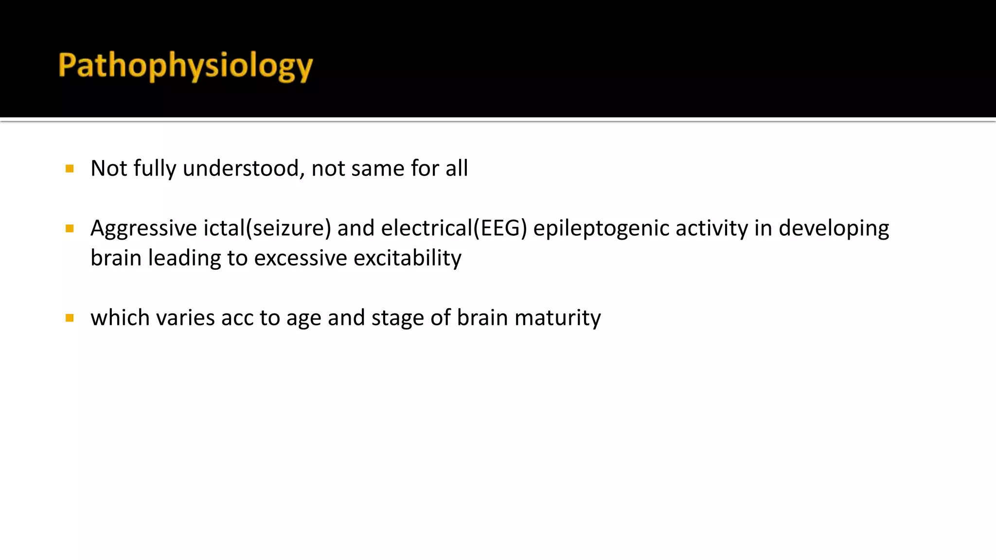
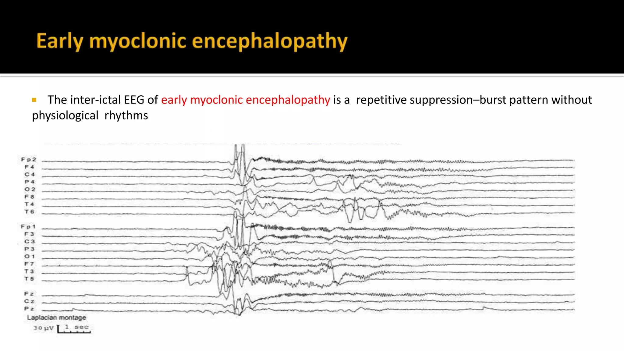
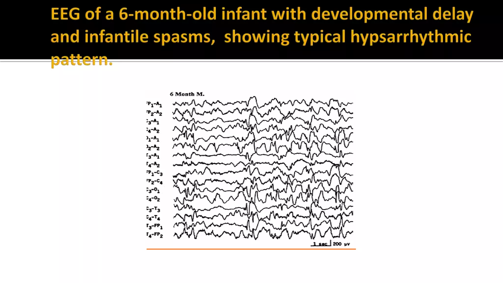
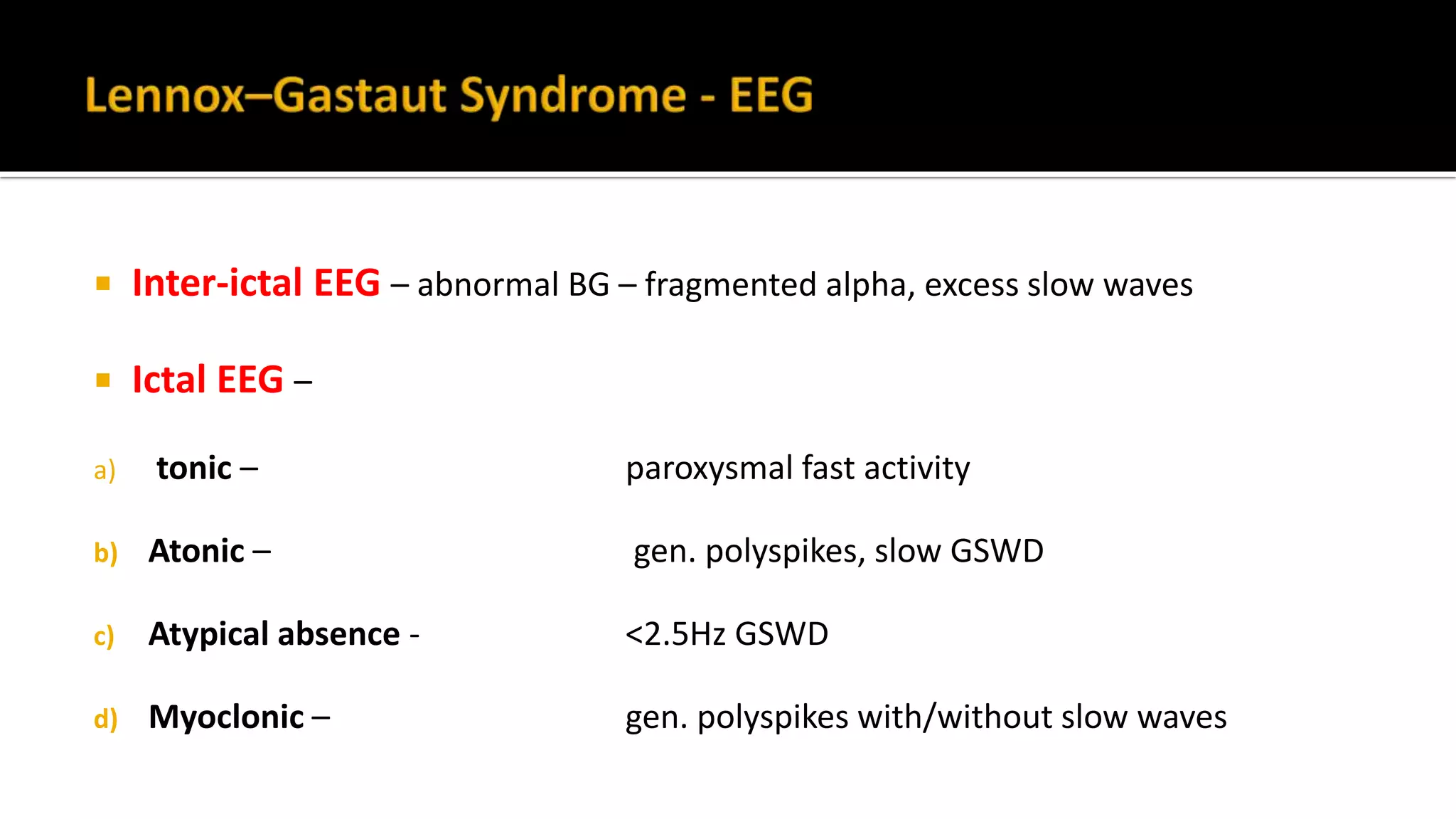
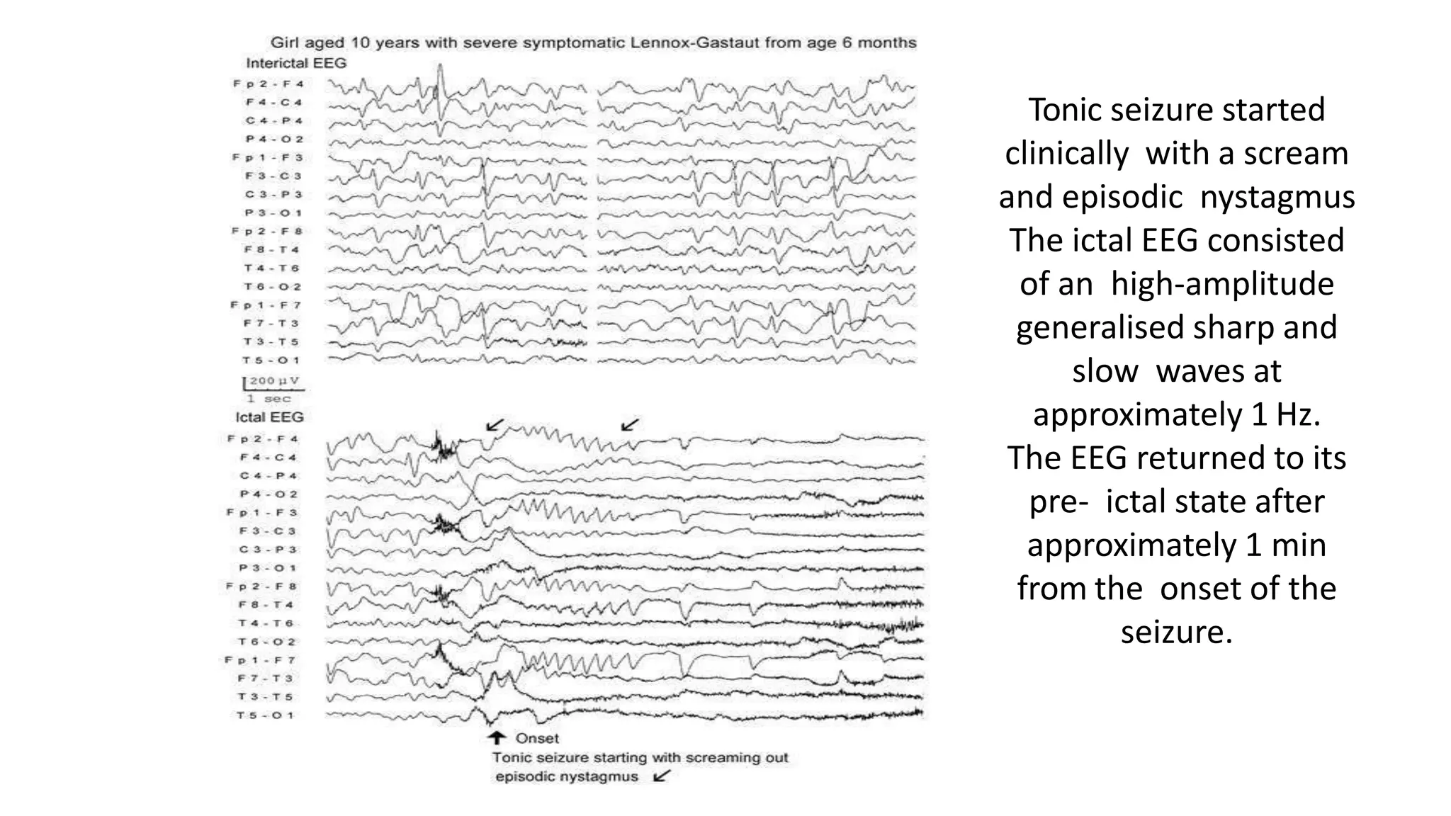
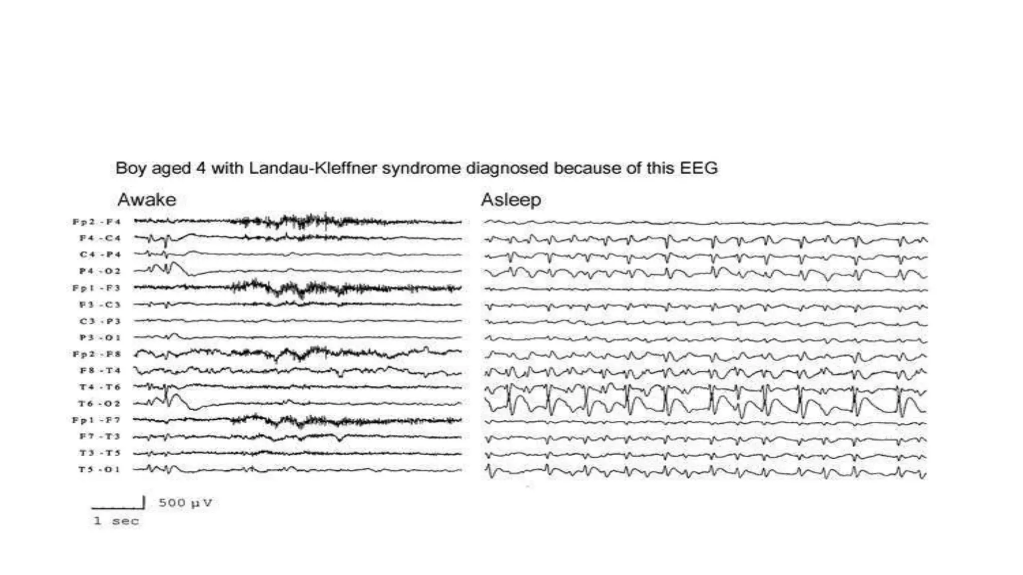
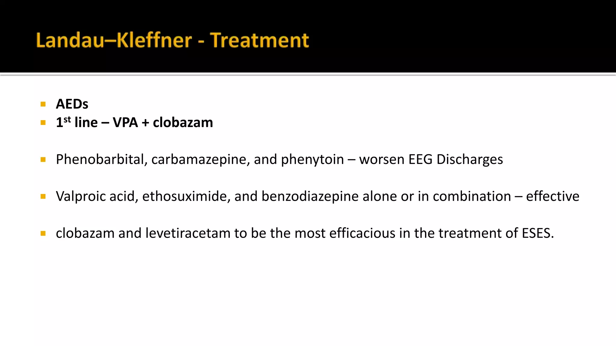
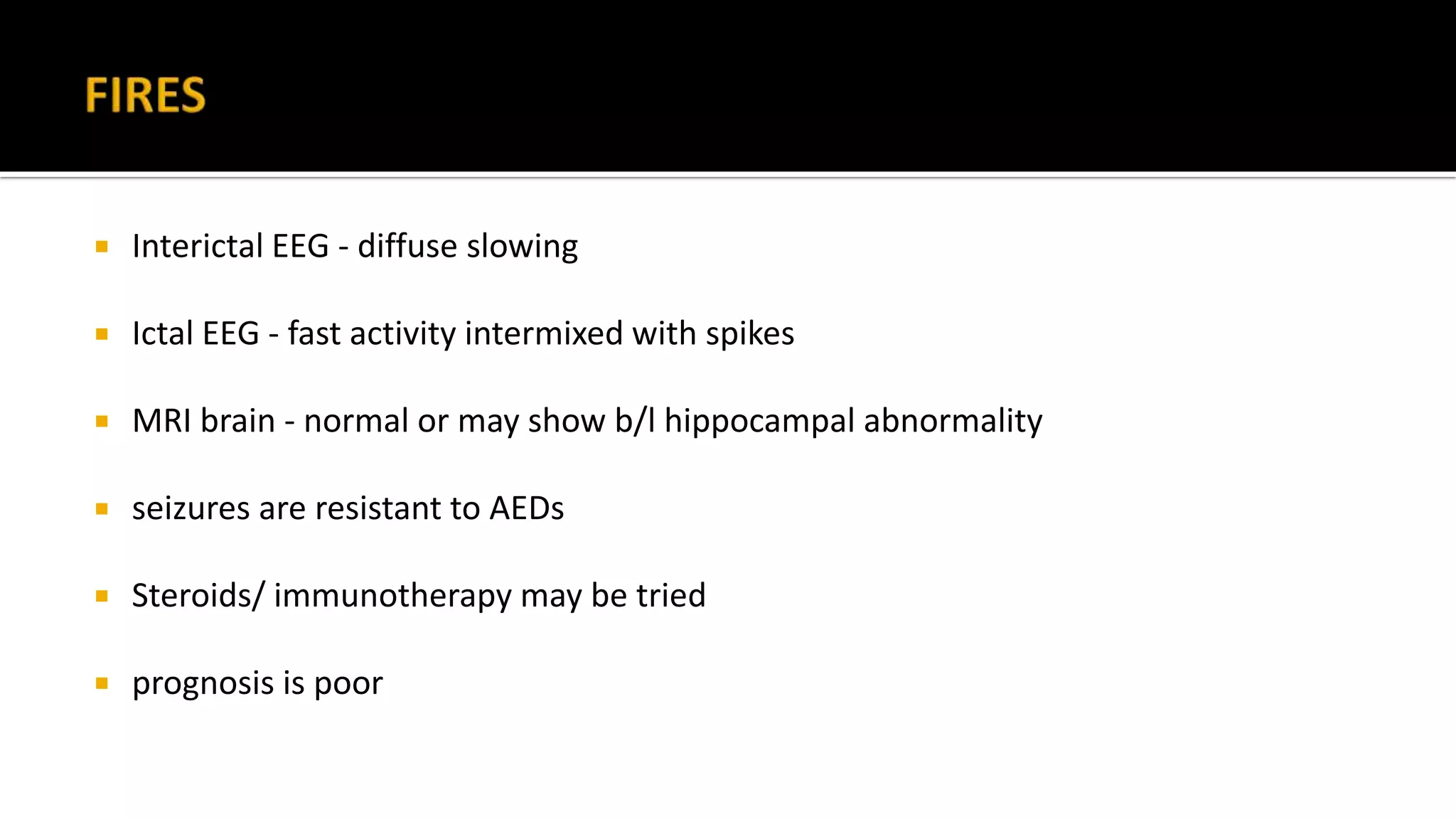
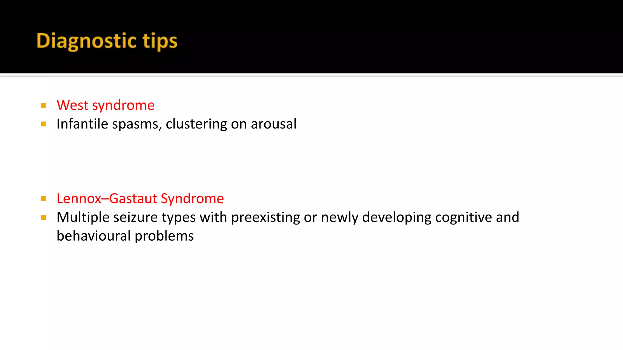
Dr. Rahi kiran.B
Senior resident
Dept. of Neurology
GMC Kota
 A group of heterogeneous brain disorders occurring at a critical period of brain
development, where frequent abnormal ictal and/or interictal EEG epileptiform
activity is mainly responsible for behavioral, cognitive and motor regression.
 (1) electrographic EEG paroxysmal activity that is often aggressive,
 (2) seizures that are usually multi-form and intractable,
 (3) cognitive, behavioral, and neurological deficits that may be relentless,
 (4) sometimes early death.
 Not fully understood, not same for all
 Aggressive ictal(seizure) and electrical(EEG) epileptogenic activity in developing
brain leading to excessive excitability
 which varies acc to age and stage of brain maturity
 In neonates – burst-suppression
 Infancy – hypsarrythmia
 Child – GSWD
 As age advances, these also change
Epileptic Encephalopathy Syndromes in
neonates
 Onset < age 3 m
 Male = female
 Etiology - Inborn errors of metabolism - mc-nonketotic hyperglycinemia,
methylmalonic acidemia.
 Triad of intractable seizures – Erratic myoclonus f/b simple focal seizures f/b
Tonic seizures
 Erratic myoclonus - shift typically from one part of the body to another in a random,
asynchronous fashion.
 simple focal seizures –
 Tonic seizures - a diffuse tonic contraction, usually extending to the extremities.
 Psychomotor devt – abnormal from onset
 Interictal EEG - suppression–burst activity,
evolves into atypical hypsarrhythmia after 3 to 5 months of life,
accentuated during sleep
 Myoclonus – no EEG expression, follows burst
 CT and MR - related to etiology,
grossly normal or have asymmetrical enlargement of one hemisphere,
dilatation of the corresponding lateral ventricle, or cortical atrophy
 the serum levels of amino acids should be determined, especially glycine and glycerol
 The inter-ictal EEG of early myoclonic encephalopathy is a repetitive suppression–burst pattern without
physiological rhythms
 The prognosis is poor - >50% die within weeks or months.
 no effective therapy – pyridoxine trial can be tried
 ILAE Definition
 Very early onset, within the first few months of life, frequent tonic spasms and a
suppression–burst EEG pattern in both the waking and sleeping states.
 Onset - 10 days to 3 months of age (occ. Intrauterine).
 Etiology – mc – malformations, no familial cases
(hemimegaloencephaly, porencephaly, Aicardi syn, focal cortical dysplasia)
 Tonic spasms - forward tonic flexion lasting 1–10 s that is singular or in long clusters
10–300 times every 24 h.
 Alternating hemiconvulsions or GTCS are exceptional.
 Myoclonic seizures are rare.
 Burst suppression with pseudorhythmic periodicity,
 disappears in 6 months - Most survivors evolve to West syndrome with
hypsarrhythmia, or later to LGS.
 prognosis is poor with severe psychomotor retardation
 Psychomotor development may be slightly better if the infants do not develop West
or Lennox-Gastaut syndrome.
 Half of the children are likely to die in infancy or childhood
 No effective treatment
 vigabatrin and zonisamide – some benefit
 ACTH – not useful
 invasive surgery, such as a partial resection or complete hemispherectomy.
Epileptic Encephalopathy Syndromes in
Infancy
 frequent partial seizures of multifocal onset, with autonomic or motor involvement
 The interictal EEG reveals multifocal epileptiform activity and slowing, diffuse
slowing of the background activity. asymmetry between different recordings
 The ictal EEG confirms multifocal onsets,
 No burst-suppression
 difficult to control with standard AEDs.
 Bromides, stiripentol, and clonazepam may be helpful
 Triad of infantile spasms, arrest of psychomotor development, and hypsarrhythmia.
 Onset -age of 3 and 12 months.
 Male(60%) > female
 Etiology – symptomatic(80%), idiopathic
 cerebral malformations, infection, hemorrhage, hypoxicischemic injury, metabolic
disorders, and genetic conditions,such as Down syndrome, TS
 Epileptic spasms - Clusters of sudden, brief (0.2–2 s), bilateral tonic contractions of
the axial and limb muscles involving extension and/or flexion
 Occurs in clusters – 1-30 / day, each with 20 – 150 attacks
 Precipitating factors- twilight state,sudden loud noises or tactile
stimulation,feeding.
 Spasms – flexor – extensor – mc
Flexor – salaam spasms
Extensor – like moro reflex
 Psychomotor delay – from the onset
 interictal -Gibbs and Gibbs –hypsarrhythmic EEG.(2/3 rds)
 Disorganized pattern with asynchronous, very high amplitude slowing and frequent
multifocal spike and sharp wave discharges
 Sleep – REM – normal
 NREM – more synchronous
 By 2-4yrs - LGS
 Ictal EEG – mc – high voltage, gen. slow wave
episodic,low amplitude slow wave
diffuse attenuation
(electrodecremental ictal EEG pattern).
Brief flexor
spasm associated
with high voltage
slow wave
discharge
 Good if treated early
 Mortality - 5%.
 60% - develop other seizure types – LGS
 50%-permanent motor disabilities
 2/3rd -severe cognitive and psychological impairment.
 5–12% - normal mental and motor development.
 First line ACTH and vigabatrin (TS)
 No role of pyridoxine
 valproate, levetiracetam, topiramate, zonisamide, lamotrigine, and benzodiazepines
 ketogenic diet is helpful in most cases
 Hemispherectomy - medically intractable
 No specific AED has been shown to affect long-term developmental outcome
 Onset – day 1 to 5yrs
 Female : male – 2: 1
 Etiology – 50 % - a/w angelman syn, 20% - birth asphyxia
 c/b – fixed, non-progressive encephalopathy
long-lasting recurrent episodic erratic myoclonic status epilepticus with
atypical absence seizures.
 interictal EEG-multifocal epileptiform discharges and background slowing
 Ictal EEG - continuous generalized slow spike and wave, or an absence pattern
 Myoclonic status – improves but with residual disability
 Treatment
 myoclonic status – BZDs
 Others - valproate with ethosuximide or clobazam
 Onset - always <1yr of life, with a peak age of 5 m.
 Male : female – 2:1
 Etiology – 50% genetic, SCN1A mutation – mostly sporadic
 Tetrad of seizures
Early onset infantile febrile clonic convulsions
Myoclonic jerks
Atypical absences
Complex focal seizures.
 In second year - more frequent and persistent, partial & myoclonic jerks start, not
a/w fever, ppted by – hot and light, gradual regression of milestones
 Precipitating Factors - Febrile illnesses , warm environment (hot baths), Photic and
pattern stimulation.
 Inter-Ictal EEG
 Normal till 18mnths age
 Initial -20% show normal BG withphotoparoxysmal discharges of spikes/
polyspikes-slowwaves.
 Within 1 year, EEG - abnormal slow background with frequent asymmetrical
paroxysms of polyspikes (GPSWD)
 Ictal EEG
 Varies according to seizuretype.
 Brain scans are usually normal.
 SCN1A” mutation - 70 per cent of children
 Treatment
 very resistant – initial - Phenobarbital, sodium valproate
 Other - stiripentol, topiramate ,clonazepam ,clobazam, LEV
 combination of sodium valproate with either topiramate or stiripentol may be the
most helpful.
 c/I – PHT,CBZ,LMT
 short course of a steroid (called prednisolone) and the ketogenic diet may also be
helpful.
Epileptic Encephalopathy Syndromes in
Childhood
 1–4% of childhood epilepsies.
Onset - Starts between 1 and 7 yrs with a peak at 3–5 years.
Male > female
Triad-
 Polymorphic intractable seizure that are mainly tonic (80%), atonic (50%)and
atypical absence seizures (70%), Myoclonic Jerks(11–28%) – sleep & awake
 Cognitive and behavioural abnormalities.
 EEG with paroxysms of fast activity and slow (less than 2.5 Hz) generalised spike-
wave discharges (GSWD).
 symptomatic - 70–78% of patients with LGS –
 encephalitis and/or meningitis,
 tuberous sclerosis,
 brain malformations
(e.g., cortical dysplasias),
 Idiopathic –
▪ normal psychomotor development occurred prior to the onset of symptoms,
▪ no underlying disorders or definite presumptive causes are present,
▪ no neurologic or neuroradiologic abnormalities are found.
birth injury
hypoxia-ischemia injury,
frontal lobe lesions
trauma.
 ILAE inclusion criteria –
 Atleast 2 seizure types – tonic, atonic, atypical absence(mandatory)
 EEG – GSWD with episodic fast activity
 Impaired intellectual functioning
 Not imp. – onset age, imaging, etiology
 Tonic seizures – 80 – 100% mc & most characteristic,
Symmetrical, brief, awake & NREM, not in REM
 Atonic – 50%
 Atypical absence – 2/3rd
 Myoclonic jerks – 11-28%
 Drop attacks -
 Inter-ictal EEG – abnormal BG – fragmented alpha, excess slow waves
 Ictal EEG –
a) tonic – paroxysmal fast activity
b) Atonic – gen. polyspikes, slow GSWD
c) Atypical absence - <2.5Hz GSWD
d) Myoclonic – gen. polyspikes with/without slow waves
Tonic seizure started
clinically with a scream
and episodic nystagmus
The ictal EEG consisted
of an high-amplitude
generalised sharp and
slow waves at
approximately 1 Hz.
The EEG returned to its
pre- ictal state after
approximately 1 min
from the onset of the
seizure.
 10–30% of cases develop from West syndrome or other epileptic encephalopathies.
 Bad prognosis
 5% - die
 90%- seizures in adult life also
 Almost all – cognitive impairment- more in symptomatic cases
 AEDs – First line – all types - VPA, clobazam,
zonisamide, rufinamide,
topiramate & felbamate (serious adverse effects)
 Lamotrigine – all but myoclonic, LEV – all but tonic
 PHT – tonic clonazepam – myoclonic
 Drop attacks – most difficult to treat- topiramate, felbamate,
Lamotrigine, rufinamide
 Atonic ,Atypical absence, Myoclonic – better
 ACTH & steroids – in SE
 ketogenic diet-refractory to AED
 Surgical-
 corpus callosotomy – resistant drop attacks
 vagus nerve stimulation – tonic & atonic
 focal cortical resection
LGS EM-AS of Doose
type Focal Generalised
Main types Tonic, atonic, atypical absence Myoclonic, atonic
tonic common never
atypical absence common uncommon
Devt. Before onset abnormal normal
West syndrome common uncommon
Background EEG abnormal normal
 “Partly reversible, epileptic encephalopathy of childhood manifesting with acquired
verbal auditory agnosia and fluctuating course of the linguistic disturbances that
occur together with other cognitive and neuropsychological behavioural
abnormalities.”
 Age at onset : 2–8 yrs with a peak at 5–7 yrs.
 male-to female ratio, 2 : 1
 Etiology - unknown
 Linguistic abnormalities-
 normal developmental milestones before aphasia symptoms
 All pts will have language abnormalities, 3/4th seizures
 First symptom – verbal auditory agnosia, later all types of aphasia
 Imp. Feature - fluctuating course
 Cognitive & Behavioural problems- 3/4th – ADHD
 Seizures – 3/4th , nocturnal,
 Types not well defined - (a/e tonic)
 Posterior temporal lobe foci of sharp-slow waves, multifocal and bi-synchronous
 CSWS also occur(not needed for diagnosis) – also persist in REM sleep
 Length of CSWS has strict correlation with language recovery
 Seizures,EEG abnormalities and language disturbance usually remit by 15yrs of age.
 50% of patients relatively normal life with 10–20% achieving complete
normalisation.
 50% left with permanent sequelae that may be very severe.
 AEDs
 1st line – VPA + clobazam
 Phenobarbital, carbamazepine, and phenytoin – worsen EEG Discharges
 Valproic acid, ethosuximide, and benzodiazepine alone or in combination – effective
 clobazam and levetiracetam to be the most efficacious in the treatment of ESES.
 corticosteroids & ACTH – if AED fail
 IVIG & ketogenic diet – no proven benefit
 surgical intervention with multiple subpial transections (MSTs) – mainly in cases
with secondary b/l synchrony
Continuous spike-andwave during sleep. All discharges vanished after receiving ACTH
 Partly reversible, age-related childhood epileptic encephalopathy c/b “subclinical”
spikes and waves occurring almost continuously during slow sleep and appearing
every night for a variable length of time in children
 triad of:
 EEG CSWS
 Seizures(except tonic seizures).
 Neuropsychological and motor impairment.
 Onset of seizures - 2 m to12 yrs, with a peak at 4–5 yrs.
 Male : female – 2/3rd
 seizure types- u/l or b/l clonic seizures, GTCS, absence, partial motor seizures.
 Tonic seizures, however, never occur.
 About 60% of patients also exhibit several types of Seizures
 The majority of affected children never return to normal levels, particularly in the
verbal area and attention
 MRI brain – abnormal in many
 Inter-ictal – fronto or centro-temporal slow spikes
 The typical EEG changes appear 1 year to 2 years after the first seizure and are
associated with behavioral deterioration.
 characteristic feature-continuous B/L spike-wave discharges on the EEG during
NREM sleep(cause Unknown)
 SWI – spike wave index - > 50 % NREM
> 85 % - poor prognosis
< 85% - better performance
 I stage is before the discoveryof CSWS
 The EEG shows multi-focal spikes and bisynchronous generalised
 sharp or spike-wavedischarges.
 First seizure is usually nocturnal
 II stage is when CSWS isfound
 Increase in seizures and the appearance or deterioration of
 neuropsychological symptoms (frontal& temporal)that prompt a sleep EEG.
 Tonic seizures never occur
 Continuous bilateral and diffuse slowspikes-waves of 1.5–2 Hz during NREM
sleep is the defining EEG patternof ECSWS.
 III stage is after clinical and EEG remission starts –
2-7yrs from onset, seizures remit in all pts, EEG normalises, left with residual deficits
 Seizures remit in all by 10 – 15yrs age – irrespective of etiology
Good prognosis
 Cognition & behaviour – incomplete recovery
 Early treatment - better
 may or may not respond - benzodiazepines, valproate, ethosuximide,
carbamazepine, phenytoin.
 Only benzodiazepines and adrenocorticotrophic hormone have been reported to
suppress the electrical Status
 TOC - benzodiazepines + valproate
 long-lasting improvement of the language function - multiple subpial transections
of the region of focal epileptic discharges
LKS CSWS
Seizures 3/4th All
Language Verbal auditory agnosia Expressive
Bahavioral Common All
CSWS onEEG 80% 100%
Spike localisation Temporal Frontal
Normal life 50% 25%
Other Severe Epileptic Encephalopathies
 Both sexes are equally affected
 Etiology- chronic viral infection, immune response to
infection,immune-mediated causes.
 typically shows –
 1. Focal motor seizures or epilepsia partialis continua (EPC)
 2. Generalized seizures contralateral to the ictal and interictal epileptiform activity
 3. Unilateral hemispheric cortical brain atrophy on MRI (abnormal signal T2/FLAIR
and atrophy or hyperintense signal of the ipsilateral caudate head)
 MRI - to exclude other causes of focal seizures
 EEG -
 response to treatment is very poor
 “A previously normal child, aged 2–15 years, within a few days of developing a FOU,
develops an acute prolonged perisylvian refractory convulsive status epilepticus,
persisting for more than 1 month”
 Later develops chronic pharmacoresistant epilepsy and cognitive dysfunction
 Cause unknown, may be immune-mediated.
 Interictal EEG - diffuse slowing
 Ictal EEG - fast activity intermixed with spikes
 MRI brain - normal or may show b/l hippocampal abnormality
 seizures are resistant to AEDs
 Steroids/ immunotherapy may be tried
 prognosis is poor
 Early Myoclonic Encephalopathy- Suspected if –
 Early Continuous shifting Segmental and erratic myoclonus f/b focal seizures,
rarely tonic seizures
 burst short, suppression long on EEG
 Ohtahara Syndrome
 Early Tonic seizures, focal seizures
 Burst suppression EEG with Pseudo-rhythmic appearance
 burst long, suppression short on EEG
 Dravet Syndrome
 Intractable myoclonic jerks and MR within 1-2 yrs from onset along with
febrile seizures –
 prolonged > 15min
 unilateral
 mainly clonic
 frequent
 ppted by low fever
 early onset (<1yr)
 West syndrome
 Infantile spasms, clustering on arousal
 Lennox–Gastaut Syndrome
 Multiple seizure types with preexisting or newly developing cognitive and
behavioural problems
Epileptic Encephalopathies
Neonates
(Burst suppression on EEG)
Infants
(Hypsarrythmia on EEG) Children
(GSWD on EEG)
EME Ohtahara syn West synDravet syn LGS LKS CSWS
LKS CSWS
Seizures 3/4th All
Language Verbal
auditory
agnosia
Expressive
Bahavioral Common All
CSWS
onEEG
80% 100%
Spike
localisation
Temporal Frontal
Normal life 50% 25%
Infantile spasms
Hypsarrythmia
30 %  LGS
DOC–ACTH +
vigabatrin
Multiple
seizures-tonic,
atonic, atypical absence
EEG-GSWD with
paroxysmal fast
activity
SCN1A mut –
70%
Atypical
febrile seizures
Stiripentol-
recently approved
MC cause-
Inborn errors of
metabolism
Imp – erratic
myoclonus, no
tonic spasams
MC-structural
abnormalities
Imp-tonic
seizures, rarely
myoclonus
 The spectrum from neonatal to early childhood is c/b variable clinical and EEG
features – reflecting developmental age rather than underlying etiology.
 Early treatment & prevention of seizure activity gives better cognitive outcomes and
quality of life
 As these are drug resistant, further studies are needed
 Khan S, Baradie RA. Epileptic Encephalopathies: An Overview Hindawi Publishing
Corporation,Epilepsy Research and Treatment. Volume 2012, 8 pages
doi:10.1155/2012/403592
 Bradley’s textbook of Neurology, 7th edition
 Covanis A. Epileptic encephalopathies (including severe epilepsy syndromes).
Epilepsia, 53(Suppl. 4):114–126, 2012 doi: 10.1111/j.1528-1167.2012.03621.x
 eMedicine@medscape:Epileptic and Epileptiform Encephalopathies

Epileptic encephalopathies

  • 1.
    Dr. Rahi kiran.B Seniorresident Dept. of Neurology GMC Kota
  • 2.
     A groupof heterogeneous brain disorders occurring at a critical period of brain development, where frequent abnormal ictal and/or interictal EEG epileptiform activity is mainly responsible for behavioral, cognitive and motor regression.  (1) electrographic EEG paroxysmal activity that is often aggressive,  (2) seizures that are usually multi-form and intractable,  (3) cognitive, behavioral, and neurological deficits that may be relentless,  (4) sometimes early death.
  • 3.
     Not fullyunderstood, not same for all  Aggressive ictal(seizure) and electrical(EEG) epileptogenic activity in developing brain leading to excessive excitability  which varies acc to age and stage of brain maturity
  • 4.
     In neonates– burst-suppression  Infancy – hypsarrythmia  Child – GSWD  As age advances, these also change
  • 6.
  • 7.
     Onset <age 3 m  Male = female  Etiology - Inborn errors of metabolism - mc-nonketotic hyperglycinemia, methylmalonic acidemia.
  • 8.
     Triad ofintractable seizures – Erratic myoclonus f/b simple focal seizures f/b Tonic seizures  Erratic myoclonus - shift typically from one part of the body to another in a random, asynchronous fashion.  simple focal seizures –  Tonic seizures - a diffuse tonic contraction, usually extending to the extremities.  Psychomotor devt – abnormal from onset
  • 9.
     Interictal EEG- suppression–burst activity, evolves into atypical hypsarrhythmia after 3 to 5 months of life, accentuated during sleep  Myoclonus – no EEG expression, follows burst  CT and MR - related to etiology, grossly normal or have asymmetrical enlargement of one hemisphere, dilatation of the corresponding lateral ventricle, or cortical atrophy  the serum levels of amino acids should be determined, especially glycine and glycerol
  • 10.
     The inter-ictalEEG of early myoclonic encephalopathy is a repetitive suppression–burst pattern without physiological rhythms
  • 11.
     The prognosisis poor - >50% die within weeks or months.  no effective therapy – pyridoxine trial can be tried
  • 12.
     ILAE Definition Very early onset, within the first few months of life, frequent tonic spasms and a suppression–burst EEG pattern in both the waking and sleeping states.  Onset - 10 days to 3 months of age (occ. Intrauterine).  Etiology – mc – malformations, no familial cases (hemimegaloencephaly, porencephaly, Aicardi syn, focal cortical dysplasia)
  • 13.
     Tonic spasms- forward tonic flexion lasting 1–10 s that is singular or in long clusters 10–300 times every 24 h.  Alternating hemiconvulsions or GTCS are exceptional.  Myoclonic seizures are rare.
  • 14.
     Burst suppressionwith pseudorhythmic periodicity,  disappears in 6 months - Most survivors evolve to West syndrome with hypsarrhythmia, or later to LGS.
  • 17.
     prognosis ispoor with severe psychomotor retardation  Psychomotor development may be slightly better if the infants do not develop West or Lennox-Gastaut syndrome.  Half of the children are likely to die in infancy or childhood
  • 18.
     No effectivetreatment  vigabatrin and zonisamide – some benefit  ACTH – not useful  invasive surgery, such as a partial resection or complete hemispherectomy.
  • 19.
  • 20.
     frequent partialseizures of multifocal onset, with autonomic or motor involvement  The interictal EEG reveals multifocal epileptiform activity and slowing, diffuse slowing of the background activity. asymmetry between different recordings  The ictal EEG confirms multifocal onsets,  No burst-suppression  difficult to control with standard AEDs.  Bromides, stiripentol, and clonazepam may be helpful
  • 21.
     Triad ofinfantile spasms, arrest of psychomotor development, and hypsarrhythmia.  Onset -age of 3 and 12 months.  Male(60%) > female  Etiology – symptomatic(80%), idiopathic  cerebral malformations, infection, hemorrhage, hypoxicischemic injury, metabolic disorders, and genetic conditions,such as Down syndrome, TS
  • 22.
     Epileptic spasms- Clusters of sudden, brief (0.2–2 s), bilateral tonic contractions of the axial and limb muscles involving extension and/or flexion  Occurs in clusters – 1-30 / day, each with 20 – 150 attacks  Precipitating factors- twilight state,sudden loud noises or tactile stimulation,feeding.  Spasms – flexor – extensor – mc Flexor – salaam spasms Extensor – like moro reflex  Psychomotor delay – from the onset
  • 23.
     interictal -Gibbsand Gibbs –hypsarrhythmic EEG.(2/3 rds)  Disorganized pattern with asynchronous, very high amplitude slowing and frequent multifocal spike and sharp wave discharges  Sleep – REM – normal  NREM – more synchronous  By 2-4yrs - LGS
  • 24.
     Ictal EEG– mc – high voltage, gen. slow wave episodic,low amplitude slow wave diffuse attenuation (electrodecremental ictal EEG pattern).
  • 27.
    Brief flexor spasm associated withhigh voltage slow wave discharge
  • 29.
     Good iftreated early  Mortality - 5%.  60% - develop other seizure types – LGS  50%-permanent motor disabilities  2/3rd -severe cognitive and psychological impairment.  5–12% - normal mental and motor development.
  • 30.
     First lineACTH and vigabatrin (TS)  No role of pyridoxine  valproate, levetiracetam, topiramate, zonisamide, lamotrigine, and benzodiazepines  ketogenic diet is helpful in most cases  Hemispherectomy - medically intractable  No specific AED has been shown to affect long-term developmental outcome
  • 31.
     Onset –day 1 to 5yrs  Female : male – 2: 1  Etiology – 50 % - a/w angelman syn, 20% - birth asphyxia  c/b – fixed, non-progressive encephalopathy long-lasting recurrent episodic erratic myoclonic status epilepticus with atypical absence seizures.
  • 32.
     interictal EEG-multifocalepileptiform discharges and background slowing  Ictal EEG - continuous generalized slow spike and wave, or an absence pattern  Myoclonic status – improves but with residual disability  Treatment  myoclonic status – BZDs  Others - valproate with ethosuximide or clobazam
  • 33.
     Onset -always <1yr of life, with a peak age of 5 m.  Male : female – 2:1  Etiology – 50% genetic, SCN1A mutation – mostly sporadic
  • 34.
     Tetrad ofseizures Early onset infantile febrile clonic convulsions Myoclonic jerks Atypical absences Complex focal seizures.  In second year - more frequent and persistent, partial & myoclonic jerks start, not a/w fever, ppted by – hot and light, gradual regression of milestones  Precipitating Factors - Febrile illnesses , warm environment (hot baths), Photic and pattern stimulation.
  • 35.
     Inter-Ictal EEG Normal till 18mnths age  Initial -20% show normal BG withphotoparoxysmal discharges of spikes/ polyspikes-slowwaves.  Within 1 year, EEG - abnormal slow background with frequent asymmetrical paroxysms of polyspikes (GPSWD)  Ictal EEG  Varies according to seizuretype.
  • 36.
     Brain scansare usually normal.  SCN1A” mutation - 70 per cent of children  Treatment  very resistant – initial - Phenobarbital, sodium valproate  Other - stiripentol, topiramate ,clonazepam ,clobazam, LEV  combination of sodium valproate with either topiramate or stiripentol may be the most helpful.  c/I – PHT,CBZ,LMT  short course of a steroid (called prednisolone) and the ketogenic diet may also be helpful.
  • 37.
  • 38.
     1–4% ofchildhood epilepsies. Onset - Starts between 1 and 7 yrs with a peak at 3–5 years. Male > female Triad-  Polymorphic intractable seizure that are mainly tonic (80%), atonic (50%)and atypical absence seizures (70%), Myoclonic Jerks(11–28%) – sleep & awake  Cognitive and behavioural abnormalities.  EEG with paroxysms of fast activity and slow (less than 2.5 Hz) generalised spike- wave discharges (GSWD).
  • 39.
     symptomatic -70–78% of patients with LGS –  encephalitis and/or meningitis,  tuberous sclerosis,  brain malformations (e.g., cortical dysplasias),  Idiopathic – ▪ normal psychomotor development occurred prior to the onset of symptoms, ▪ no underlying disorders or definite presumptive causes are present, ▪ no neurologic or neuroradiologic abnormalities are found. birth injury hypoxia-ischemia injury, frontal lobe lesions trauma.
  • 40.
     ILAE inclusioncriteria –  Atleast 2 seizure types – tonic, atonic, atypical absence(mandatory)  EEG – GSWD with episodic fast activity  Impaired intellectual functioning  Not imp. – onset age, imaging, etiology
  • 41.
     Tonic seizures– 80 – 100% mc & most characteristic, Symmetrical, brief, awake & NREM, not in REM  Atonic – 50%  Atypical absence – 2/3rd  Myoclonic jerks – 11-28%  Drop attacks -
  • 42.
     Inter-ictal EEG– abnormal BG – fragmented alpha, excess slow waves  Ictal EEG – a) tonic – paroxysmal fast activity b) Atonic – gen. polyspikes, slow GSWD c) Atypical absence - <2.5Hz GSWD d) Myoclonic – gen. polyspikes with/without slow waves
  • 43.
    Tonic seizure started clinicallywith a scream and episodic nystagmus The ictal EEG consisted of an high-amplitude generalised sharp and slow waves at approximately 1 Hz. The EEG returned to its pre- ictal state after approximately 1 min from the onset of the seizure.
  • 45.
     10–30% ofcases develop from West syndrome or other epileptic encephalopathies.  Bad prognosis  5% - die  90%- seizures in adult life also  Almost all – cognitive impairment- more in symptomatic cases
  • 46.
     AEDs –First line – all types - VPA, clobazam, zonisamide, rufinamide, topiramate & felbamate (serious adverse effects)  Lamotrigine – all but myoclonic, LEV – all but tonic  PHT – tonic clonazepam – myoclonic  Drop attacks – most difficult to treat- topiramate, felbamate, Lamotrigine, rufinamide  Atonic ,Atypical absence, Myoclonic – better
  • 47.
     ACTH &steroids – in SE  ketogenic diet-refractory to AED  Surgical-  corpus callosotomy – resistant drop attacks  vagus nerve stimulation – tonic & atonic  focal cortical resection
  • 48.
    LGS EM-AS ofDoose type Focal Generalised Main types Tonic, atonic, atypical absence Myoclonic, atonic tonic common never atypical absence common uncommon Devt. Before onset abnormal normal West syndrome common uncommon Background EEG abnormal normal
  • 49.
     “Partly reversible,epileptic encephalopathy of childhood manifesting with acquired verbal auditory agnosia and fluctuating course of the linguistic disturbances that occur together with other cognitive and neuropsychological behavioural abnormalities.”  Age at onset : 2–8 yrs with a peak at 5–7 yrs.  male-to female ratio, 2 : 1  Etiology - unknown
  • 50.
     Linguistic abnormalities- normal developmental milestones before aphasia symptoms  All pts will have language abnormalities, 3/4th seizures  First symptom – verbal auditory agnosia, later all types of aphasia  Imp. Feature - fluctuating course
  • 51.
     Cognitive &Behavioural problems- 3/4th – ADHD  Seizures – 3/4th , nocturnal,  Types not well defined - (a/e tonic)
  • 52.
     Posterior temporallobe foci of sharp-slow waves, multifocal and bi-synchronous  CSWS also occur(not needed for diagnosis) – also persist in REM sleep  Length of CSWS has strict correlation with language recovery
  • 54.
     Seizures,EEG abnormalitiesand language disturbance usually remit by 15yrs of age.  50% of patients relatively normal life with 10–20% achieving complete normalisation.  50% left with permanent sequelae that may be very severe.
  • 55.
     AEDs  1stline – VPA + clobazam  Phenobarbital, carbamazepine, and phenytoin – worsen EEG Discharges  Valproic acid, ethosuximide, and benzodiazepine alone or in combination – effective  clobazam and levetiracetam to be the most efficacious in the treatment of ESES.
  • 56.
     corticosteroids &ACTH – if AED fail  IVIG & ketogenic diet – no proven benefit  surgical intervention with multiple subpial transections (MSTs) – mainly in cases with secondary b/l synchrony
  • 57.
    Continuous spike-andwave duringsleep. All discharges vanished after receiving ACTH
  • 58.
     Partly reversible,age-related childhood epileptic encephalopathy c/b “subclinical” spikes and waves occurring almost continuously during slow sleep and appearing every night for a variable length of time in children  triad of:  EEG CSWS  Seizures(except tonic seizures).  Neuropsychological and motor impairment.  Onset of seizures - 2 m to12 yrs, with a peak at 4–5 yrs.  Male : female – 2/3rd
  • 59.
     seizure types-u/l or b/l clonic seizures, GTCS, absence, partial motor seizures.  Tonic seizures, however, never occur.  About 60% of patients also exhibit several types of Seizures  The majority of affected children never return to normal levels, particularly in the verbal area and attention  MRI brain – abnormal in many
  • 60.
     Inter-ictal –fronto or centro-temporal slow spikes  The typical EEG changes appear 1 year to 2 years after the first seizure and are associated with behavioral deterioration.  characteristic feature-continuous B/L spike-wave discharges on the EEG during NREM sleep(cause Unknown)  SWI – spike wave index - > 50 % NREM > 85 % - poor prognosis < 85% - better performance
  • 62.
     I stageis before the discoveryof CSWS  The EEG shows multi-focal spikes and bisynchronous generalised  sharp or spike-wavedischarges.  First seizure is usually nocturnal  II stage is when CSWS isfound  Increase in seizures and the appearance or deterioration of  neuropsychological symptoms (frontal& temporal)that prompt a sleep EEG.  Tonic seizures never occur  Continuous bilateral and diffuse slowspikes-waves of 1.5–2 Hz during NREM sleep is the defining EEG patternof ECSWS.  III stage is after clinical and EEG remission starts – 2-7yrs from onset, seizures remit in all pts, EEG normalises, left with residual deficits
  • 64.
     Seizures remitin all by 10 – 15yrs age – irrespective of etiology Good prognosis  Cognition & behaviour – incomplete recovery  Early treatment - better
  • 65.
     may ormay not respond - benzodiazepines, valproate, ethosuximide, carbamazepine, phenytoin.  Only benzodiazepines and adrenocorticotrophic hormone have been reported to suppress the electrical Status  TOC - benzodiazepines + valproate  long-lasting improvement of the language function - multiple subpial transections of the region of focal epileptic discharges
  • 66.
    LKS CSWS Seizures 3/4thAll Language Verbal auditory agnosia Expressive Bahavioral Common All CSWS onEEG 80% 100% Spike localisation Temporal Frontal Normal life 50% 25%
  • 67.
    Other Severe EpilepticEncephalopathies
  • 68.
     Both sexesare equally affected  Etiology- chronic viral infection, immune response to infection,immune-mediated causes.
  • 69.
     typically shows–  1. Focal motor seizures or epilepsia partialis continua (EPC)  2. Generalized seizures contralateral to the ictal and interictal epileptiform activity  3. Unilateral hemispheric cortical brain atrophy on MRI (abnormal signal T2/FLAIR and atrophy or hyperintense signal of the ipsilateral caudate head)
  • 70.
     MRI -to exclude other causes of focal seizures  EEG -  response to treatment is very poor
  • 71.
     “A previouslynormal child, aged 2–15 years, within a few days of developing a FOU, develops an acute prolonged perisylvian refractory convulsive status epilepticus, persisting for more than 1 month”  Later develops chronic pharmacoresistant epilepsy and cognitive dysfunction  Cause unknown, may be immune-mediated.
  • 72.
     Interictal EEG- diffuse slowing  Ictal EEG - fast activity intermixed with spikes  MRI brain - normal or may show b/l hippocampal abnormality  seizures are resistant to AEDs  Steroids/ immunotherapy may be tried  prognosis is poor
  • 73.
     Early MyoclonicEncephalopathy- Suspected if –  Early Continuous shifting Segmental and erratic myoclonus f/b focal seizures, rarely tonic seizures  burst short, suppression long on EEG  Ohtahara Syndrome  Early Tonic seizures, focal seizures  Burst suppression EEG with Pseudo-rhythmic appearance  burst long, suppression short on EEG
  • 74.
     Dravet Syndrome Intractable myoclonic jerks and MR within 1-2 yrs from onset along with febrile seizures –  prolonged > 15min  unilateral  mainly clonic  frequent  ppted by low fever  early onset (<1yr)
  • 75.
     West syndrome Infantile spasms, clustering on arousal  Lennox–Gastaut Syndrome  Multiple seizure types with preexisting or newly developing cognitive and behavioural problems
  • 76.
    Epileptic Encephalopathies Neonates (Burst suppressionon EEG) Infants (Hypsarrythmia on EEG) Children (GSWD on EEG) EME Ohtahara syn West synDravet syn LGS LKS CSWS LKS CSWS Seizures 3/4th All Language Verbal auditory agnosia Expressive Bahavioral Common All CSWS onEEG 80% 100% Spike localisation Temporal Frontal Normal life 50% 25% Infantile spasms Hypsarrythmia 30 %  LGS DOC–ACTH + vigabatrin Multiple seizures-tonic, atonic, atypical absence EEG-GSWD with paroxysmal fast activity SCN1A mut – 70% Atypical febrile seizures Stiripentol- recently approved MC cause- Inborn errors of metabolism Imp – erratic myoclonus, no tonic spasams MC-structural abnormalities Imp-tonic seizures, rarely myoclonus
  • 77.
     The spectrumfrom neonatal to early childhood is c/b variable clinical and EEG features – reflecting developmental age rather than underlying etiology.  Early treatment & prevention of seizure activity gives better cognitive outcomes and quality of life  As these are drug resistant, further studies are needed
  • 78.
     Khan S,Baradie RA. Epileptic Encephalopathies: An Overview Hindawi Publishing Corporation,Epilepsy Research and Treatment. Volume 2012, 8 pages doi:10.1155/2012/403592  Bradley’s textbook of Neurology, 7th edition  Covanis A. Epileptic encephalopathies (including severe epilepsy syndromes). Epilepsia, 53(Suppl. 4):114–126, 2012 doi: 10.1111/j.1528-1167.2012.03621.x  eMedicine@medscape:Epileptic and Epileptiform Encephalopathies