Downloaded 231 times


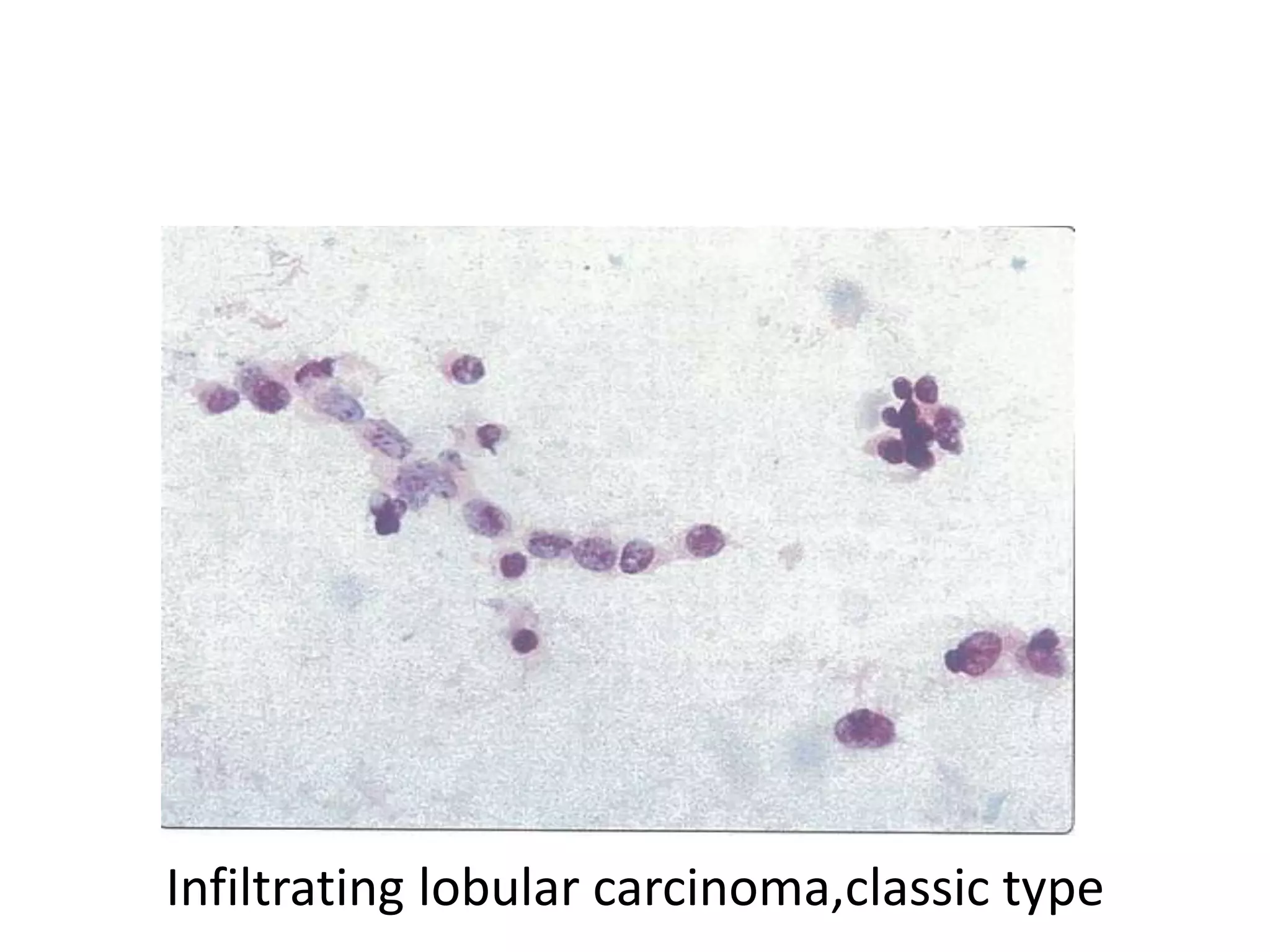

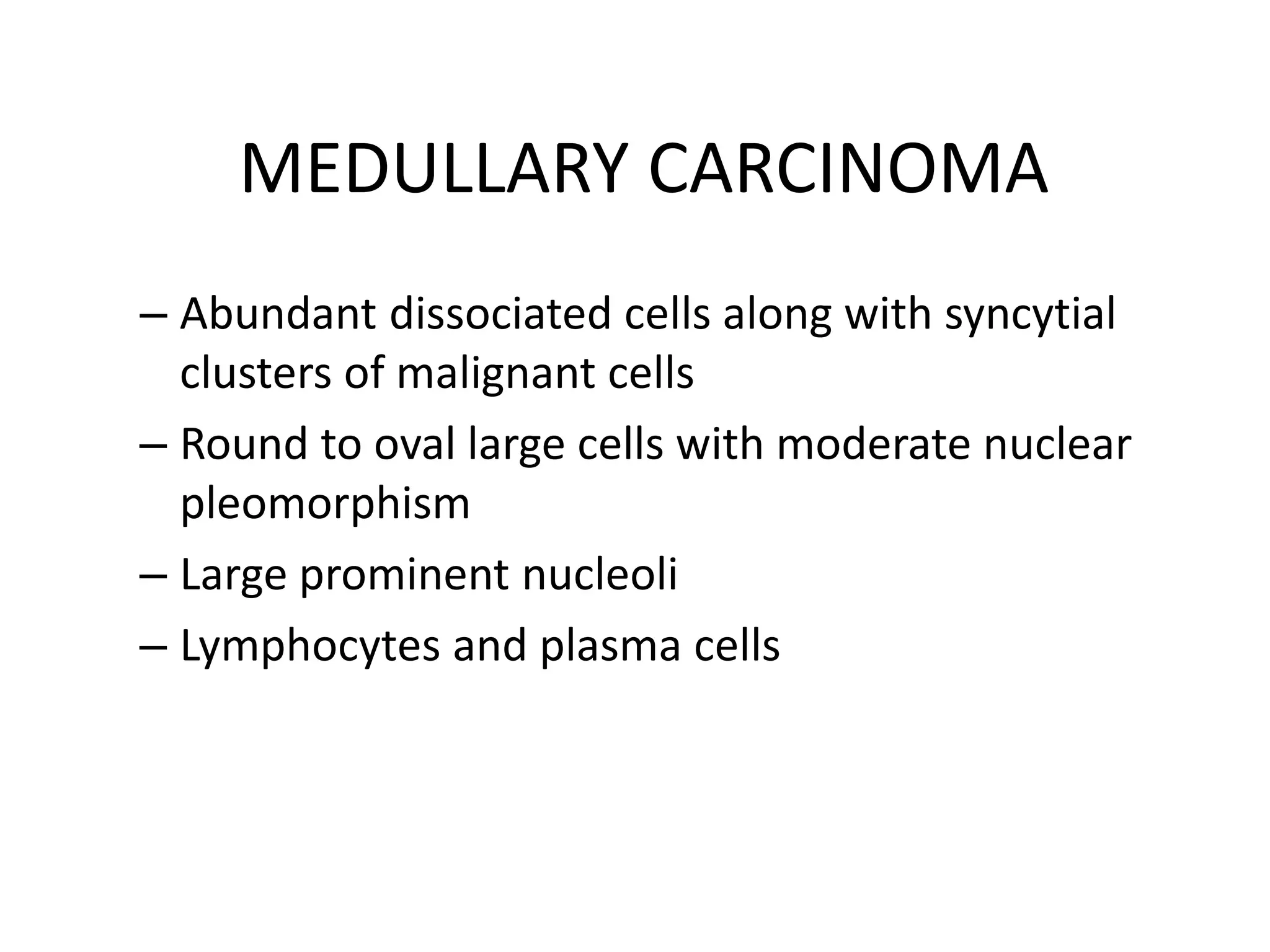


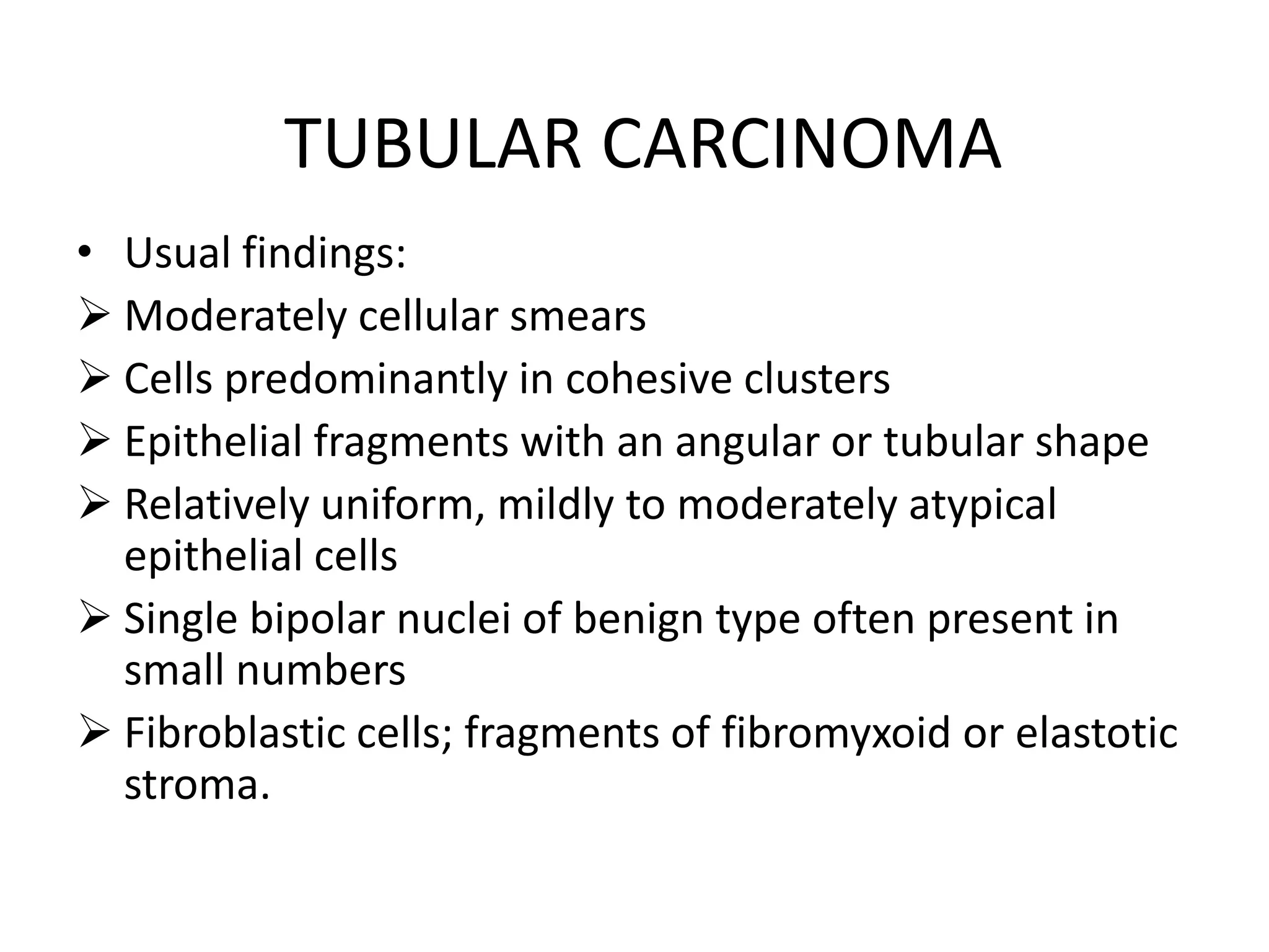

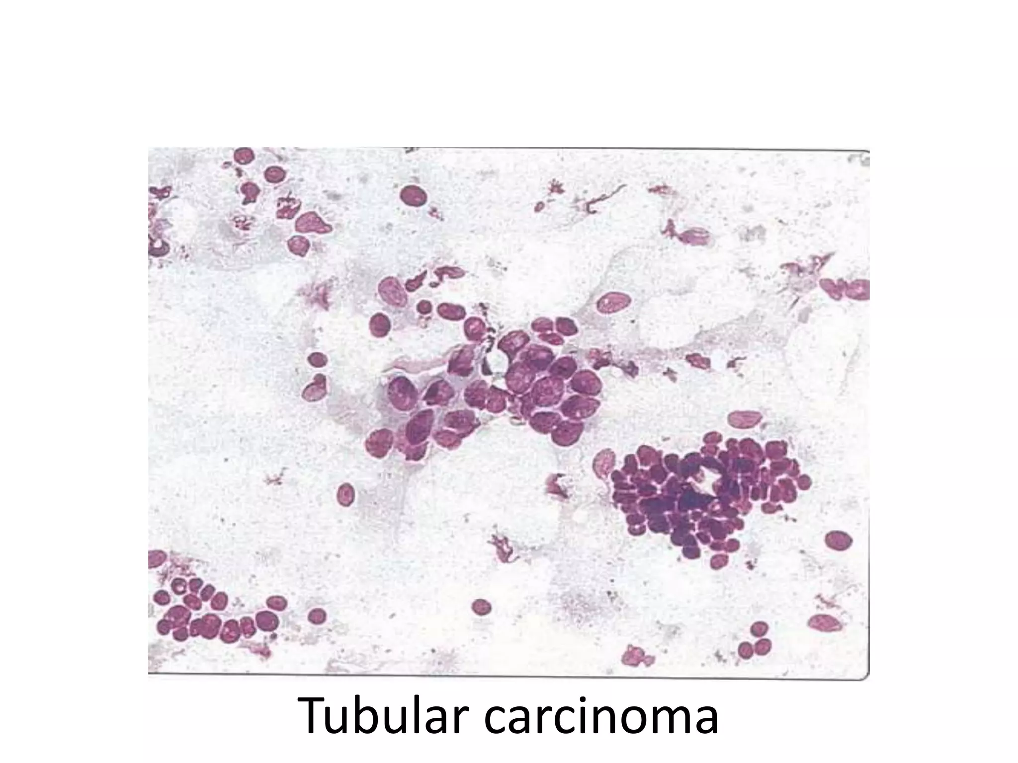


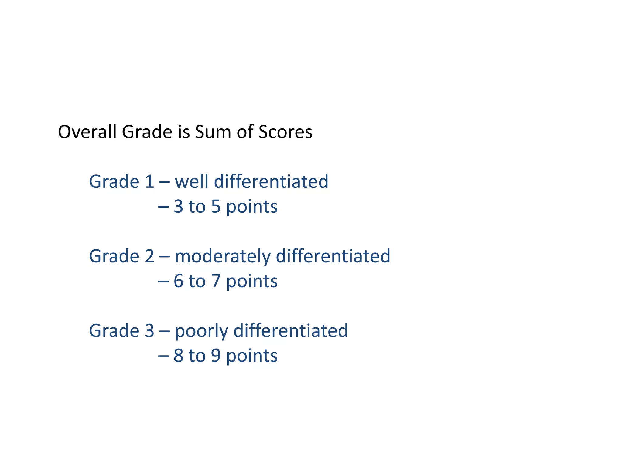


This document provides information on fine needle aspiration cytology (FNAC) findings for different types of breast cancers and lesions. It describes the typical cellular appearance and characteristics seen on FNAC for normal breast tissue, ductal carcinoma, lobular carcinoma, medullary carcinoma, mucinous carcinoma, tubular carcinoma, and metaplastic carcinoma. It also outlines the Scarff-Bloom-Richardson grading system used to assess breast cancer prognosis based on histopathological analysis of tumor cells and tissue structure.


![Apporach to lung biopsy [Auto-saved].pptx latest](https://cdn.slidesharecdn.com/ss_thumbnails/apporachtolungbiopsyauto-saved-251211225655-93258539-thumbnail.jpg?width=640&height=640&fit=bounds)


