Downloaded 146 times
![Background
• Priapism is defined as an abnormal
persistent erection of the penis. It is an
involuntary prolonged erection unrelated
to sexual stimulation and unrelieved by
ejaculation. As with many medical
emergencies, the saying "time is tissue"
holds true for priapism. This condition is a
true urologic emergency, and early
intervention allows the best chance for
functional recovery.[1]](https://image.slidesharecdn.com/priapism-160122115128/85/Priapism-2-320.jpg)


![– Erection: Duration of longer than 4 hours is
consistent with priapism.
– Duration of pain
– Similar prior episodes
– Genitourinary (GU) trauma
– Medical history (eg, sickle cell disease [SCD]):
Onset occurs during sleep, when relative
oxygenation decreases.](https://image.slidesharecdn.com/priapism-160122115128/85/Priapism-15-320.jpg)


![• Uncommonly, both low- and high-flow
priapism are idiopathic in nature.
• Secondary causes of low-flow priapism
are as follows:
– Thromboembolic/hypercoagulable states
• Sickle cell anemia (SCD) - Polycythemia: A recent
study found that, in unscreened children with SCD,
priapism was the first presentation in 0.5% of
cases.[3]](https://image.slidesharecdn.com/priapism-160122115128/85/Priapism-26-320.jpg)


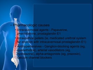
![• Psychotropics - Phenothiazine, butyrophenones,
hypnotics (eg, mesoridazine, perphenazine),
trazodone, selective serotonin reuptake inhibitors
(eg, fluoxetine, sertraline)[4]
• Anticoagulants - Heparin, warfarin
• Recreational drugs - Cocaine, marijuana, ethanol
• Hormones - Gonadotropin-releasing hormone
(GnRH), tamoxifen, testosterone](https://image.slidesharecdn.com/priapism-160122115128/85/Priapism-30-320.jpg)
![• Herbal medicine - Ginkgo biloba with concurrent
use of antipsychotic agents[5]
• Miscellaneous medications - Metoclopramide,
omeprazole, penile injection of cocaine, epidural
infusion of morphine and bupivacaine[6]
• Secondary causes of high-flow priapism
are as follows:
– GU trauma
• Straddle injury
• Intracavernous injections with direct cavernosal](https://image.slidesharecdn.com/priapism-160122115128/85/Priapism-31-320.jpg)


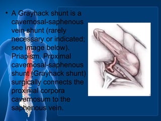
![• already esent.[11]
Priapism. Proximal
cavernosal-spongiosum shunt (Quackel
shunt) surgically connects the proximal
corpora cavernosa to the corpora
spongiosum](https://image.slidesharecdn.com/priapism-160122115128/85/Priapism-42-320.jpg)

Priapism is an abnormal, persistent erection unrelated to sexual stimulation. There are two types - low-flow (ischemic) and high-flow (non-ischemic). Low-flow priapism is more common and results from failure of venous outflow, trapping blood in the penis. If not treated promptly, it can lead to fibrosis and erectile dysfunction. Causes include sickle cell disease, medications, and trauma. Treatment involves differentiating the type and using medical or surgical methods to resolve the erection like irrigation, drugs, or shunt placement. Prognosis depends on duration, with early intervention providing the best chance of functional recovery.